Abstract
The purposes of the study were to determine whether there are differences in texture analysis parameters between tonsil cancers and normal tonsils, and to correlate texture analysis with 18F-FDG PET/CT to investigate the relationship between texture analysis and metabolic parameters. Sixty-four patients with squamous cell carcinoma of the palatine tonsil were included. A ROI was drawn, including all slices, to involve the entire tumor. The contralateral normal tonsil was used for comparison with the tumors. Texture analysis parameters, mean, standard deviation (SD), entropy, mean positive pixels, skewness, and kurtosis were obtained using commercially available software. Parameters were compared between the tumor and the normal palatine tonsils. Comparisons were also performed among early tonsil cancer, advanced tonsil cancer, and normal tonsils. An ROC curve analysis was performed to assess discrimination of tumor from normal tonsils. Correlation between texture analysis and 18F-FDG PET/CT was performed. Compared to normal tonsils, the tumors showed a significantly lower mean, higher SD, higher entropy, lower skewness, and higher kurtosis on most filters (p<0.001). On comparisons among normal tonsils, early cancers, and advanced tonsil cancers, SD and entropy showed significantly higher values on all filters (p<0.001) between early cancers and normal tonsils. The AUC from the ROC analysis was 0.91, obtained from the entropy. A mild correlation was shown between texture parameters and metabolic parameters. The texture analysis parameters, especially entropy, showed significant differences in contrast-enhanced CT results between tumor and normal tonsils, and between early tonsil cancers and normal tonsils. Texture analysis can be useful as an adjunctive tool for the diagnosis of tonsil cancers.
Introduction
Palatine tonsils are lymphoid tissues that comprise the anterior tonsillar pillar, tonsillar fossa, and posterior tonsillar pillar [1, 2]. Squamous cell carcinoma accounts for about 90% of tonsil cancer [3]. Patients with advanced tonsil cancer usually present with large oropharyngeal masses and cervical nodal metastasis. However, early tonsil cancer sometimes can be difficult to identify on contrast-enhanced CT because it can have the same appearance as normal lymphoid tissue [4]. Accordingly, the detection of tonsil cancer can be difficult on conventional CT. Therefore, preoperative additional imaging can be needed for the detection of tonsil cancer.
18F-FDG PET/CT has been widely used and reported to be valuable in the detection of tonsil cancer, showing higher sensitivity in detecting primary tumors than CT and MR imaging [5]. However, it has the limitation of radiation exposure and false-negative readings. Diffusion weighted image (DWI) has been used to differentiate normal tonsils from tumors [2, 4]. Bathia et al. [4] reported that tonsil cancer shows higher mean apparent diffusion coefficient (ADC) than normal tonsils. Histogram analysis of DWI also showed that the standard deviation of the overall curve is a useful parameter for the detection of occult palatine tonsil cancer [2]. However, the clinical value of histogram analysis of ADC maps remains challenging in daily clinical practice.
Recently, texture analysis has been introduced and applied in lung [6, 7], esophageal [8], colorectal [9], and head and neck cancers [10–12]. It quantifies the tumor heterogeneity using mathematical calculations of spatial patterns or arrangement of pixel intensities [13]. In head and neck cancers, it is usually used for treatment assessment [11, 12] and correlates with human papillomavirus (HPV) status [10, 14]. Until now, it has not been used for the discrimination of tonsil cancer from normal tonsils. Therefore, the purpose of the study was to determine whether there are differences in texture analysis parameters between tonsil cancers and normal tonsils. Additionally, this study aimed to correlate texture analysis with 18F-FDG PET/CT to investigate the relationship between texture analysis and metabolic parameters and to enhance the understanding of texture analysis results.
Materials and methods
The Hanyang University Hospital institutional review board and Hanyang University Guri Hospital institutional review board approved the study, and informed consent was waived in accordance with the requirements of a retrospective study. This study included 64 consecutive patients with histopathologically confirmed unilateral tonsillar cancer (38 men, 26 women; age range, 48–88 years; mean age, 59.13 years). Diagnosis was made with tonsillectomy in 44 cases and with biopsy in 20 cases. Patients underwent pretreatment contrast-enhanced neck CT (CECT) between March 2005 and July 2019 in a tertiary care hospital (Hanyang University Hospital) and a secondary care hospital (Hanyang University Guri Hospital). Tumor, node, and metastasis were staged according to the 8th edition of the AJCC staging system. All patients underwent both a CECT and a 18F-FDG PET/CT. And a gap between CECT and a 18F-FDG PET/CT was less than 2 weeks.
CT imaging and texture analysis
CT imaging of 41 patients in Hanyang University Hospital was performed using a 100-s delay, 120 kVp, 200 mAs, 2-mm slice thickness reconstruction (Brilliance 64, Philips Healthcare, Best, The Netherlands; SOMATOM Definition Flash, Siemens Healthcare, Erlangen, Germany). CT imaging of 23 patients at Hanyang University Guri Hospital was performed using the same protocols with 64- or 128-channel scanner systems (SOMATOM Definition DS and SOMATOM Definition Edge, Siemens Healthcare, Erlangen, Germany).
Two neuroradiologists with 7-year and 3-year experiences in the head and neck area performed texture analysis using commercially available software (TexRAD, Cambridge, UK). The freehand region of interest (ROI) was drawn along the outer border of the primary tumor and normal contralateral palatine tonsil on multiple consecutive axial slices to perform volumetric CT texture measurements. Slices with dental artifacts were excluded from the measurements. TexRAD software uses filtration-histogram methods [15], in which image filtering highlights image and object features of a particular size according to the type of filter used. In the spatial scaling factor (SSF, 0, 2, 3, 4, 5, 6), SSF 0 and 2 are defined as fine, SSF 3 and 4 as medium, and SSF 5 and 6 as coarse. Fine filters show enhancement of tissue parenchymal features, whereas medium and coarse filters show enhanced vascular features [16]. The texture analysis-derived parameters were obtained: mean, standard deviation (SD), entropy (texture irregularities and tumor heterogeneity), mean of positive pixels (MPP), kurtosis (peakedness of pixels), and skewness (asymmetry of pixel distribution).
18F-FDG PET/CT image acquisition
All images were obtained with a PET/CT system (GE Discovery, GE healthcare, Waukesha, USA and Siemens, Germany). Patients fasted for at least 6 h before 18F-FDG PET/CT. Blood glucose level was measured prior to FDG injection and was confirmed to be <180 mg/dL in all patients. Approximately 5.18 MBq/kg 18F-FDG was intravenously injected 50 min before imaging. First, low-dose CT (120 kVp, tube current modulation) was performed, and a PET scan was obtained from the skull base to the proximal thighs, with an acquisition time of 2.5 min per bed position in three-dimensional mode. PET images were reconstructed with ordered-subset expectation maximization with attenuation correction using vendor-provided software (VUE Point High Definition, GE Healthcare, Milwaukee, WI, USA).
18F-FDG PET/CT image analysis
All PET/CT images were transferred to in-house software (MIM software). The VOI was drawn in the tumor and normal tonsils. Automatically, the metabolic parameters were obtained: standardized uptake values (SUVmax and SUVmean) and total lesion glycolysis (TLG) on the tumor side and normal side. The SUVmaxT/N was additionally obtained after normalization by dividing by the normal side values.
Statistical analysis
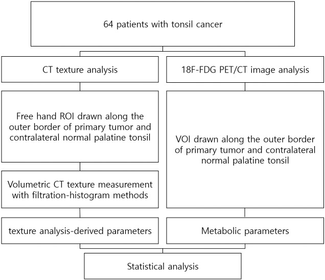
The Kolmogorov–Smirnov test was used to assess the normality of data. Differences in general categorical patient characteristics were analyzed using the χ2 test or Fisher’s exact test, as appropriate. The Mann–Whitney U test was performed to assess the difference between normal and palatine tonsils. The differences among the three groups were evaluated using one-way analysis of variance with post hoc analysis or the nonparametric Kruskal-Wallis test with the Mann–Whitney U test. The inter-reader agreement was assessed using the intraclass correlation coefficient (ICC). Receiver operating characteristic (ROC) curve analysis was performed for discrimination of tumors from normal tonsils. Spearman’s correlation was obtained to evaluate the linear correlation between texture analysis and metabolic parameters from 18F-FDG PET/CT. The block diagram of the work is shown in Fig 1.
Fig 1. The block diagram of the work.
All statistical calculations were performed using SPSS version 23 (IBM Corporation, Armonk, NY, USA) and MedCalc version 18 (MedCalc, Ostend, Belgium); p < 0.05 was considered to be statistically significant.
Results
General tumor characteristics
There were 14 cancers in the T1 stage, 34 in the T2 stage, 5 in the T3 stage, and 11 in the T4 stage. Forty-eight cancers were early, and 16 were advanced. There were 34 HPV (+) and 18 HPV (-) patients, and 12 with undetermined HPV status. Patient demographic information and staging of the tumors are summarized in Table 1.
Table 1. Baseline patient and tumor characteristics.
| Total (n = 64) | Early stage (n = 48) | Advanced stage (n = 16) | |
|---|---|---|---|
| Age | 62.32 ± 10.93 | 61.09 ± 11.45 | 65.94 ± 8.44 |
| Sex | |||
| male | 54 | 39 | 15 |
| female | 10 | 9 | 1 |
| HPV | |||
| positive | 34 | 25 | 9 |
| negative | 18 | 16 | 2 |
| untested | 12 | 7 | 5 |
| T stage | |||
| T1 | 14 | 14 | NA |
| T2 | 34 | 34 | NA |
| T3 | 5 | NA | 5 |
| T4 | 11 | NA | 11 |
| N stage | |||
| N0 | 13 | 11 | 2 |
| N1 | 16 | 15 | 1 |
| N2 | 29 | 21 | 8 |
| N3 | 6 | 1 | 5 |
HPV: Human papilloma virus, NA: not applicable.
CT texture analysis
The tumor side exhibited a significantly lower mean value with SSF 2–6 than the normal side (p<0.001). The tumor side showed significantly higher SD and higher entropy than the normal side at all filter values (p<0.001). The tumor side exhibited significantly lower skewness with SSF 0–4 than the normal side (p<0.001, <0.001, <0.001, and = 0.003, respectively). The tumor side showed significantly higher kurtosis than the normal side at all filter values (p = 0.001, <0.001, <0.001, <0.001, <0.001, and = 0.012, respectively).
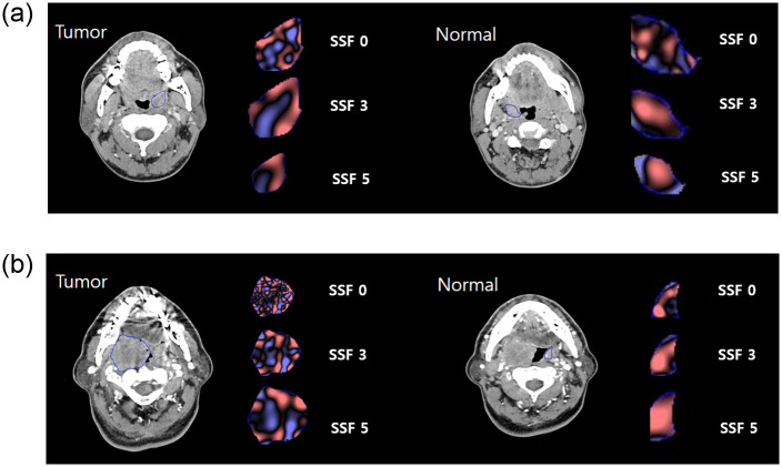
Fig 2 shows the texture analysis-derived features of tonsil cancer, in both early and advanced tumors, compared with contralateral normal tonsils. For the 3 group comparisons, SD, entropy, and kurtosis showed significant differences at all filters (all p<0.001 for SD and entropy; p = 0.005, <0.001, <0.001, <0.001, = 0.001, and = 0.043, respectively, for kurtosis). The mean with SSF 2–6 (p<0.001), and the skewness with SSF 0–4 and 6 (p<0.001, <0.001, <0.001, = 0.012, and = 0.044, respectively), exhibited significant differences. For the comparison between early stage tumors and normal tonsils, early stage tumors showed significantly higher SD, entropy, and kurtosis than normal tonsils at all filter values (all p<0.001 for SD and entropy; p = 0.002, <0.001, <0.001, <0.001, <0.001, and = 0.021 for kurtosis, respectively). Early stage tumors showed significantly lower mean than normal tonsils for SSF 2–6 (p<0.001, <0.001, <0.001, <0.001, and = 0.001, respectively). Early stage tumors showed significantly lower skewness than normal tonsils with SSF 0–4 (p<0.001, <0.001, <0.001, and = 0.021, respectively). In the comparison between the early and advanced tumor groups, only the mean for SSF 3–5 was significantly different (p = 0.032, = 0.022, and = 0.014, respectively). In comparison between the advanced tumor group and the normal tonsil, the mean with SSF 2–6 showed significant differences (p<0.001). SD and entropy exhibited significant differences at all filters (p <0.001, 0.002, <0.001, <0.001, <0.001, <0.001, respectively for SD, and all p<0.001 for entropy). Skewness of SSF 0–3 and 6 showed significant differences (p = 0.006, = 0.002, <0.001, and = 0.043, respectively). Kurtosis of SSF 2–5 showed significant differences (p<0.001, <0.001, <0.001, and = 0.010). All texture analysis parameter values and p values are summarized in Tables 2 and 3. Inter-reader measurement reliability for texture analysis parameters was almost perfect (ICC, 0.81–0.99).
Fig 2. Imaging appearance and texture analysis features of early stage (a) and advanced stage (b) tonsil cancer.
ROI for tonsil cancer and contralateral normal tonsils are demonstrated. Representative texture images with SSF 0, 3, 5 and coarse filters are shown. The red color in texture images indicates positive pixel values, and the blue color indicates negative pixel values, which are made after filtration according to filters of different sizes. The tumor side is more heterogeneous than the normal side and tends to have more negative pixel values. This feature is more noticeable with the SSF 0, fine filter.
Table 2. Comparisons of texture analysis-derived parameters between normal tonsil and tonsil cancer.
| Mean | SD | Entropy | |||||||
| SSF | Tumor (n = 64) | Normal (n = 64) | p value | Tumor (n = 64) | Normal (n = 64) | p value | Tumor (n = 64) | Normal (n = 64) | p value |
| 0 | 84.3 | 83.43 | 0.672 | 25.38 | 17.83 | <0.001* | 4.59 | 4.17 | <0.001* |
| 2 | 8.62 | 19.97 | <0.001* | 61.39 | 50.28 | <0.001* | 5.5 | 5.17 | <0.001* |
| 3 | 14.35 | 31.26 | <0.001* | 60.46 | 49.61 | <0.001* | 5.48 | 5.17 | <0.001* |
| 4 | 18.15 | 35.95 | <0.001* | 58.86 | 46.59 | <0.001* | 5.45 | 5.1 | <0.001* |
| 5 | 21.45 | 36.22 | <0.001* | 58.42 | 42.94 | <0.001* | 5.44 | 5.02 | <0.001* |
| 6 | 23.05 | 39.02 | <0.001* | 57.29 | 40.92 | <0.001* | 5.44 | 4.99 | <0.001* |
| MPP | Skewness | Kurtosis | |||||||
| SSF | Tumor (n = 64) | Normal (n = 64) | p value | Tumor (n = 64) | Normal (n = 64) | p value | Tumor (n = 64) | Normal (n = 64) | p value |
| 0 | 85.79 | 83.72 | 0.306 | -0.88 | -0.31 | <0.001* | 2.62 | 1.08 | 0.001* |
| 2 | 48.57 | 47.53 | *0.365 | -0.29 | 0.2 | <0.001* | 1.27 | 0.38 | <0.001* |
| 3 | 51.06 | 53.22 | *0.750 | -0.33 | 0.3 | <0.001* | 0.98 | -0.2 | <0.001* |
| 4 | 53 | 55.29 | *0.804 | -0.31 | -0.14 | 0.003* | 0.79 | 0.1 | <0.001* |
| 5 | 53.32 | 56.07 | *0.990 | -0.31 | -0.29 | 0.801 | 0.4 | -0.11 | <0.001* |
| 6 | 53.46 | 57.92 | *0.712 | -0.31 | -0.41 | 0.141 | 0.16 | -0.14 | 0.012* |
* Statistically significant.
SD standard deviation, MPP mean positive pixel, SSF spatial scaling factor.
Table 3. Comparisons of texture analysis-derived parameters among normal tonsil, early tonsil cancer, and advanced tonsil cancer.
| SSF | Mean | SD | Entropy | MPP | Skewness | Kurtosis | |
|---|---|---|---|---|---|---|---|
| p value | 0 | 0.635 | <0.001* | <0.001* | 0.444 | <0.001* | 0.005* |
| 2 | <0.001* | <0.001* | <0.001* | 0.569 | <0.001* | <0.001* | |
| 3 | <0.001* | <0.001* | <0.001* | 0.763 | <0.001* | <0.001* | |
| 4 | <0.001* | <0.001* | <0.001* | 0.771 | 0.012* | <0.001* | |
| 5 | <0.001* | <0.001* | <0.001* | 0.809 | 0.861 | 0.001* | |
| 6 | <0.001* | <0.001* | <0.001* | 0.833 | 0.044* | 0.043* | |
| Early vs normal | 0 | 0.997 | <0.001* | <0.001* | 0.786 | <0.001* | 0.002* |
| 2 | <0.001* | <0.001* | <0.001* | 0.312 | <0.001* | <0.001* | |
| 3 | <0.001* | <0.001* | <0.001* | 0.930 | <0.001* | <0.001* | |
| 4 | <0.001* | <0.001* | <0.001* | 0.953 | 0.021* | <0.001* | |
| 5 | <0.001* | <0.001* | <0.001* | 0.899 | 0.909 | <0.001* | |
| 6 | 0.001* | <0.001* | <0.001* | 0.661 | 0.749 | 0.021* | |
| Early vs advanced | 0 | 0.669 | 0.556 | 0.927 | 0.727 | 0.871 | 0.745 |
| 2 | 0.072 | 0.988 | 1.000 | 0.515 | 0.420 | 0.471 | |
| 3 | 0.032* | 0.871 | 0.935 | 0.438 | 0.281 | 0.871 | |
| 4 | 0.022* | 0.614 | 0.442 | 0.369 | 0.973 | 0.858 | |
| 5 | 0.014* | 0.525 | 0.372 | 0.321 | 0.974 | 0.914 | |
| 6 | 0.20 | 0.520 | 0.567 | 0.485 | 0.214 | 0.975 | |
| Advanced vs normal | 0 | 0.622 | <0.001* | <0.001* | 0.432 | 0.006* | 0.43 |
| 2 | <0.001* | 0.002* | <0.001* | 0.829 | 0.002* | <0.001* | |
| 3 | <0.001* | <0.001* | <0.001* | 0.532 | <0.001* | <0.001* | |
| 4 | <0.001* | <0.001* | <0.001* | 0.613 | 0.099 | <0.001* | |
| 5 | <0.001* | <0.001* | <0.001* | 0.819 | 0.878 | 0.010* | |
| 6 | <0.001* | <0.001* | <0.001* | 0.971 | 0.043* | 0.111 |
* Statistically significant.
SD standard deviation, MPP mean positive pixel, SSF spatial scaling factor.
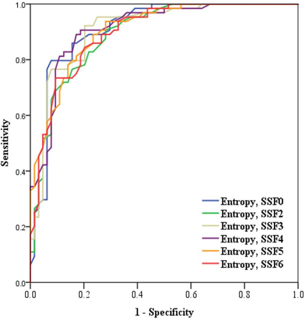
The AUC for entropy with SSF 0 and 4 was the highest with 0.91, differentiating tonsil cancer from normal tonsils (Fig 3). For the entropy with SSF 0, the sensitivity was 79.7% and the specificity was 92.2% at the cutoff value of 4.45. For the entropy with SSF 4, the sensitivity was 90.6% and the specificity was 88.2% at the cutoff value of 5.3. For differentiation of early tonsil cancer from normal tonsils, entropy was the most accurate with an AUC of 0.90, with SSF 0, 3, and 4. For differentiation of advanced tonsil cancer from normal tonsils, entropy was the most accurate with an AUC of 0.94 with SSF 0, 4, and 5.
Fig 3. The ROC curve analysis was performed to differentiate tonsil cancer from normal tonsils.
The AUC for entropy with SSF 0 and 4 was the highest at 0.91.
The correlations between texture analysis and 18F-FDG PET/CT are shown in S1 Table. There was mild significant correlation between mean and SUVmax (r = 0.28, p = 0.04) and between MPP and SUVmax (r = 0.27, p = 0.048) at SSF 0. There were also significant correlations between mean and SUVmean (r = 0.3, p = 0.03) and between MPP and SUVmean (r = 0.29, p = 0.03) at SSF 0. Mean showed a negative correlation with TLG at SSF 2, 3, 4, and 5 (r = -0.34, -0.35, -0.34, and -0.31, respectively; p = 0.01). After normalization with the contralateral normal tonsil, entropy showed a linear correlation with SUVmaxT/N at SSF 2 (r = 0.27, p = 0.04). In addition, entropy showed a mild correlation with SUVmeanT/N at SSF 0, 2, 3, and 4 (r = 0.30, 0.36, 0.33, and 0.30; p = 0.03, 0.01, 0.02, 0.03, respectively).
Discussion
In this study, we found that texture analysis can be useful for differentiating tumors from normal tonsils. Among the texture analysis-derived parameters, entropy was the most helpful in distinguishing early tonsil cancer from normal tonsils. In cases of early tonsil cancer, which could be difficult to detect with visual analysis on contrast-enhanced CT, the entropy derived from the texture analysis could be useful for the diagnosis of tonsil cancer. There was a mild correlation between texture analysis parameters and FDG uptake on 18F-FDG PET/CT, which suggests that the texture analysis parameters relate to the physiologic state of the tumor.
Tumor heterogeneity is a well-recognized histopathologic feature of malignancy, reflecting high cell density, necrosis, hemorrhage, and myxoid change [6, 17]. High tumoral heterogeneity is associated with adverse biology, aggressive clinical course, and increased resistance to treatment [18]. A recent study on oropharyngeal cancer showed a correlation between texture and HPV status, showing a lower value of entropy and SD in the HPV-positive group [10]. For the correlation between texture and treatment response, texture analysis parameters are known to be associated with local failure in patients with head and neck cancer with chemoradiotherapy. CT texture features correlated with TNM stages in gastric cancer and esophageal cancer, indicating that tumors with higher T or N stages have higher levels of heterogeneity-related features [19, 20]. Furthermore, several texture features can discriminate between high- and low-grade lung cancers [21, 22]. Meyer et al. reported that CT entropy was correlated with hypoxic related pathological parameter, hypoxia-inducible factor-1-alpha expression [23]. This study was concordant with previous studies that showed high entropy in malignant tumors [10].
Texture analysis is a computer-based, image processing technique that allows for the mathematical detection of changes in pixel density, which may be visually imperceptible [6]. It can assess the tumoral heterogeneity by analyzing the distribution and relationship of the pixel gray level in the images. It can maximize the information obtained from routinely acquired diagnostic images in current clinical practice without additional acquisition of images or an invasive procedure. However, the filtration histogram-technique is required owing to the CT photon-noise, which can influence the radiologist’s impression of image quality and mask the biologic heterogeneity. By using filters, CT texture analysis reduces the effect of photon noise and enhances the biological heterogeneity [7]. Fine filters usually enhance tissue parenchymal features, and medium to coarse filters enhance vascular features [24].
Tonsil cancer usually demonstrates asymmetric enlargement on contrast-enhanced CT. In cases of early tonsil cancer, the cancer density usually appears similar to that of normal tonsils. Therefore, the visual analysis sometimes fails to depict the mass in the tonsil because there is no difference in enhancement between tonsil cancer and normal palatine tonsils. In these cases, the patients are classified as having a malignancy of unknown origin presenting with cervical nodal metastasis. These patients undergo biopsies of the tonsil, and tongue base to determine the origin of the primary tumor [25]. Accordingly, the hypothesis was that computer-based assessment of texture analysis could be useful in detecting tonsil cancer, especially in the early stage. The study results showed that entropy was the most significant parameter for detecting tonsil cancer, compared with normal palatine tonsils. Entropy refers to the randomness of pixel intensity. Therefore, in cases of early tonsil cancer, which could be difficult to detect with visual analysis on contrast-enhanced CT, the entropy derived from the texture analysis could be useful for the diagnosis of tonsil cancer.
It can also be difficult to differentiate tonsil cancer from normal lymphoid tissue using magnetic resonance imaging. DWI is an MR technique that has the potential to improve the detection of head and neck cancer [26]. It is well known that malignant tumors have lower ADC values than benign lesions. Multiple studies have provided ADC threshold values, but they have showed a broad spectrum [27–29]. A recent meta-analysis reported that DWI/ADC alone cannot be used as an imaging biomarker of malignancy in head and neck cancer [30].
The correlation analysis to evaluate the relationship between texture analysis parameter and metabolic parameter on 18F-FDG PET/CT was performed. Generally, FDG uptake by tumors on 18F-FDG PET/CT shows a correlation with malignancy and reflects the physiologic condition of the tumor [31]. Texture analysis parameters are already known to be related to tumor biology. Texture analysis showed a mild linear correlation with the tumor uptake on 18F-FDG PEC/CT in this study. This result was concordant with the study by Ganeshan et al [32], which showed a strong correlation between SUVmax or SUVmean and entropy in esophageal cancer.
This study has several limitations. First, the texture analysis was performed in only patients with tonsil cancers. To verify the texture analysis based on tumor detection, further studies with patients with head and neck cancer could be needed with prospective and multicenter studies. Second, a correlation between texture parameters and histopathologic markers, such as angiogenesis or hypoxia, was not performed. To better understand the tumor biology, a future correlation study with histopathologic markers is needed. Finally, despite the scan protocols were same, there might be a potential influence of different CT scanner on the texture analysis.
Conclusion
In conclusion, entropy was the most useful parameter to distinguish tonsil cancer from normal tonsils. Texture analysis showed a mild linear correlation with tumor uptake on 18F-FDG PET/CT. Therefore, as a measurement method of tumor heterogeneity, texture analysis relates to the physiologic condition of the tumor. In cases of early tonsil cancer, which could be difficult to detect with visual analysis on contrast-enhanced CT, the entropy derived from the texture analysis could be useful for the diagnosis of tonsil cancer.
Supporting information
(PDF)
Abbreviations
- ADC
Apparent diffusion coefficient
- CT
Computed tomography
- FDG
Fluorodeoxyglucose
- MRI
Magnetic resonance imaging
- PET/CT
Positron-emission tomography-computed tomography
- ROC
Receiver operating characteristic
- ROI
Region of interest
- SD
standard deviation
Data Availability
Data cannot be shared publicly because of data contain potentially identifying or sensitive patient information. Data are available from the Hanyang University Hospital and Hanyang University Guri Hospital Institutional Data Access / Ethics Committee (contact via irb@hyumc.com and hyirbguri@gmail.com) for researchers who meet the criteria for access to confidential data.
Funding Statement
The author(s) received no specific funding for this work.
References
- 1.Trotta BM, Pease CS, Rasamny JJ, Raghavan P, Mukherjee S. Oral cavity and oropharyngeal squamous cell cancer: key imaging findings for staging and treatment planning. Radiographics. 2011;31(2):339–54. Epub 2011/03/19. doi: 10.1148/rg.312105107 . [DOI] [PubMed] [Google Scholar]
- 2.Choi YJ, Lee JH, Kim HO, Kim DY, Yoon RG, Cho SH, et al. Histogram analysis of apparent diffusion coefficients for occult tonsil cancer in patients with cervical nodal metastasis from an unknown primary site at presentation. Radiology. 2016;278(1):146–55. doi: 10.1148/radiol.2015141727 [DOI] [PubMed] [Google Scholar]
- 3.Stambuk HE, Karimi S, Lee N, Patel SG. Oral cavity and oropharynx tumors. Radiol Clin North Am. 2007;45(1):1–20. Epub 2006/12/13. doi: 10.1016/j.rcl.2006.10.010 . [DOI] [PubMed] [Google Scholar]
- 4.Bhatia KS, King A, Yeung D, Mo F, Vlantis AC, Yu K, et al. Can diffusion-weighted imaging distinguish between normal and squamous cell carcinoma of the palatine tonsil? The British journal of radiology. 2010;83(993):753–8. doi: 10.1259/bjr/58331222 [DOI] [PMC free article] [PubMed] [Google Scholar]
- 5.Lee JR, Kim JS, Roh J-L, Lee JH, Baek JH, Cho K-J, et al. Detection of occult primary tumors in patients with cervical metastases of unknown primary tumors: comparison of 18F FDG PET/CT with contrast-enhanced CT or CT/MR imaging—prospective study. Radiology. 2015;274(3):764–71. doi: 10.1148/radiol.14141073 [DOI] [PubMed] [Google Scholar]
- 6.Ganeshan B, Panayiotou E, Burnand K, Dizdarevic S, Miles K. Tumour heterogeneity in non-small cell lung carcinoma assessed by CT texture analysis: a potential marker of survival. European radiology. 2012;22(4):796–802. doi: 10.1007/s00330-011-2319-8 [DOI] [PubMed] [Google Scholar]
- 7.Ganeshan B, Goh V, Mandeville HC, Ng QS, Hoskin PJ, Miles KA. Non-small cell lung cancer: histopathologic correlates for texture parameters at CT. Radiology. 2013;266(1):326–36. Epub 2012/11/22. doi: 10.1148/radiol.12112428 . [DOI] [PubMed] [Google Scholar]
- 8.Yip C, Landau D, Kozarski R, Ganeshan B, Thomas R, Michaelidou A, et al. Primary esophageal cancer: heterogeneity as potential prognostic biomarker in patients treated with definitive chemotherapy and radiation therapy. Radiology. 2014;270(1):141–8. Epub 2013/08/30. doi: 10.1148/radiol.13122869 . [DOI] [PubMed] [Google Scholar]
- 9.Ng F, Ganeshan B, Kozarski R, Miles KA, Goh V. Assessment of primary colorectal cancer heterogeneity by using whole-tumor texture analysis: contrast-enhanced CT texture as a biomarker of 5-year survival. Radiology. 2013;266(1):177–84. doi: 10.1148/radiol.12120254 [DOI] [PubMed] [Google Scholar]
- 10.Lee JY, Han M, Kim KS, Shin S-J, Choi JW, Ha EJ. Discrimination of HPV status using CT texture analysis: tumour heterogeneity in oropharyngeal squamous cell carcinomas. Neuroradiology. 2019;61(12):1415–24. doi: 10.1007/s00234-019-02295-w [DOI] [PubMed] [Google Scholar]
- 11.Zhang H, Graham CM, Elci O, Griswold ME, Zhang X, Khan MA, et al. Locally advanced squamous cell carcinoma of the head and neck: CT texture and histogram analysis allow independent prediction of overall survival in patients treated with induction chemotherapy. Radiology. 2013;269(3):801–9. Epub 2013/08/06. doi: 10.1148/radiol.13130110 . [DOI] [PubMed] [Google Scholar]
- 12.Kuno H, Qureshi MM. CT Texture Analysis Potentially Predicts Local Failure in Head and Neck Squamous Cell Carcinoma Treated with Chemoradiotherapy. 2017;38(12):2334–40. doi: 10.3174/ajnr.A5407 . [DOI] [PMC free article] [PubMed] [Google Scholar]
- 13.Lubner MG, Smith AD, Sandrasegaran K, Sahani DV, Pickhardt PJ. CT Texture Analysis: Definitions, Applications, Biologic Correlates, and Challenges. Radiographics. 2017;37(5):1483–503. Epub 2017/09/13. doi: 10.1148/rg.2017170056 . [DOI] [PubMed] [Google Scholar]
- 14.Buch K, Fujita A, Li B, Kawashima Y, Qureshi MM, Sakai O. Using Texture Analysis to Determine Human Papillomavirus Status of Oropharyngeal Squamous Cell Carcinomas on CT. AJNR Am J Neuroradiol. 2015;36(7):1343–8. Epub 2015/04/04. doi: 10.3174/ajnr.A4285 . [DOI] [PMC free article] [PubMed] [Google Scholar]
- 15.Miles KA, Ganeshan B, Hayball MP. CT texture analysis using the filtration-histogram method: what do the measurements mean? Cancer Imaging. 2013;13(3):400. doi: 10.1102/1470-7330.2013.9045 [DOI] [PMC free article] [PubMed] [Google Scholar]
- 16.Yip C, Landau D, Kozarski R, Ganeshan B, Thomas R, Michaelidou A, et al. Primary Esophageal Cancer: Heterogeneity as Potential Prognostic Biomarker in Patients Treated with Definitive Chemotherapy and Radiation Therapy. Radiology. 2014;270(1):141–8. doi: 10.1148/radiol.13122869 . [DOI] [PubMed] [Google Scholar]
- 17.Ganeshan B, Goh V, Mandeville HC, Ng QS, Hoskin PJ, Miles KA. Non–small cell lung cancer: histopathologic correlates for texture parameters at CT. Radiology. 2013;266(1):326–36. doi: 10.1148/radiol.12112428 [DOI] [PubMed] [Google Scholar]
- 18.Dagogo-Jack I, Shaw AT. Tumour heterogeneity and resistance to cancer therapies. Nat Rev Clin Oncol. 2018;15(2):81–94. Epub 2017/11/09. doi: 10.1038/nrclinonc.2017.166 . [DOI] [PubMed] [Google Scholar]
- 19.Liu S, Shi H, Ji C, Zheng H, Pan X, Guan W, et al. Preoperative CT texture analysis of gastric cancer: correlations with postoperative TNM staging. Clin Radiol. 2018;73(8):756.e1–.e9. Epub 2018/04/08. doi: 10.1016/j.crad.2018.03.005 . [DOI] [PubMed] [Google Scholar]
- 20.Liu S, Zheng H, Pan X, Chen L, Shi M, Guan Y, et al. Texture analysis of CT imaging for assessment of esophageal squamous cancer aggressiveness. J Thorac Dis. 2017;9(11):4724–32. Epub 2017/12/23. doi: 10.21037/jtd.2017.06.46 . [DOI] [PMC free article] [PubMed] [Google Scholar]
- 21.Bae JM, Jeong JY, Lee HY, Sohn I, Kim HS, Son JY, et al. Pathologic stratification of operable lung adenocarcinoma using radiomics features extracted from dual energy CT images. Oncotarget. 2017;8(1):523–35. Epub 2016/11/24. doi: 10.18632/oncotarget.13476 . [DOI] [PMC free article] [PubMed] [Google Scholar]
- 22.Liu Y, Liu S, Qu F, Li Q, Cheng R, Ye Z. Tumor heterogeneity assessed by texture analysis on contrast-enhanced CT in lung adenocarcinoma: association with pathologic grade. Oncotarget. 2017;8(32):53664–74. Epub 2017/09/09. doi: 10.18632/oncotarget.15399 . [DOI] [PMC free article] [PubMed] [Google Scholar]
- 23.Meyer HJ, Hamerla G, Höhn AK, Surov A. CT Texture Analysis-Correlations With Histopathology Parameters in Head and Neck Squamous Cell Carcinomas. Front Oncol. 2019;9:444. Epub 2019/06/14. doi: 10.3389/fonc.2019.00444 [DOI] [PMC free article] [PubMed] [Google Scholar]
- 24.Deng Y, Soule E, Samuel A, Shah S, Cui E, Asare-Sawiri M, et al. CT texture analysis in the differentiation of major renal cell carcinoma subtypes and correlation with Fuhrman grade. European Radiology. 2019;29(12):6922–9. doi: 10.1007/s00330-019-06260-2 [DOI] [PubMed] [Google Scholar]
- 25.Strojan P, Ferlito A, Medina JE, Woolgar JA, Rinaldo A, Robbins KT, et al. Contemporary management of lymph node metastases from an unknown primary to the neck: I. A review of diagnostic approaches. Head Neck. 2013;35(1):123–32. Epub 2011/10/29. doi: 10.1002/hed.21898 . [DOI] [PubMed] [Google Scholar]
- 26.Thoeny HC, De Keyzer F, King AD. Diffusion-weighted MR imaging in the head and neck. Radiology. 2012;263(1):19–32. Epub 2012/03/23. doi: 10.1148/radiol.11101821 . [DOI] [PubMed] [Google Scholar]
- 27.Das A, Bhalla AS, Sharma R, Kumar A, Thakar A, Vishnubhatla SM, et al. Can Diffusion Weighted Imaging Aid in Differentiating Benign from Malignant Sinonasal Masses?: A Useful Adjunct. Polish journal of radiology. 2017;82:345–55. Epub 2017/07/26. doi: 10.12659/PJR.900633 . [DOI] [PMC free article] [PubMed] [Google Scholar]
- 28.Li S, Cheng J, Zhang Y, Zhang Z. Differentiation of benign and malignant lesions of the tongue by using diffusion-weighted MRI at 3.0 T. Dentomaxillofac Radiol. 2015;44(7):20140325. Epub 2015/04/01. doi: 10.1259/dmfr.20140325. [DOI] [PMC free article] [PubMed] [Google Scholar]
- 29.Wang J, Takashima S, Takayama F, Kawakami S, Saito A, Matsushita T, et al. Head and neck lesions: characterization with diffusion-weighted echo-planar MR imaging. Radiology. 2001;220(3):621–30. Epub 2001/08/30. doi: 10.1148/radiol.2202010063 . [DOI] [PubMed] [Google Scholar]
- 30.Surov A, Meyer HJ, Wienke A. Apparent Diffusion Coefficient for Distinguishing Between Malignant and Benign Lesions in the Head and Neck Region: A Systematic Review and Meta-Analysis. Front Oncol. 2019;9:1362. Epub 2020/01/24. doi: 10.3389/fonc.2019.01362. [DOI] [PMC free article] [PubMed] [Google Scholar]
- 31.Orlhac F, Soussan M, Maisonobe JA, Garcia CA, Vanderlinden B, Buvat I. Tumor texture analysis in 18F-FDG PET: relationships between texture parameters, histogram indices, standardized uptake values, metabolic volumes, and total lesion glycolysis. J Nucl Med. 2014;55(3):414–22. Epub 2014/02/20. doi: 10.2967/jnumed.113.129858 . [DOI] [PubMed] [Google Scholar]
- 32.Ganeshan B, Skogen K, Pressney I, Coutroubis D, Miles K. Tumour heterogeneity in oesophageal cancer assessed by CT texture analysis: preliminary evidence of an association with tumour metabolism, stage, and survival. Clin Radiol. 2012;67(2):157–64. Epub 2011/09/29. doi: 10.1016/j.crad.2011.08.012 . [DOI] [PubMed] [Google Scholar]


