Abstract
Purpose:
To introduce a new color imaging technique using improved settings of red, green, and blue channels for improved delineation of retinal damage in patients with solar retinopathy.
Method:
A retrospective case series of patients with poor vision secondary to solar retinopathy were analyzed. All patients underwent visual acuity, refraction, and dilated fundus examination. A spectral domain–optical coherence tomography of the macula and color fundus imaging using optimized red, green, and blue color setting was performed. Patients were reviewed over a 6-month period. The data were analyzed for statistical significance using an independent t test and a receiver operating characteristic curve.
Results:
In total, 20 eyes of 10 patients were included between 2009 and 2017. The mean age was 24.9 ± 18.1 years. Best corrected visual acuity at first consultation was 0.78 ± 0.11 and after 6 months was 0.83 ± 0.09. Spectral domain–optical coherence tomography demonstrated retinal abnormalities at the myoid zone, ellipsoid zone, and the outer segment of photoreceptors. Receiver operating characteristic curve analysis showed an improving effect (area under the curve = 0.62; 95% confidence interval = 0.42–0.79). The color channels parameters, which improve visualization of the lesions were found to be 67-0.98-255 for the R-guided setting, 19-0.63-121 for the B-guided setting, and 7-1.00-129 for the G-guided setting. The ideal red, green, and blue setting was in 24-0.82-229.
Conclusion:
The use of a new setting of red, green, and blue channels could improve the diagnosis and monitoring of solar retinopathy, hence improving patient care.
Keywords: Retinal light toxicity, retina, retinal pathology/research, ocular trauma (includes Shaken Baby), pediatric ophthalmology, choroidal/retinal inflammation, uvea
Introduction
Solar retinopathy is a rare ocular disease primarily as a result of direct observation of the sun.1,2 The adverse effects of solar radiation have been described in studies by physicians and scientists for more than two centuries.3,4 The earliest investigations of light-related damage of the retina were conducted by Deutschmann4 and Widemark.3 It has also been observed that repetitive long-term exposure to bright ambient light can result in a subtle, cumulative retinal damage at the level of photoreceptors and retinal pigment epithelium (RPE).5 Chronic retinal phototoxicity caused by indirect exposure to bright ambient light has also been reported after prolonged exposure to solar radiation reflected from snow.6 Symptoms are usually bilateral, yet may be asymmetric.7 The characteristic symptoms include blurred vision, central or paracentral scotoma, chromatopsia, metamorphopsia, photophobia, and headache.8 Fundus exam has revealed central foveal changes in the form of a small yellowish-white spot and surrounding gray, granular pigmentation.1,9,10 Over time, the spot may evolve and appear as a sharply circumscribed, red spot at the area of fovea.11 Histopathological studies have demonstrated that both, the RPE layer and the outer segments of the photoreceptor layer are most susceptible to solar damages.12 Simultaneous analysis of the results from optical coherence tomography (OCT) and short wavelength-fundus autofluorescence (SW-FAF) improve the diagnosis of this condition.13 In this study, we evaluated a new setting of red (R), green (G), and blue (B) channels for fundal imaging to optimize the visualization of solar retinopathy lesions.
Methods
This retrospective case series adhered to the 2013 tenets of declaration of Helsinki. All the patients enrolled provided informed consent for participation in the study. Patients with a diagnosis of solar retinopathy, due to a history of prolonged sun gazing (more than 1 min), were included within the study.14 Exclusion criteria were visual reduction prior to the episode of sun exposure, previous retinotoxic drug administration, inherited macular dystrophies, previous intraocular surgery, or trauma. Patients were recruited from the medical retina service at Paul Stradins Clinical University Hospital, Riga, Latvia from March 2009 to February 2017. Each patient underwent a comprehensive ocular examination with visual acuity (VA), refraction, and dilated biomicroscopic examination of the retina. Additional investigations included a macular spectral domain–optical coherence tomography (SD-OCT; Heidelberg Engineering, Heidelberg, Germany) and a fundus color photo with the Zeiss FF450 plus IR Fundus Camera (Carl Zeiss, Germany) with an excitation filter setting of 510–580 nm and barrier filter setting of 650–735 nm. The patients were reviewed over a 6-month period. The area of the lesions observed in the color photos were optimized using differing settings of the R, G, and B channels to improve and maximize the visualization of affected area. This was performed manually initially with the R channel, followed by the G and B channels. The modification of each channel ranged from 0 to 250; 0 meaning the color component is turned off and 255 meaning the color component is at full intensity. Therefore, on the red, green, and blue (RGB) model color 0,0,0 is an entirely black image and a color setting of 255,0,0 is the brightest red, a setting of 0,255,0 is the brightest green and a setting of 0,0,255 is considered the brightest blue. The color setting of each channel was gradually altered until an optimum value, which resulted in the maximum visualization of the lesion’s details, for example, sharpness and extensions of margins. Eventually a new RGB setting was defined. Continuous variables were described as mean values and their standard deviation. A Student’s t-test of independent samples was used to determine statistical significance. A relationship between variables was analyzed using Spearmen correlation coefficient (rs). To understand if differences were statistically meaningful, receiver operating characteristic (ROC) curve area (0.5–0.7 = small effect size, 0.7–0.8 = medium effect size, 0.8–1 = large effect size) for t-test was used. A two-tailed t-test p-value <0.05 was considered statistically significant. Statistical analyses were performed using IBM SPSS Statistics (version 23 for Windows, IBM Corporation, Somers, NY, USA).
Results
In total, 20 eyes of 10 patients were included in the study. The mean age was 24.9 ± 18.1 years. Eight patients had solar retinopathy as a result of direct observation of a solar eclipse, one patient directly observed a high beam and one patient had directly gazed at the sun for a prolonged period.
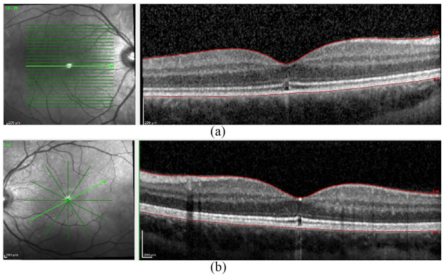
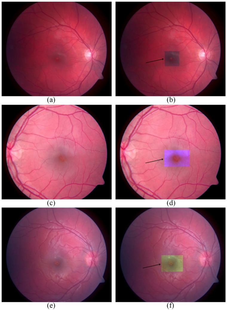
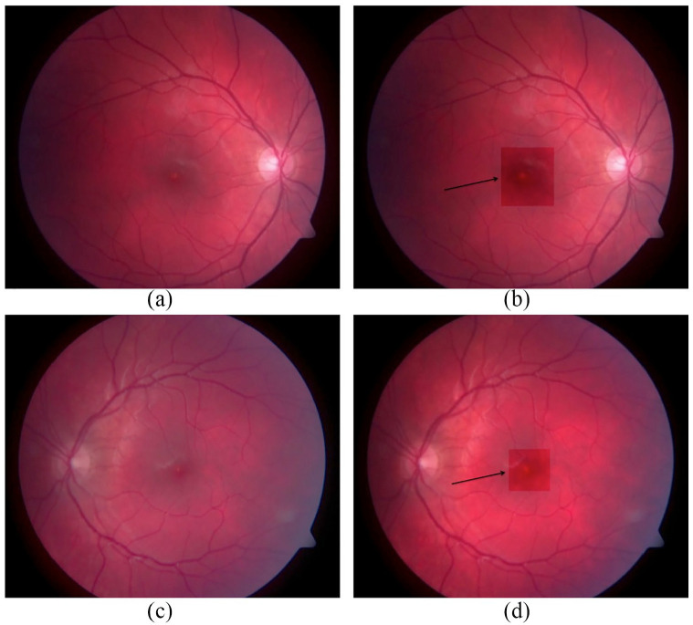
There was a statistically significant (p < 0.001) difference between best corrected visual acuity (BCVA) at the first consultation (day 0; 0.78 ± 0.11) and after a 6-month period (0.83 ± 0.09; Figure 1(a)). All eyes were noted to have a small, yellow lesion in the center of the fovea at day 0 that resolved at the 6-month follow-up visit. At presentation, SD-OCT showed a disruption of the continuity of the retianl layers in the fovea (Figure 2(a) and (b)) and damage to the myoid zone, ellipsoid zone, and outer segment of photoreceptors.15 These changes had partially resolved in all cases at 6 months. The statistical effect size calculation using ROC curve analysis showed improving effect (area under curve (AUC) = 0.62; 95% CI: 0.42–0.79; Figure 1(b)). There was an positive correlation between vision improvement and area of the lesion (rs = 0.65; p = 0.001; Figure 1(c)). SD-OCT in combination with fundus imaging, with gradually improved settings of R, G, and B color channels, resulted in a improved delineation of the lesions after solar exposure (Figure 2(a) and (b)). The R channel guided setting that demonstrated an enhanced demarcation of the lesion was 67-0.98-255. It helped to determine the margin more effectively in comparison to the images that were not adjusted (Figure 3(a) and (b)). In the instances where changes of the R channel setting did not improve the quality of the lesion, better results were obtained with a B-guided channel setting of 19-0.63-121 (Figure 3(c) and (d)) or a G-guided channels setting of 7-1.00-129 (Figure 3(e) and (f)). To avoid adjusting R, G, and B color setting independently, we discovered that a RGB value of 24-0.82-229 collectively provided the optimum setting (Figure 4(a)–(d)).
Figure 1.
Best corrected visual acuity (BCVA), receiver operating curve (ROC), change observed in visual acuity, and area of lesion: (a) a significant difference was observed in the BCVA between the first visit and at 6 months, (b) an improvement was seen after the analysis of the ROC values, and (c) a positive correlation was demonstrated between vision improvement and area of lesion.
Figure 2.
Optical coherence tomography (OCT images): (a) right eye and (b) left eye of a 22-year old man displaying lesions in the ellipsoid zone.
Figure 3.
Fundus images with amended settings of R, G, and B channels: (a) right eye fundal image without alteration, (b) right eye fundal image with a R-guided channel setting of 67-0.98-255, (c) left eye without alteration, (d) left eye with a B-guided channel setting of 19-0.63-121, (e) right eye without alteration, and (f) right eye with a G-guided channel setting of 7-1.00-129. The arrows indicate the images with amended settings.
Figure 4.
Fundus images with the suggested RGB setting of 24-0.82-229. (a) A fundus image showing the central foveal defect in (a) right eye and (b) left eye. The fundus image after using the suggested RGB setting of 24-0.82-229 of the (c) right eye and (d) left eye of the same patient. The arrows indicate the images with amended settings.
Discussion
Solar retinopathy is likely to be a combination of thermal and photochemical reactions or thermally enhanced photochemical damage.16 Typically the condition is bilateral with signs of asymmetry.11 Alongside the use of SD-OCT, this study demonstrates how a color fundus image with new settings of R, G, and B channels successfully improves the identification of damaged retinal areas. An additional advantage of this modified color image setting is that it could be readily implemented in centers where a SD-OCT is not available. In acute solar retinopathy visual loss may resolve with time, usually from 3 to 6 months, and patients may regain good VA.17 In this retrospective case series, six patients continued to suffer from visual deficiencies with persistent central or paracentral scotoma. This is likely to be as a result of irreversible solar damage to the ellipsoid zone and outer segment of photoreceptors led to a clinically significant retinal atrophy as previously reported.17,18 The lesion may depend on the intensity, duration, and light spectrum of the solar exposure or other font of UV, ocular pigmentation, clarity of the ocular media, and environmental conditions. Interestingly, the visual recovery was better in patients with larger lesions. We speculate that the larger area of the lesion resulted in a more significant loss of vision in the acute phase, while smaller lesions are compensated for by residual retinal tissue. A limitation of the study is that it was not possible to predict the most appropriate channel to improve the delineation of every case. This could possibly depend on the anatomical differences of the eyes or different degrees of retinal damage due to differing levels of energy exposure. In addition, the settings of the channels were investigated manually. This could be improved in the future with the creation of a software that would be able to automatically detect the area of lesion. A further limitation was that we did not test the modified setting in images captured with other cameras as these were not readily available. To simplify and to give a single setting, easier to use in all cases, a RGB value of 24-0.82-229 was found to gain images of better quality in all solar retinopathy eyes. In conclusion, once solar retinopathy is suspected, a fundus imaging with this novel modified setting of RGB channels could enhance the detection of the structure. This does not detract from the need for education in prevention of solar retinopathy through appropriate eye protection.
Footnotes
Authors’ Note: The work has not been published previously.
Declaration of conflicting interests: The author(s) declared no potential conflicts of interest with respect to the research, authorship, and/or publication of this article.
Funding: The author(s) received no financial support for the research, authorship, and/or publication of this article.
ORCID iD: Davide Borroni  https://orcid.org/0000-0001-6952-5647
References
- 1.Wu CY, Jansen ME, Andrade J, et al. Acute solar retinopathy imaged with adaptive optics, optical coherence tomography angiography, and en face optical coherence tomography. JAMA Ophthalmol 2018; 136(1): 82–85. [DOI] [PMC free article] [PubMed] [Google Scholar]
- 2.Abdellah MM, Mostafa EM, Anber MA, et al. Solar maculopathy: prognosis over one year follow up. BMC Ophthalmol 2019; 19(1): 201. [DOI] [PMC free article] [PubMed] [Google Scholar]
- 3.Widmark J.Ueber glendung der netzhauit. Skand Arch 1893; 4: 281–295. [Google Scholar]
- 4.Deutschmann R.Elinige Erfaliruniigeni fiber die verwenidung des Jodoforms inl der Auigeniheilkuniide. Albrecht uon Graefes Arch Klin Exp Ophthalmol 1882; 28: 214–224. [Google Scholar]
- 5.Czepita M, Machalinska A, Czepita D.Near-infrared fundus autoflorescence imaging in solar retinopathy. GMS Ophthalmol Cases 2017; 7: Doc05. [DOI] [PMC free article] [PubMed] [Google Scholar]
- 6.Shukla D.Optical coherence tomography and autofluorescence findings in chronic phototoxic maculopathy secondary to snow-reflected solar radiation. Indian J Ophthalmol 2015; 63(5): 455–457. [DOI] [PMC free article] [PubMed] [Google Scholar]
- 7.Begaj T, Schaal S.Sunlight and ultraviolet radiation-pertinent retinal implications and current management. Surv Ophthalmol 2018; 63(2): 174–192. [DOI] [PubMed] [Google Scholar]
- 8.Ricks C, Montoya A, Pettey J.The ophthalmic fallout in Utah after the Great American Solar Eclipse of 2017. Clin Ophthalmol 2018; 12: 1853–1857. [DOI] [PMC free article] [PubMed] [Google Scholar]
- 9.Gonzalez Martin-Moro J, Hernandez Verdejo JL, Zarallo Gallardo J. Photic maculopathy: a review of the literature (ii). Arch Soc Esp Oftalmol 2018; 93(11): 542–550. [DOI] [PubMed] [Google Scholar]
- 10.Sheemar A, Takkar B, Temkar S, et al. Solar retinopathy: the yellow dot and the rising sun. BMJ Case Rep 2017; 2017: bcr-2017-222690. [DOI] [PMC free article] [PubMed] [Google Scholar]
- 11.Hunter JJ, Morgan JI, Merigan WH, et al. The susceptibility of the retina to photochemical damage from visible light. Prog Retin Eye Res 2012; 31(1): 28–42. [DOI] [PMC free article] [PubMed] [Google Scholar]
- 12.Hope-Ross MW, Mahon GJ, Gardiner TA, et al. Ultrastructural findings in solar retinopathy. Eye 1993; 7(Pt 1): 29–33. [DOI] [PubMed] [Google Scholar]
- 13.Kristian P, Timkovic J, Cholevik D, et al. [Solar maculopathy after watching the partial solar eclipse]. Cesk Slov Oftalmol 2015; 71(5): 253–258. [PubMed] [Google Scholar]
- 14.Moran S, O’Donoghue E.Solar retinopathy secondary to sungazing. BMJ Case Rep 2013; 2013: bcr2012008402. [DOI] [PMC free article] [PubMed] [Google Scholar]
- 15.Staurenghi G, Sadda S, Chakravathy U, et al. Proposed lexicon for anatomic landmarks in normal posterior segment spectral-domain optical coherence tomography: the IN*OCT consensus. Ophthalmology 2014; 121(8): 1572–1578. [DOI] [PubMed] [Google Scholar]
- 16.Kung YH, Wu TT, Sheu SJ.Subtle solar retinopathy detected by fourier-domain optical coherence tomography. J Chin Med Assoc 2010; 73(7): 396–398. [DOI] [PubMed] [Google Scholar]
- 17.Soderberg PG.Optical radiation and the eyes with special emphasis on children. Prog Biophys Mol Biol 2011; 107(3): 389–392. [DOI] [PubMed] [Google Scholar]
- 18.dell’Omo R, Konstantopoulou K, Wong R, et al. Presumed idiopathic outer lamellar defects of the fovea and chronic solar retinopathy: an OCT and fundus autofluorescence study. Br J Ophthalmol 2009; 93(11): 1483–1487. [DOI] [PubMed] [Google Scholar]


