Abstract
Objective
We aimed to determine the differences in stage at diagnosis of cervical cancer among Korean women according to screening history.
Methods
Using linkage data from the Korean Central Cancer Registry and Korean National Cancer Screening Program (KNCSP), we included 18,388 women older than 30 years who were newly diagnosed with cervical cancer between 2013 and 2014 and examined their screening history. Between individuals, age group and socioeconomic status were matched to control for potential confounders.
Results
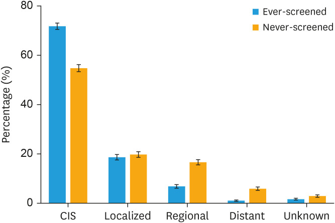
Significantly more cases of carcinoma in situ (CIS) were diagnosed in the ever-screened (71.77%) group than in the never-screened group (54.78%), while localized, regional, distant, and unknown stage were more frequent in the never-screened group. Women in the ever-screened group were most likely to be diagnosed with CIS than with invasive cervical cancer (adjusted odds ratio [aOR]=2.40; 95% confidence interval [CI]=2.18–2.65). The aOR for being diagnosed with CIS was highest among women who were screened 3 times or more (aOR=5.10; 95% CI=4.03–6.45). The ORs were highest for women screened within 24 months of diagnosis and tended to decrease with an increasing time since last screening (p-trend <0.01).
Conclusion
The KNCSP for cervical cancer was found to be positively associated with diagnosis of cervical cancers at earlier stages among women aged 30 years or older. The benefit of screening according to time was highest for women screened within 24 months of diagnosis.
Keywords: Uterine Cervical Neoplasms, Mass Screening, Papanicolaou Test, Early Detection of Cancer, Neoplasm Staging
INTRODUCTION
Cervical cancer is a well-known example of a preventable cancer due to its slow progression, the availability of cost-effective screening tests for early detection, and widespread access to the human papillomavirus (HPV) vaccine. Since 1999, the Korean National Cancer Screening Program (KNCSP) has provided biennial Pap smear tests for cervical cancer screening free-of-charge to all women aged 30 years and older [1]. In 1999, the incidence and mortality of invasive cervical cancer (ICC) in Korea were higher than those in other developed countries [2]. However, with the implementation of the KNCSP, much improvement has been made: From 1999 to 2015, the age-standardized incidence rate (ASIR) of ICC gradually decreased from 18.6 to 9.1 per 100,000 women [2], with an annual percentage change (APC) of −3.9% [3]. Similarly, the age-standardized mortality rate also continuously decreased from 2.87 to 2.00 per 100,000 women (APC=−5.2% in 2003–2015 period) [3]. The ASIR of ICC followed a decreasing trend from 19.3 in 1993 to 10.5 per 100,000 women in 2009 (APC=−4.2), whereas the ASIR increased from 7.5 to 19.0 per 100,000 women during this period (APC=5.7) [4].
Changes in the incidences of ICC and carcinoma in situ (CIS) appear to be closely related to cervical cancer screening. One previous cohort study conducted in 2009 using the National Health Insurance (NHI) Corporation Study found that only women who underwent Pap smear testing 2 or more times showed 60% and 53% reduced risks of ICC and CIS, respectively, compared to those who never did [5]. A protective effect for screening was also found among women who underwent screening one time, although the difference was not significant. While these results supported the importance of periodic cervical cancer screening, the study did not provide any evidence on how effective regular Pap smear tests are in detecting CIS and early-stage ICC.
Therefore, in order to evaluate the effectiveness of early detection of cervical cancer through screening, the present study was conducted to estimate the odds of being diagnosed with CIS and early-stage ICC among women who had participated in the KNCSP and women who had no screening history. Additionally, we investigated the association between stage at diagnosis of ICC or CIS and number of Pap smear tests and time from the most recent Pap smear test.
MATERIALS AND METHODS
1. Study population
This study included all women aged 30 years and older who were diagnosed with ICC (C53) or CIS of the cervix uteri (D06) between 2013 and 2014, as recorded in the Korean Central Cancer Registry (KCCR) [6]. Before 2016, the KNCSP had provided biennial cervical cancer screening free-of-charge for all women aged 30 years and older, and there was no upper age limit for screening [7]. Therefore, in this study, we excluded women less than 30 years of age to account for recommendation guidelines (n=1,432).
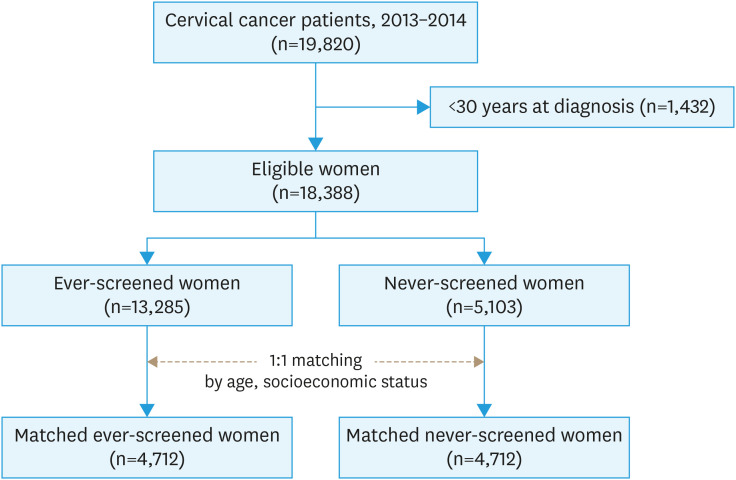
Among the 18,388 women with either CIS or ICC, 13,285 women had undergone screening via the KNCSP at least once (ever-screened) between January 1, 2002 and December 31, 2014, and 5,103 women had never done so (never-screened) (Fig. 1). To control for possible confounders, we conducted 1:1 individual matching between never- and ever-screened groups according to age and socioeconomic status. Insurance type was considered as a measure of socioeconomic status since the monthly premium of a beneficiary is calculated according to his/her income and assets. Insurance type was classified as follows: (a) Medical Aid Program (MAP) recipients (extremely poor people who received livelihood assistant and were unable to pay for healthcare or insurance); (b) NHI Service beneficiaries with a premium of 50% or less; and (c) NHI Service beneficiaries with a premium higher than 50%. Finally, 4,712 women in the never-screened group were matched with 4,712 women in the ever-screened group. The need for informed consent was waived. This study was approved by the Institutional Review Board (IRB) of the National Cancer Center, Korea (IRB No. NCCNC2015-0095).
Fig. 1. Flow chart of the study population.
2. Measures
Information on cervical cancer diagnosis, diagnosis date, disease code according to the International Classification of Diseases 10th revision, histological classification, anatomic site, and the stage of diagnosed cancer was obtained from the KCCR database. Tumor stage at diagnosis in the KCCR was recorded as localized, regional, distant, or unknown, following the Surveillance, Epidemiology, and End Results (SEER) staging system published by the National Cancer Institute [8]. CIS was defined as non- or pre invasive (International Federation of Gynecology and Obstetrics [FIGO] Stage 0). Localized tumors were invasive cancer confined to the cervix uteri or uterus (FIGO stage I). Regional tumors referred to those that extend beyond the cervix uteri, invading surrounding tissue and/or regional lymph nodes (FIGO stage II–III). Distant cancers were defined as those with metastasis to distant sites or lymph nodes. Unknown stage was assigned to cases for which sufficient evidence with which to adequately assign a stage was lacking.
The KCCR database was linked with the KNSCP database using unique identification numbers for each participant to ascertain information on cancer screening. The KNCSP database contains information on demographic characteristics (age, health insurance status), date of screening test, and screening results of all women invited to participate in the KNCSP for cervical cancer screening from 2002 to 2014. Using the KNSCP database, we assessed cervical cancer screening history, screening frequency, and time from last screening to cancer diagnosis to examine the effect of cervical cancer screening on stage at diagnosis. Screening frequency was categorized as once, twice, and three times or never-screened. Time from last screening to cancer diagnosis was grouped as ≤11 months, 12–24 months, 24–36 months, and ≥36 months.
3. Statistical analyses
We conducted statistical analysis for both matched and un-matched datasets. Demographic characteristics, tumor characteristics, and stage at diagnosis of cervical cancer were compared between ever- and never-screened groups using the χ2-test. Conditional and unconditional logistic regression was performed to calculate odds ratios (ORs) and 95% confidence intervals (CIs) for being diagnosed with CIS in the matched and un-matched datasets, respectively. We also conducted subgroup analyses according to age group, socioeconomic status, screening frequency, and time from screening test to diagnosis. SAS software (version 9.3; SAS Institute Inc., Cary, NC, USA) was used for all statistical analyses.
RESULTS
1. Characteristics of the study population
The demographics characteristics of the matched and un-matched datasets are shown in Table 1. In the un-matched dataset, there were more women aged 30–39 years and older than 80 years in the never-screened group. CIS was significantly more frequent in the ever-screened women than in the never-screened women (p<0.001).
Table 1. Demographic and tumor characteristics of ever-screened and never-screened cervical cancer patients in the National Cancer Screening Program.
| Characteristics | Matched (n=9,424) | Un-matched (n=18,388) | |||||
|---|---|---|---|---|---|---|---|
| Never (n=4,712) | Ever (n=4,712) | p-value | Never (n=5,103) | Ever (n=13,285) | p-value | ||
| Age at diagnosis (yr) | 1.00 | <0.01 | |||||
| 30–39 | 2,161 (45.86) | 1,210 (25.68) | 2,475 (48.50) | 2,892 (21.77) | |||
| 40–49 | 1,210 (25.68) | 588 (12.48) | 1,210 (23.71) | 4,483 (33.74) | |||
| 50–59 | 588 (12.48) | 281 (5.96) | 588 (11.52) | 2,783 (20.95) | |||
| 60–69 | 281 (5.96) | 291 (6.18) | 281 (5.51) | 1,715 (12.91) | |||
| 70–79 | 291 (6.18) | 181 (3.84) | 291 (5.70) | 1,145 (8.62) | |||
| ≥80 | 181 (3.84) | 1,210 (25.68) | 258 (5.06) | 267 (2.01) | |||
| Socioeconomic status | 1.00 | 0.04 | |||||
| NHI (premium >50%) | 1,844 (39.13) | 1,844 (39.13) | 1,943 (38.08) | 5,308 (39.95) | |||
| NHI (premium ≤50%) | 2,711 (57.53) | 2,711 (57.53) | 2,987 (58.53) | 7,446 (56.05) | |||
| MAP recipients | 157 (3.33) | 157 (3.33) | 173 (3.39) | 531 (4.00) | |||
| Tumor stage* | <0.01 | <0.01 | |||||
| CIS | 2,581 (54.78) | 3,382 (71.77) | 2,819 (55.24) | 8,847 (66.59) | |||
| Localized | 931 (19.76) | 879 (18.65) | 996 (19.52) | 2,715 (20.44) | |||
| Regional | 784 (16.64) | 323 (6.85) | 821 (16.09) | 1,178 (8.87) | |||
| Distant | 278 (5.90) | 50 (1.06) | 307 (6.02) | 286 (2.15) | |||
| Unknown | 138 (2.93) | 78 (1.66) | 160 (3.14) | 259 (1.95) | |||
Values are presented as number (%).
CIS, carcinoma in situ; MAP, Medical Aid Program; NHI, National Health Insurance.
*Tumor stage definitions adapted from the Surveillance, Epidemiology, and End Results Cancer Statistics Review: CIS, neoplasm confined to the cervix without invading the surrounding stroma; localized, neoplasm confined entirely to the cervix without serosal involvement; regional, neoplasm that extends beyond the limits of the cervix of uteri, invading the surrounding tissue; distant, neoplasm that spreads to parts of the body remote from the primary tumor; and unknown, neoplasm with insufficient or unavailable information to assign a stage.
2. Distribution of stages at diagnosis
The distributions of stage at diagnosis of cervical cancer (using matched dataset) are visually presented in Fig. 2. There were significantly more cases diagnosed with CIS in the ever-screened than in the never-screened group, while localized, regional, distant, and unknown stages were more frequent in the never-screened group (p<0.001) (Table 1).
Fig. 2. Distribution of stages at cervical cancer diagnosis in patients according to history of cervical cancer screening via the National Cancer Screening Program.
CIS, carcinoma in situ.
3. Screening frequency and time from last screening to cancer diagnosis
The proportions of the study population according to screening frequency and time from last screening to cancer diagnosis are described in Table 2 and Tables S1 and S2. In the matched dataset of never- and ever-screened women, 10.52% of women had undergone 3 or more Pap smear tests; while in the un-matched dataset, 24.71% of women had undergone 3 or more Pap smear tests. Regarding lengths of time between last screening and cancer diagnosis, 35.35% and 51.05% of women had been screened within 11 months in the matched and un-matched datasets, respectively.
Table 2. Stage distribution according to screening frequency and time from last screening to cancer diagnosis.
| Variables | Matched dataset (n=9,424) | Un-matched dataset (n=18,388) | |||||
|---|---|---|---|---|---|---|---|
| Diagnosed with CIS | Diagnosed at higher stages* | Total | Diagnosed with CIS | Diagnosed at higher stages* | Total | ||
| Screening frequency | |||||||
| Never | 2,581 (43.28) | 2,131 (61.57) | 4,712 (50.00) | 2,819 (24.16) | 2,284 (33.98) | 5,103 (27.75) | |
| Once | 1,885 (31.61) | 713 (20.60) | 2,598 (27.57) | 3,527 (30.23) | 1,903 (28.31) | 5,430 (29.53) | |
| Twice | 826 (13.85) | 297 (8.58) | 1,123 (11.92) | 2,234 (19.15) | 1,078 (16.04) | 3,312 (18.01) | |
| Three times or more | 671 (11.27) | 320 (9.25) | 991 (10.51) | 1,282 (26.45) | 666 (21.68) | 1,948 (24.71) | |
| Time from last screening to cancer diagnosis | |||||||
| Never | 2,581 (27.39) | 2,131 (22.61) | 4,712 (50.00) | 2,819 (15.33) | 2,284 (12.42) | 5,103 (27.75) | |
| ≤11 mo | 2,484 (26.36) | 847 (8.99) | 3,331 (35.35) | 6,539 (35.56) | 2,848 (15.49) | 9,387 (51.05) | |
| 12–23 mo | 458 (4.86) | 145 (1.54) | 603 (6.40) | 1,046 (5.69) | 507 (2.76) | 1,553 (8.45) | |
| 24–35 mo | 196 (2.08) | 93 (0.99) | 289 (3.07) | 500 (2.72) | 282 (1.53) | 782 (4.25) | |
| ≥36 mo | 244 (2.59) | 245 (2.60) | 489 (5.19) | 762 (6.53) | 801 (11.92) | 1,563 (8.50) | |
| Total | 5,963 (63.27) | 3,461 (36.73) | 9,424 (100) | 11,666 (63.44) | 6,722 (36.56) | 18,388 (100) | |
Values are presented as number (%).
CIS, carcinoma in situ.
*Higher stages include localized, regional, and distant stage.
4. Effect of screening with Pap smear tests on stage at cervical cancer diagnosis
ORs were calculated to investigate associations between screening history for cervical cancer and stage at diagnosis of cervical cancer (Table 3). Overall, in the matched dataset, ever-screened women were significantly more likely to be diagnosed with CIS than with ICC compared to never-screened women (adjusted OR [aOR]=2.40; 95% CI=2.18–2.65). Specifically, the OR for being diagnosed with CIS was highest among patients who were aged 80 years or older (aOR=6.86; 95% CI=3.10–15.15). Among MAP recipients, the OR for being diagnosed with CIS was more than 4 times higher in ever-screened women than in never-screened women. We conducted further analyses to examine associations between screening frequency and time from screening to diagnosis and diagnosis at CIS stage. Significant trends were observed in the number of screening tests and time from screening to diagnosis (p<0.001). Women who had undergone 3 or more Pap smear tests were more likely to be diagnosed with CIS (aOR=5.10; 95% CI=4.03–6.45) than with ICC compared to those who had never been screened. Similarly, women with a recent Pap smear test (≤11 months) were more likely to be diagnosed with CIS than with ICC (aOR=2.51; 95% CI=2.24–2.82). Interestingly, the OR of CIS detection was highest for women who had been screened within 12–23 months (aOR=2.89; 95% CI=2.16–3.85) and tended to decrease gradually thereafter. Women who had undergone a Pap smear test after more than 36 months before diagnosis still showed benefits from screening, compared to those who had never been screened, even though the effect size was smaller (aOR=1.55; 95% CI=1.15–2.08).
Table 3. Association between cervical cancer screening history and being diagnosed with CIS*.
| Variables | Matched | Un-matched | |||
|---|---|---|---|---|---|
| OR‡ | 95% CI | OR‡ | 95% CI | ||
| Overall | 2.40 | 2.18–2.65 | 2.43 | 2.25–2.63 | |
| Age at cancer diagnosis (yr)† | |||||
| 30–39 | 1.51 | 1.29–1.76 | 1.42 | 1.24–1.63 | |
| 40–49 | 2.28 | 1.91–2.71 | 2.27 | 1.99–2.59 | |
| 50–59 | 4.05 | 3.07–5.33 | 3.70 | 3.03–4.51 | |
| 60–69 | 4.78 | 3.15–7.23 | 4.64 | 3.45–6.24 | |
| 70–79 | 5.62 | 3.53–8.94 | 7.31 | 5.02–10.65 | |
| ≥80 | 6.86 | 3.10–15.15 | 4.99 | 2.90–8.58 | |
| Socioeconomic status† | |||||
| NHIS (premium >50%) | 1.90 | 1.62–2.22 | 2.03 | 1.79–2.30 | |
| NHIS (premium ≤50%) | 2.68 | 2.36–3.05 | 2.64 | 2.39–2.92 | |
| MAP recipients | 4.79 | 2.69–8.51 | 4.37 | 2.86–6.67 | |
| Screening frequency | |||||
| Never-screened | 1.00 | Reference | 1.00 | Reference | |
| Once | 1.65 | 1.45–1.87 | 1.78 | 1.63–1.94 | |
| Twice | 2.87 | 2.33–3.54 | 2.60 | 2.35–2.88 | |
| Three times or more | 5.10 | 4.03–6.45 | 3.92 | 3.55–4.33 | |
| p for trend | <0.01 | <0.01 | |||
| Time from last screening to cancer diagnosis | |||||
| Never-screened | 1.00 | Reference | 1.00 | Reference | |
| ≤11 mo | 2.51 | 2.24–2.82 | 2.77 | 2.55–3.00 | |
| 12–23 mo | 2.89 | 2.16–3.85 | 2.26 | 1.99–2.58 | |
| 24–35 mo | 2.11 | 1.43–3.10 | 2.05 | 1.74–2.42 | |
| ≥36 mo | 1.55 | 1.15–2.08 | 1.34 | 1.19–1.52 | |
| p for trend | <0.01 | <0.01 | |||
CI, confidence interval; CIS, carcinoma in situ; MAP, Medical Aid Program; NHIS, National Health Insurance Service; OR, odds ratio.
*Conditional and unconditional logistic regression models were used for the matched and un-matched datasets, respectively; †OR of detecting CIS in the ever-screened vs. never-screened groups in a subgroup; ‡ORs compare the odds of being diagnosed with CIS in ever-screened group to the odds of being diagnosed with invasive cervical cancer in ever-screened group.
The results of the analysis with un-matched data were also similar to the matched results, except for results in women aged 70–79 years and for the time from last screening to cancer diagnosis. In the un-matched dataset, ever-screened patients aged 70–79 years were more likely to be diagnosed with CIS, and the OR for being diagnosed with CIS was highest for the time interval of 11 months since the last Pap smear.
Further, ORs were calculated for both CIS or localized ICC, and ever-screened women were significantly more likely to be detected with CIS and localized ICC than with regional and distant stage compared to never-screened women (Table S3).
DISCUSSION
Using population-based data for the KNCSP for cervical cancer, we investigated the effect of Pap smear testing on stages of cervical cancer at diagnosis in Korean women. In this study, after matching the cases and controls according to age and socioeconomic status, we found that ever-screened women were most likely to be diagnosed with CIS than with ICC.
A similar down-staging effect and a decrease in cervical cancer incidence after implementation of a screening program have been reported in other countries [9,10,11,12,13,14]. Even so, there are still cases where inadequate uptake and insufficient quality of a screening program can hinder attaining an improvement in disease status in the population [15,16]. In Korea, even though cervical cancer screening rates were low in 2002 (30.8%), they have increased gradually since then (40.9% in 2012) [17]: the lifetime screening rate of cervical cancer was much higher (74.8%) [18]. The sensitivity and specificity of Pap smear tests in 2014 were 91.2% and 97.7%, respectively [19]. We believe that the quality of the Pap smear test might have contributed to the observed effects in this study. Our findings are in line with previous nested case-control results in the United Kingdom, which found that regular screening was associated with a 67% (95% CI=62%–73%) reduction in stage 1A cancer and a 95% (95% CI=94%–97%) reduction in stage 3 or worse cervical cancer [14]. Irregular screening also provided protective effects against invasive cancers, compared with those who had never undergone screening.
Screening intervals should be considered based on the balance between benefits, harms, and costs of screening. In this study, we evaluated who might benefit from cervical cancer screening according to time from last screening to cancer diagnosis. Compared with never-screened patients, ORs for having CIS were highest for women who had last been screened within 24 months of diagnosis and tended to decrease with an increasing time since last screening. Even though, time since last screening to cancer diagnosis and screening interval may differ in the exact sense, similar results were observed for organized screening programs in other countries. In a study in England, a screening interval of 2 to 2.5 years was found to provide the most protective benefits from invasive cancer in women of ages from 20 to 69 years; a 3-year screening interval was only significantly associated with a reduction in ICC among women aged 40–59 years [20]. Similarly, cervical cancer screening performed 3–36 months prior to the date of diagnosis was found to have a protective benefit against ICC only in women aged 40–59 years [21]; however, screening was still protective against cervical cancer deaths in all women older than 30 years [11]. In our study, we found that women who had last been screened within 3 years faced higher odds of being diagnosed at the CIS stage rather than with ICC stage, compared to never-screened women. We then further analyzed times since last screening of more than 3 years and found that a time interval of more than 5 years since last screening no longer had a protective effect on early detection of cervical cancer (Tables S2 and S4). However, due to the small sample size in each stratum (less than 1% of the study population), the results of these subgroups showed high variance and were subject to bias. Nevertheless, this finding supports that even if women have ever been screened in their lifetime, infrequent screening (more than 5 years) offers no further protective effect over never being screened.
Another finding from this study is that women who had more screening tests were more likely to be diagnosed at an early-stage. With a higher screening frequency, the odds of being diagnosed at CIS rather than with ICC was higher (Table 3 and Table S5). Meanwhile, there were a few women who had been screened more than 5 times (2.15% of the study population in the matched dataset), which led to a wider CI, as observed in Table S5. However, this finding still provides more evidence on the effectiveness of frequent screening rather than sporadic screening.
This study has several limitations. First, stage at diagnosis is not the ultimate outcome measure for evaluating the effectiveness of a screening program. Possible screening-related bias (e.g., length time bias) can affect stage at diagnosis. For example, a higher proportion of localized cervical cancers in the screening group might be associated with a tendency for screening to detect slow-growing lesions with a better prognosis and to miss fast-growing lesions with poorer survival. Thus, using mortality as an outcome of interest can provide more accurate estimation of the effects of a screening program. However, in the case of cervical cancer, precursors lesions of cervical cancer can be detected through screening, and treatment of these precancerous lesions can prevent the development of ICC and reduce cervical cancer incidence. Additionally, 5-year survival rates of cervical cancer are much different by stage at diagnosis. In Korea, 5-years survival rates (2006–2010) for localized, regional, and distant were 91.1%, 70.9%, and 25.8%, respectively [22]. Therefore, stage at diagnosis would be an appropriate intermediate outcome measure of use in assessing the effectiveness of a screening program.
Second, our results might be underestimated because we could not exclude symptomatic women who attended screening due to self-conscious symptoms. These women might be more likely to be diagnosed at advance stage but categorized as ever-screened women. Moreover, opportunistic screening information is not available within the KNCSP database. Even though cervical cancer screening services are provided free-of-charge, a part of the population still prefers utilizing opportunistic screening [23], and these participants might have been categorized to the never-screened group in this study, which again might have resulted in underestimation of the effectiveness of the screening test.
Third, we used information on stage at diagnosis from the KCCR, which only assigns stages according to the SEER staging system. Data on other staging systems, such as the American Joint Committee on Cancer TNM classification or FIGO, are not available. Therefore, we are not able to classify the stages in detail (e.g., stage IB2, IB3).
Finally, we could not control for potential bias and confounders, such as selection bias or HPV vaccination. Evaluating the screening program without considering these confounders might provide an overestimation of the magnitude of observed effects. Nevertheless, HPV vaccination rates in Korea were low (≤12.6%) at the start of the study (2013) [24], and HPV vaccination was only included in the National Immunization Program since 2016. Therefore, the effect of HPV vaccination in the population in 2013 would likely be small. Also, we matched the case and control groups according to age groups and socioeconomic status to minimize the potential for confounding effects. Even though Pap smear tests were provided free to every eligible women, screening rates differed by socioeconomic status [17]. Only 28.2% of eligible MAP recipients underwent screening test, while 44.7% of NHI beneficiaries with a premium over 50% underwent screening in 2012 [17].
Despite these limitations, by linking 2 adequate and reliable national data resources, the KNCSP and KCCR, we were able to classify incident cases according to screening history and to evaluate the effectiveness of the screening program on early detection. In conclusion, we found that women who had ever been screened for cervical cancer were more likely to be diagnosed at earlier stages, compared with never-screened patients. A significant increase in the protective effects (down-staging and reduced incidence) of screening against cervical cancer was observed as the number of screening tests increased and as the time from last screening to diagnosis decreased. The benefit of cervical cancer screening according to time since last screening was highest for women screened within 24 months of diagnosis and tended to decrease with an increasing time since last screening, although the benefits were still present up to 36 months. Further study is warranted to evaluate the performance of the KNCSP in reducing cervical cancer mortality and to provide more accurate measure of effectiveness.
Footnotes
Funding: This study was supported by a Grant-in-Aid for Cancer Research and Control from the National Cancer Center, Korea (#1910231).
Conflict of Interest: No potential conflict of interest relevant to this article was reported.
- Conceptualization: B.C.N., H.S., S.M., J.J.K., L.M.C., C.K.S.
- Data curation: B.C.N., J.J.K.
- Formal analysis: B.C.N., H.S.
- Funding acquisition: C.K.S.
- Investigation: B.C.N., H.S., S.M., J.J.K., C.K.S.
- Methodology: B.C.N., S.M., J.J.K., J.K.W., L.M.C., C.K.S.
- Project administration: C.K.S.
- Resources: J.K.W., C.K.S.
- Supervision: C.K.S.
- Validation: C.K.S.
- Writing - original draft: B.C.N., H.S.
- Writing - review & editing: B.C.N., J.J.K., J.K.W., L.M.C., C.K.S.
SUPPLEMENTARY MATERIALS
Stages at diagnosis according to screening frequency
Stages at diagnosis according to time since last screening
Association between cervical cancer screening history and being diagnosed at early stages (CIS and localized stage)*
ORs of being diagnosed with carcinoma in situ according to time since last screening*
ORs of being diagnosed with carcinoma in situ according to screening frequency*
References
- 1.Lee YH, Choi KS, Lee HY, Jun JK. Current status of the national cancer screening program for cervical cancer in Korea, 2009. J Gynecol Oncol. 2012;23:16–21. doi: 10.3802/jgo.2012.23.1.16. [DOI] [PMC free article] [PubMed] [Google Scholar]
- 2.Park Y, Vongdala C, Kim J, Ki M. Changing trends in the incidence (1999–2011) and mortality (1983–2013) of cervical cancer in the Republic of Korea. Epidemiol Health. 2015;37:e2015024. doi: 10.4178/epih/e2015024. [DOI] [PMC free article] [PubMed] [Google Scholar]
- 3.Jung KW, Won YJ, Kong HJ, Lee ES Community of Population-Based Regional Cancer Registries. Cancer statistics in Korea: incidence, mortality, survival, and prevalence in 2015. Cancer Res Treat. 2018;50:303–316. doi: 10.4143/crt.2018.143. [DOI] [PMC free article] [PubMed] [Google Scholar]
- 4.Oh CM, Jung KW, Won YJ, Shin A, Kong HJ, Jun JK, et al. Trends in the incidence of in situ and invasive cervical cancer by age group and histological type in Korea from 1993 to 2009. PLoS One. 2013;8:e72012. doi: 10.1371/journal.pone.0072012. [DOI] [PMC free article] [PubMed] [Google Scholar]
- 5.Jun JK, Choi KS, Jung KW, Lee HY, Gapstur SM, Park EC, et al. Effectiveness of an organized cervical cancer screening program in Korea: results from a cohort study. Int J Cancer. 2009;124:188–193. doi: 10.1002/ijc.23841. [DOI] [PubMed] [Google Scholar]
- 6.Won YJ, Sung J, Jung KW, Kong HJ, Park S, Shin HR, et al. Nationwide cancer incidence in Korea, 2003–2005. Cancer Res Treat. 2009;41:122–131. doi: 10.4143/crt.2009.41.3.122. [DOI] [PMC free article] [PubMed] [Google Scholar]
- 7.Kim Y, Jun JK, Choi KS, Lee HY, Park EC. Overview of the national cancer screening programme and the cancer screening status in Korea. Asian Pac J Cancer Prev. 2011;12:725–730. [PubMed] [Google Scholar]
- 8.Young JL, Jr, Roffers SD, Ries LAG, Fritz AG, Hurlbut AA. SEER summary staging manual - 2000: codes and coding instructions. Bethesda, MD: National Cancer Institute; 2001. [Google Scholar]
- 9.Miller JW, Royalty J, Henley J, White A, Richardson LC. Breast and cervical cancers diagnosed and stage at diagnosis among women served through the National Breast and Cervical Cancer Early Detection Program. Cancer Causes Control. 2015;26:741–747. doi: 10.1007/s10552-015-0543-2. [DOI] [PMC free article] [PubMed] [Google Scholar]
- 10.Serraino D, Gini A, Taborelli M, Ronco G, Giorgi-Rossi P, Zappa M, et al. Changes in cervical cancer incidence following the introduction of organized screening in Italy. Prev Med. 2015;75:56–63. doi: 10.1016/j.ypmed.2015.01.034. [DOI] [PubMed] [Google Scholar]
- 11.Vicus D, Sutradhar R, Lu Y, Kupets R, Paszat L Ontario Cancer Screening Research Network. Association between cervical screening and prevention of invasive cervical cancer in Ontario: a population-based case-control study. Int J Gynecol Cancer. 2015;25:106–111. doi: 10.1097/IGC.0000000000000305. [DOI] [PubMed] [Google Scholar]
- 12.Pettersson BF, Hellman K, Vaziri R, Andersson S, Hellström AC. Cervical cancer in the screening era: who fell victim in spite of successful screening programs? J Gynecol Oncol. 2011;22:76–82. doi: 10.3802/jgo.2011.22.2.76. [DOI] [PMC free article] [PubMed] [Google Scholar]
- 13.Brewer N, Pearce N, Jeffreys M, Borman B, Ellison-Loschmann L. Does screening history explain the ethnic differences in stage at diagnosis of cervical cancer in New Zealand? Int J Epidemiol. 2010;39:156–165. doi: 10.1093/ije/dyp303. [DOI] [PubMed] [Google Scholar]
- 14.Landy R, Pesola F, Castañón A, Sasieni P. Impact of cervical screening on cervical cancer mortality: estimation using stage-specific results from a nested case-control study. Br J Cancer. 2016;115:1140–1146. doi: 10.1038/bjc.2016.290. [DOI] [PMC free article] [PubMed] [Google Scholar]
- 15.Ojamaa K, Innos K, Baburin A, Everaus H, Veerus P. Trends in cervical cancer incidence and survival in Estonia from 1995 to 2014. BMC Cancer. 2018;18:1075. doi: 10.1186/s12885-018-5006-1. [DOI] [PMC free article] [PubMed] [Google Scholar]
- 16.Nowakowski A, Cybulski M, Buda I, Janosz I, Olszak-Wąsik K, Bodzek P, et al. Cervical cancer histology, staging and survival before and after implementation of organised cervical screening programme in Poland. PLoS One. 2016;11:e0155849. doi: 10.1371/journal.pone.0155849. [DOI] [PMC free article] [PubMed] [Google Scholar]
- 17.Suh M, Song S, Cho HN, Park B, Jun JK, Choi E, et al. Trends in participation rates for the national cancer screening program in Korea, 2002–2012. Cancer Res Treat. 2017;49:798–806. doi: 10.4143/crt.2016.186. [DOI] [PMC free article] [PubMed] [Google Scholar]
- 18.Suh M, Choi KS, Park B, Lee YY, Jun JK, Lee DH, et al. Trends in cancer screening rates among Korean men and women: results of the Korean National Cancer Screening Survey, 2004–2013. Cancer Res Treat. 2016;48:1–10. doi: 10.4143/crt.2014.204. [DOI] [PMC free article] [PubMed] [Google Scholar]
- 19.Lee JH, Kim H, Choi H, Jeong H, Ko Y, Shim SH, et al. Contributions and limitations of national cervical cancer screening program in Korea: a retrospective observational study. Asian Nurs Res (Korean Soc Nurs Sci) 2018;12:9–16. doi: 10.1016/j.anr.2017.12.002. [DOI] [PubMed] [Google Scholar]
- 20.Sasieni P, Adams J, Cuzick J. Benefit of cervical screening at different ages: evidence from the UK audit of screening histories. Br J Cancer. 2003;89:88–93. doi: 10.1038/sj.bjc.6600974. [DOI] [PMC free article] [PubMed] [Google Scholar]
- 21.Vicus D, Sutradhar R, Lu Y, Elit L, Kupets R, Paszat L, et al. The association between cervical cancer screening and mortality from cervical cancer: a population based case-control study. Gynecol Oncol. 2014;133:167–171. doi: 10.1016/j.ygyno.2014.02.037. [DOI] [PubMed] [Google Scholar]
- 22.Jung KW, Won YJ, Kong HJ, Oh CM, Shin A, Lee JS. Survival of korean adult cancer patients by stage at diagnosis, 2006–2010: national cancer registry study. Cancer Res Treat. 2013;45:162–171. doi: 10.4143/crt.2013.45.3.162. [DOI] [PMC free article] [PubMed] [Google Scholar]
- 23.Hahm MI, Chen HF, Miller T, O'Neill L, Lee H-Y. Why do some people choose opportunistic rather than organized cancer screening? The Korean National Health and Nutrition Examination Survey (KNHANES) 2010–2012. Cancer Res Treat. 2017;49:727–738. doi: 10.4143/crt.2016.243. [DOI] [PMC free article] [PubMed] [Google Scholar]
- 24.Lee SG, Jeon SY, Park O, Kim MY, Yang HI, Park EY. Vaccination coverage of adults aged above 19 years using mixed-mode random digit dialing (RDD) survey. J Korean Soc Matern Child Health. 2015;19:58–70. [Google Scholar]
Associated Data
This section collects any data citations, data availability statements, or supplementary materials included in this article.
Supplementary Materials
Stages at diagnosis according to screening frequency
Stages at diagnosis according to time since last screening
Association between cervical cancer screening history and being diagnosed at early stages (CIS and localized stage)*
ORs of being diagnosed with carcinoma in situ according to time since last screening*
ORs of being diagnosed with carcinoma in situ according to screening frequency*


