



Abstract
Cognitive impairments account for significant morbidity in schizophrenia and are present at disease onset. Controlled processes are particularly susceptible and may contribute to pervasive selective attention deficits. The present study assessed fronto-parietal attention network (FPAN) functioning during cue presentation on a visual search task in first-episode schizophrenia spectrum patients (FE) and its relation to symptom burden and community functioning. Brain activity was recorded with magnetoencephalography from 38 FE and 38 healthy controls (HC) during blocks of pop-out and serial search target detection. Activity during cue presentation was compared between groups across bilateral FPAN regions (frontal eye fields (FEF), inferior frontal gyrus (IFG), midcingulate cortex (MCC), and intraparietal sulcus (IPS)). FE exhibited greater right hemisphere IFG activity despite worse performance relative to HC. Performance and FPAN activity were not correlated in HC. Among FE, however, stronger activity within right hemisphere FEF and IFG was associated with faster responses. Stronger right IPS and left IFG activity in patients was also associated with reduced negative symptoms and improved community functioning, respectively. Increased reliance on the FPAN for task completion suggests an inefficient cognitive control network and might reflect a compensation for impaired attentional deployment during target detection, a strategy employed by those with less severe illness. These findings represent a critical step towards identifying the neural substrates of negative symptoms and impaired neurocognition at disease onset.
Keywords: First-episode, schizophrenia, attention, fronto-parietal attention network, magnetoencephalography
Introduction
The ability to focus attention on certain percepts in our environment while ignoring others, broadly defined as selective attention, is an essential cognitive function that promotes coherent, goal-directed behaviors. Disruptions in this cognitive function have been well-documented in people with schizophrenia (Fioravanti et al 2005), are associated with negative symptoms (Cornblatt et al 1985; Sanz et al 2012) and poor functional outcomes (Miley et al 2005; Vesterager et al 2012), and remain largely impervious to current pharmacologic treatments (Millan et al 2014; Miyamoto et al 2012). Importantly, deficits of selective attention are present early in the course of the illness (Fusar-Poli et al 2012; Orellana et al 2012; Ren et al 2021), providing a meaningful treatment target during a critical period for disease intervention. However, a more mechanistic understanding of the dysfunctional neural system contributing to impairments of attention is needed to support future interventional approaches.
A frequent challenge complicating attempts to characterize disruptions in attention is the overly broad conceptualization of this cognitive construct. To address this problem, the Cognitive Neuroscience Treatment Research to Improve Cognition in Schizophrenia (CNTRICS) group has provided recommendations for paradigms with high construct validity that engage facets of the attentional system with demonstrated sensitivity to disruption in psychosis (Luck et al 2012). According to these recommendations, attentional cuing tasks emphasizing executive control of attention are particularly well suited for examining cognitive control networks with the ability to broadly modulate activity across sensory-perceptual systems. Though modulatory functions are not the exclusive source of attentional deficits in psychosis, they show greater impairment than tasks examining the implementation of attention (Lesh et al 2011; Luck et al 2008).
There is a considerable body of literature detailing a distributed fronto-parietal attention network (FPAN) responsible for the control of visual attention in healthy adults that serves as a model system to interrogate selective attention deficits (Corbetta & Shulman 2002; Vossel et al 2014). While functional distinctions between dorsal (frontal eye fields (FEF) and intraparietal sulcus (IPS)) and ventral (temporoparietal junction (TPJ) and ventral frontal cortex (VFC)) systems have been well described within this network, dynamic interactions between both are critical to the allocation of attentional resources. Interestingly, studies examining differences between location and feature (e.g., color) based cues have observed considerable overlap in the FPAN activated by both including FEF, IPS, and VFC (Egner et al 2008; Giesbrecht et al 2003). The FPAN, therefore, appears to represent a more general system reflecting internal goal representations rather than one tied to a specific type of cue.
Although few studies in psychosis have examined alterations in neural activity during anticipatory cue periods on selective attention tasks, deficits during working memory encoding reliant upon similar frontal and parietal structures have been well-described (Deserno et al 2012; Schneider et al 2007). A frequently observed phenomenon in these studies is a load-dependent dysfunction with patients exhibiting increased activity during less challenging conditions relative to healthy controls and impaired activity at higher load levels (Coffman et al 2020; Kim et al 2010; Potkin et al 2009). This effect was also observed using an implicit cueing paradigm in a Go/No-go task of behavioral inhibition with increased inferior frontal gyrus activation interpreted as an inefficient compensatory mechanism in patients to meet the demands of a fairly simple task (Arce et al 2006).
Our goal in the present study was to evaluate functioning of the FPAN during color cue processing and its relationship to the implementation of attention in first-episode psychosis. Brain activity was recorded using magnetoencephalography (MEG) during a cued visual search task with varying levels of attentional demand, and structural MRI scans were obtained to provide precise anatomic localization of sensor-level activity. The less demanding task condition (pop-out) consisted of a consistent cue-target color pair across trials and emphasized a parallel search strategy whereas the more demanding condition (serial search) emphasized a controlled serial search of stimuli with a variable cue-target color across trials. Our laboratory recently identified an impairment in the deployment of attention towards target during presentation of the search array among individuals with a schizophrenia spectrum illness at disease onset (Sklar et al 2020a). In the present investigation, we therefore examined FPAN activity during the anticipatory cue period to evaluate the integrity of this cognitive control network and its relationship to selective attention impairments and disease burden at the first episode of psychosis.
Methods and Materials
Participants
Participants included in the current report were identical to the sample in a previously published manuscript (Sklar et al 2020b). Demographic and clinical assessment information is presented in Table 1. Thirty-eight healthy controls (HC) and forty-six first-episode schizophrenia spectrum patients (FE) participated. Eight FE were excluded from analysis due to poor quality neurophysiologic data (n=2) and to obtain a matching with HC on age, gender, Wechsler Abbreviated Scale of Intelligence IQ, and parental socioeconomic status (n=6).
Table 1.
Demographics clinical assessment data (Mean ±SD) by group
| HC (n=38) | FESz (n=38) | t/χ2 | p | |
|---|---|---|---|---|
| Female/Male | 13/25 | 14/24 | 0.06 | 1.0 |
| Education (years) | 14.1±3.2 | 12.4±2.4 | 2.58 | .01 |
| Age | 22.8±5.0 | 21.8±4.4 | 0.91 | .37 |
| WASIa | 108.2±10.6 | 107.3±14.2 | 0.31 | .76 |
| MATRICS-Total | 48.2±7.6 | 38.2±14.3 | 3.79 | .00 |
| SESb | 34.2±14.6 | 29.4±13.2 | 1.46 | .15 |
| PSESc | 53.0±13.7 | 47.6±15.0 | 1.62 | .11 |
| GF: Roled | 9.00±0.4 | 5.76±2.4 | 8.25 | .00 |
| GF: Sociale | 9.04±0.2 | 5.55±1.8 | 12.2 | .00 |
| SANSf/SAPSg: | ||||
| Inexpressivity | - | 10.2±6.6 | ||
| Asociality/Apathy | - | 10.8±6.3 | ||
| Reality Distortion | - | 11.0±7.0 | ||
| Disorganization | - | 4.34±5.2 | ||
| Percent Medicated | - | 65.8 | ||
| Medication (CPZh mg/day) | - | 193.3±165.5 |
Wechsler Abbreviated Scale of Intelligence;
Socioeconomic status;
Parental socioeconomic status;
Global Functioning: Role scale;
Global Functioning: Social scale;
Scale for the Assessment of Negative Symptoms;
Scale for the Assessment of Positive Symptoms;
Chlorpromazine equivalent dose
FE included individuals presenting with new-onset psychotic symptoms to any University of Pittsburgh Medical Center affiliated facility. HC were recruited from the greater Pittsburgh, PA region. All participants underwent a clinical assessment session that included a Structured Clinical Interview for DSM-IV (SCID-IV), medical screening questions to exclude individuals with a history of concussion, alcohol or substance use disorder, or neurological comorbidity, and a screening for colorblindness using pseudoisochromatic plates. All participants also completed the MATRICS Cognitive Consensus Battery (MCCB) to assess neurocognitive performance (Green et al 2004) as well as the Global Functioning: Role (GF: Role) and Global Functioning: Social (GF: Social) scales to assess community functioning (Cornblatt et al 2007). FE were assessed using Scale for the Assessment of Negative Symptoms (SANS) and Scale for the Assessment of Positive Symptoms (SAPS) to rate symptom severity prior to testing. SANS/SAPS scores were used to derive the 4-dimension symptom model (reality distortion, disorganization, inexpressivity, and apathy/asociality) validated by previous factor analyses using markers of neural dysfunction and long-term outcomes (Kotov et al 2016). Using this factor analysis, positive symptoms are comprised of reality distortion and disorganization while negative symptoms are comprised of inexpressivity and apathy/asociality.
HC with a current or past Axis I diagnosis were excluded. Only patients with a first-episode schizophrenia spectrum disorder were included in the present study. Diagnostic makeup for FE included 23 with schizophrenia, 6 with schizoaffective disorder (4 depressive and 2 bipolar type), and 9 with psychotic disorder not otherwise specified. FE participated within 2 months of first clinical contact and had less than 2 months of lifetime antipsychotic treatment with thirteen (~34%) being unmedicated at the time of testing.
All procedures were approved by the University of Pittsburgh IRB and participants provided informed consent prior to participation and were paid for participation.
Cued Attention Task
Participants completed a visual search task (Figure 1) described in pervious manuscripts (Sklar et al 2020a; Sklar 2020b). Each trial began with a centrally presented fixation cross (1°, 500ms). Fixation was followed by a centrally presented, feature-based cue (0.65°, 500ms) that indicated the target color for the upcoming search array. The search array (500ms) was made up of 6 eccentrically arranged colored annuli (subtending 0.65°, positioned 2° from fixation). Participants were instructed to covertly shift their attention towards the target annulus, identified based on its color, and indicate via button-press when a gap subsequently appeared to the right or left on the target.
Figure 1.
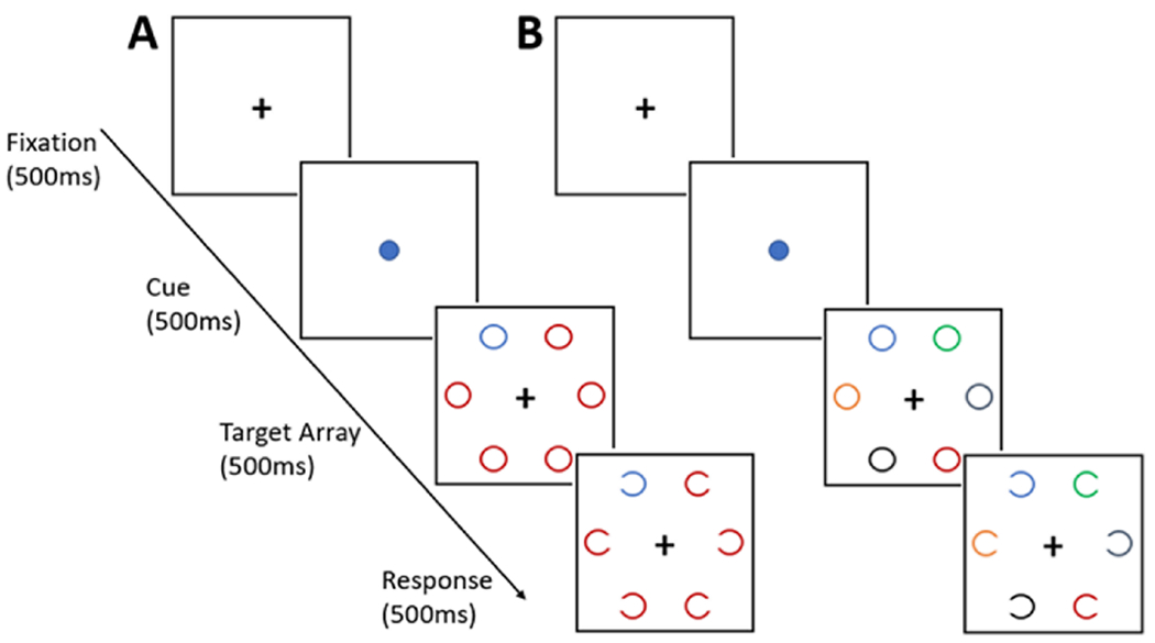
The visual search task used in the present study. In the pop-out version (A) the cue-target color remained consistent across trials and all distractors during the target array were of a uniform color. During the serial search task (B), cue-target color varied across trials and all target array distractors had unique colors. Cortical activity was examined during the Cue period.
The task consisted of 2 conditions (pop-out (PO) and serial search (SS)) with varying degrees of attentional demand. During PO, the color of the cue/target stimuli remained the same across trials and the 5 non-target annuli presented during the search array had uniform colors thereby emphasizing a parallel search strategy reliant upon bottom-up processing. In contrast, the cue/target color was variable across trials during SS and all non-target annuli in the search array had unique colors. This paradigm requires a search of individual array stimuli guided by top-down attentional systems in order to successfully locate the target annulus. Examination of the neural systems engaged by this task using lesions in non-human primates suggests that communication between cognitive control and sensory cortices is required for successful completion of the SS condition (Rossi et al 2007). Participants completed 4 task blocks, 2 of each condition with order counterbalanced across participants. Each block consisted of 144 trials.
Data Acquisition
Structural MRI scans were obtained for each participant using a Siemens Tim Trio 3T system. T1-weighted images were collected using a multi-echo 3D MP-RAGE sequence (TR/TE/TI = 2530/1.74, 3.6, 5.46, 7.32/1260 ms, flip angle = 7°, field of view (FOV) = 220 x 220 mm, 1 mm isotropic voxel size, 176 slices, GRAPPA acceleration factor = 2).
MEG data were recorded in a magnetically shielded room using an Elekta Neuromag VectorView system (Elekta Oy, Helsinki, Finland). Data was sampled from 306 sensors arranged into triplets, each with 2 orthogonal gradiometers and one magnetometer, at a rate of 1000Hz (online bandpass filter 0.1-330Hz). Head position was recorded throughout the testing session using 4 head positioning indicator coils and Neuromag MaxFilter software (http://imaging.mrc-cbu.cam.ac.Uk/meg/Maxfilter_V2.2) was used to correct for head motion during the scan. Blinks and eye movements were monitored using bipolar leads placed above and below the left eye (VEOG) and lateral to the outer canthi (HEOG), respectively, and cardiac activity was monitored using bipolar ECG leads.
MEG Data Pre-processing
Pre-processing procedures were followed to reduce the presence of artifacts from our MEG data. A temporal extension of the Signal Space Separation method (Uusitalo & Ilmoniemi 1997) was used to remove environmental magnetic artifacts from signals generated by neural activity (Taulu & Hari 2009). Data were visually inspected and the EEGLAB Toolbox (Delorme & Makeig 2004) was used to remove channels (re-interpolated following pre-processing) and segments of data corrupted by excessive noise. After applying a high-pass filter (0.5Hz; 12dB/oct), an adaptive mixture independent component analysis was preformed to isolate and remove eye-blink and cardiac activity.
Cortical Source Localization
MEG sensor data was registered to each participant’s structural MRI scan. The grey/white matter boundary was segmented using Freesurfer (http://www.surfer.nmr.mgh.harvard.edu) and tessellated into an icosahedron mesh with 5mm spacing between vertices. Subsequent processing steps were conducted using Brainstorm software (Tadel et al 2011). Sensor-level data was subjected to a low-pass (100Hz; 24dB/oct) filter and segmented into epochs constructed around the presentation of the color cue. A 300ms (−350ms to −50ms pre-cue) window was used for baseline correction and segments with data exceeding ±5pT were excluded with remaining trials used to generate participant averages. The minimum-norm estimate approach, a distributed source solution, was used to project MEG sensor data to the cortical surface. The forward solution, modeled as a series of overlapping spheres (one per sensor location), and a noise covariance matrix calculated from the baseline window were used to create a linear inverse operator using an orientation constraint of 0 (current dipoles normal to the cortex) with depth weighting applied (HÄmÄlÄinen and Hari 2002; Lin et al 2004). Cortical current density values were normalized to the baseline period in order to create a z-score for activity at each vertex.
Selection of regions of interest (ROIs) comprising the FPAN was guided by previous functional imaging work. These studies have identified a remarkably similar network for location and feature (e.g. color) based cues including the frontal eye fields (FEF), inferior frontal cortex, posterior parietal structures, and dorsal portions of the cingulate cortex (Egner et al 2008; Giesbrecht et al 2003). ROIs included in the current analysis were selected from the Destrieux parcellation available in Freesurfer (Destrieux et al 2010) based on sulci and gyri within this network exhibiting robust activity during the 500ms cue period (Figure 2). These regions included bilateral (right (RH) and left (LH) hemisphere) FEF comprised of superior and inferior pre-central sulci (Lobel et al 2001), the pars opercularis region of the inferior frontal gyrus (IFG), the intraparietal sulcus (IPS), and the middle-posterior segment of the cingulate cortex (MCC).
Figure 2.
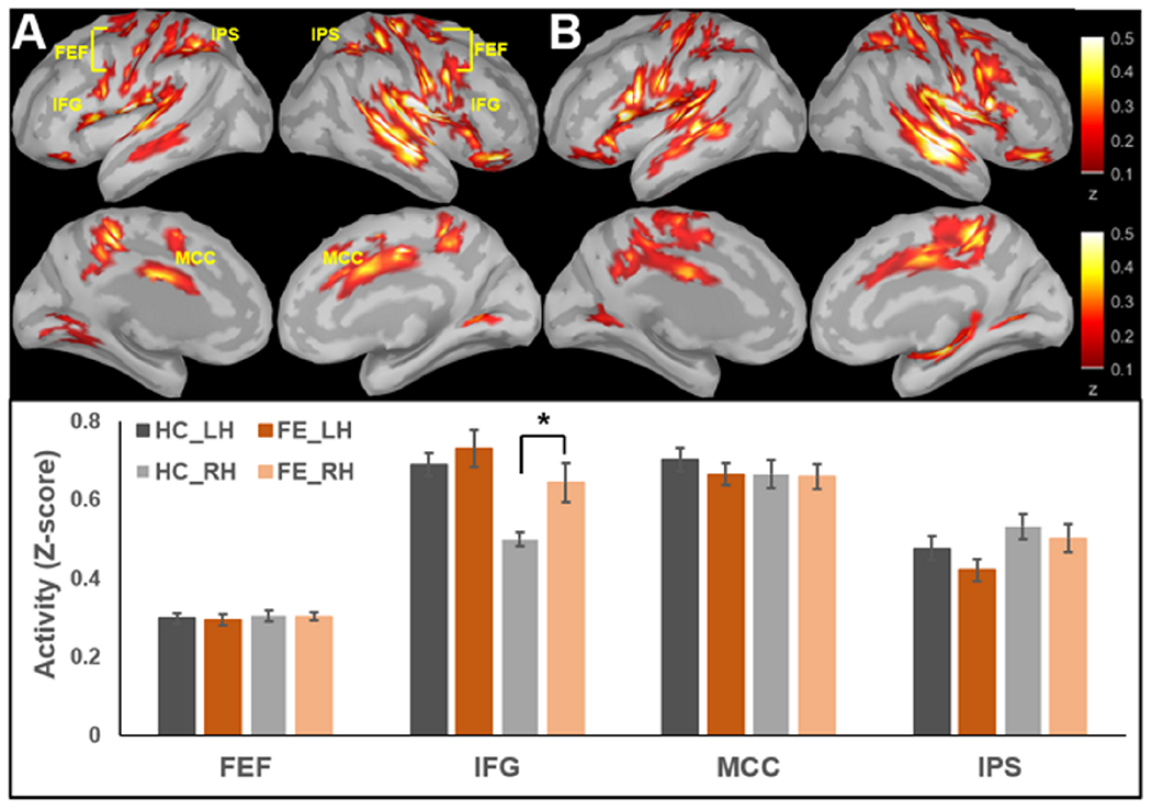
Cortical activity averaged over the 500ms cue period for HC (A) and FE (B) as well as quantification of activity within each region displayed by group. Regions of a fronto-parietal network selected for analysis based on previous literature utilizing feature-based cues. Error bars represent SEM. *p<.01.
Data Analysis
Task accuracy and response times were analyzed using a 2 (group: HC/FE) x 2 (task: PO/SS) ANOVA. Both performance measures were subjected to log transformation due to violation of normality (Shapiro-Wilk Test p>.05) prior to analysis. Cortical activity averaged over the cue period was analyzed using a 2 (group: HC/FE) x 2 (task: PO/SS) x 8 (ROI) ANOVA. Linear regressions for each ROI were conducted including measures of task performance (accuracy and response time) for both FE and HC as well as symptom burden (reality distortion, disorganization, inexpressivity, and apathy/asociality dimensions) and community functioning (GF: Role and GF: Social scales) in FE. Finally, effects of medication on outcome measures of interest were examined by including medication status as a between-group factor in analyses of FPAN activity and performance among FE Results were considered significant at p≤0.05.
Results
Task Performance
FE responded less accurately (F1,74=7.05, p<.01) and more slowly (F1,74=7.19, p<.01) compared to HC. Overall, responses were less accurate during SS compared to PO (F1,74=10.39, p<.01), though this did not differ between groups (p=.55). There was no effect of task or interaction between task and group on response times (p’s>.1).
FPAN Source Localized Activity
Cortical activity evoked during the 500ms cue period in each group is presented in Figure 2. Although overall activity across the FPAN did not differ between groups (p=.79), the pattern of activity did differ between HC and FE across ROI (Group x ROI interaction F7,518=3.00, p<.01). Specifically, FE exhibited larger RH IFG activity compared to HC (t74=−2.75, p<.01) with no differences in other ROIs (p’s>.1). No additional interactions involving group were significant (p’s>.3).
A main effect of ROI was also observed (F7,518=81.18, p<.01). Full reporting of ROI activity differences is available in Supplemental Table 1. No main effect of task or interactions involving it were observed (p’s>.2).
Relationship Between FPAN activity, Task Performance, and Clinical Measures
Regression statistics for FE are presented in Table 2. Partial regression plots depicting significant associations are presented in Figure 3. In FE, more rapid responses were associated with greater activity in RH FEF (β=−0.52, p=.03) and RH IFG (β=−0.49, p=.04). Larger LH IFG activity was also associated with bigger Global Functioning: Role scores (β=0.52, p=.03) and larger RH IPS activity was associated with lower inexpressivity scores (β=−0.44, p=.03) in FE. No significant relationships between FPAN activity and either task performance (p’s>.05) or community functioning (p’s>. 1) were observed among HC.
Table 2.
Regression statistics for each fronto-parietal attention network region.
| LH FEF R2=.24 | RH FEF R2=.27 | LH IFG R2=.33 | RH IFG R2=.27 | LH MCC R2=.18 | RH MCC R2=.20 | LH IPS R2=.12 | RH IPS R2=.46 | |
|---|---|---|---|---|---|---|---|---|
| Accuracy | −0.22 | −0.32 | −0.30 | −0.17 | 0.03 | −0.03 | −0.03 | −0.33 |
| Response Time | −0.44 | −0.52 | −0.13 | −0.49 | 0.07 | −0.03 | −0.09 | −0.37 |
| Inexpressivity | −0.33 | 0.1 | −0.15 | −0.03 | −0.05 | −0.12 | −0.13 | −0.44 |
| Asociality | −0.34 | −0.12 | −0.03 | −0.16 | 0.04 | −0.07 | −0.35 | −0.23 |
| Reality Distortion | −0.34 | −0.19 | −0.28 | −0.07 | −0.06 | 0.05 | 0.15 | 0.19 |
| Disorganization | 0.01 | −0.23 | 0.10 | −0.14 | 0.01 | −0.17 | −0.11 | 0.05 |
| GFS: Role | 0.06 | −0.08 | 0.52 | 0.20 | 0.37 | 0.41 | 0.12 | 0.28 |
| GFS: Social | −0.17 | −0.07 | 0.1 | −0.12 | 0.05 | −0.26 | −0.23 | −0.21 |
FEF: Frontal eye fields; IFG: Inferior frontal gyrus; MCC: Midcingulate cortex; IPS: interparietal sulcus; GFS: Global Functioning Scale
Boldface indicates significant coefficients (p<.05)
Figure 3.
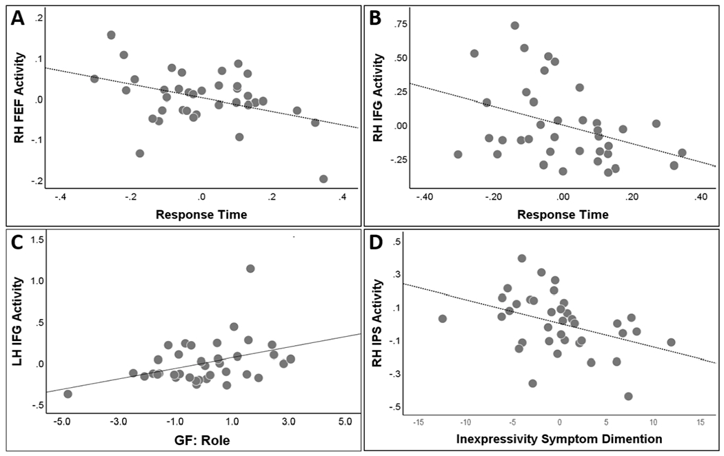
Partial regression plots depicting relationship between response time and RH FEF (A) and RH IFG (B) activity, GF: Role measure of community functioning and LH IFG activity (C), and the inexpressivity negative symptom dimension and RH IPS activity (D).
Medication Effects
There was no effect of medication status or interactions involving it on FPAN activity (p’s>.1) or behavior measures (p’s>.2).
Discussion
The present study examined functioning of the FPAN during cued visual search and its association with disease morbidity at the onset of schizophrenia spectrum illness. Using a combination of high resolution neurophysiological and structural imaging techniques, we observed increased network activity in FE, specifically within the RH IFG, despite impaired task performance relative to HC. Furthermore, associations between increased activity and improved performance among FE, a finding not present in HC, suggest a reliance on this network to meet the demands of a relatively simple selective attention task. These results suggest a cognitive control network engaged in excess of task demands to overcome deficits in the deployment of attention, a compensatory mechanism that may become overwhelmed in more complex real-world environments or during more chronic illness stages. Finally, greater disease burden was associated with an impaired ability to employ this cognitive strategy.
Group differences in cue-related network activity were restricted to the RH IFG. As a node within the ventral FPAN, this region is typically associated with bottom-up reorienting towards task-relevant stimuli (Corbetta & Shulman 2002; Vossel et al 2014). However, regions of the right inferior frontal cortex including the portion of the RH IFG examined in the current investigation represent an interface between dorsal and ventral attention networks, acting in concert with dorsal frontal and parietal regions to direct the focus of attention (Asplund et al 2010; Corradi-Dell’Acqua et al 2015). Furthermore, this region has been directly implicated in the anticipatory, top-down modulation of sensory-perceptual activity within visual cortices during stimulus color processing (Zanto et al 2010). Finally, the RH IFG is engaged in a number of cognitive control tasks involving inhibitory attentional control including gaiting of irrelevant distractors during visual search (Fockert et al 2004; Suda et al 2020). Larger RH IFG activity in FE during the current investigation can therefore be conceptualized as an enhanced utilization of cortical resources dedicated to the control of attention. While this finding appears to contradict previous reports of functional and structural abnormalities of the IFG in schizophrenia (Harms et al 2010; Iwashiro et al 2012; Jeong et al 2009), this conflict might be explained by disease chronicity with relatively preserved IFG function at illness onset.
Contrary to the intended design of the visual search task, there was no significant difference in FPAN recruitment between PO and SS. In addition, the lack of any relationship between FPAN activity and performance among HC emphasizes the relative simplicity of the task. In contrast, FE exhibited significant correlations between two frontal regions within this network and response time, indicating an increased reliance on an executive control network to complete an otherwise simple task. Interestingly, an inefficient use of cognitive resources was also observed in our recent analysis of attentional deployment following presentation of the stimulus array in which FE exhibited an increased reliance on parietal structures during the SS condition (Sklar et al 2020a). In contrast, HC relied more heavily on sensory-perceptual cortices to carry out this process. Together, these results are consistent with findings of a load-dependent difference in executive network activity between HC and people with schizophrenia in the working memory literature used to support the “inverted U” model of cognitive efficiency (Coffman et al 2020, Kim et al 2010; Potkin et al 2009). An alternative interpretation of the larger RH IFG activity is that it reflects an increased focus of attention on singleton items such as the centrally presented color cue in people with schizophrenia, a phenomenon referred to as “hyperfocusing” (Luck et al 2019).
Beyond revealing a difference in cognitive strategy relative to HC, FPAN activity in FE and its associations with their clinical assessments underscore the impact of cognitive dysfunction on disease burden during this early illness stage. Despite a lack of any significant correlation with positive symptom dimensions, weaker cue-related FPAN activity within RH IPS and LH IFG was associated with greater inexpressivity dimension scores and lower GF: Role scores, respectively. This pattern is consistent with previous reports documenting robust associations between attention impairments and both negative symptoms and impaired global functioning in schizophrenia with relatively weak or absent associations with core positive symptoms of the disorder (Chen & Faraone 2020; Curtin et al 2019). While the causal relationships underlying these correlations remain speculative, previous regression analyses suggest that negative symptoms act as mediators of the relationship between neurocognitive deficit and impaired community functioning (Ventura et al 2009; Meyer et al 2014). Regardless of the directional nature of these interactions, identifying the neural substrates of negative symptoms and impaired neurocognition at disease onset for future targeted interventions would be of considerable clinical value.
Limitations
There are limitations to be considered when interpreting results from the present investigation and to be addressed by future work. First, as previously noted, our manipulation of attentional load did not have as substantial an effect as desired, limiting our ability to assess efficiency of the FPAN across a meaningful range of task complexity. A task employing a more complex cue-target design would be better suited for this purpose. Second, our participants engaged several structures outside the previously defined color-cue FPAN, most notably in temporal regions. This temporal lobe recruitment might have resulted from participants verbalizing the color of the cue at the moment of its presentation, though this possibility cannot be addressed with the data at hand. Finally, the cue delay period used in the current paradigm was fairly short (500ms). A longer epoch would have provided a better opportunity to capture dynamic spectral changes, particularly within lower frequency bands with longer period lengths, to assess effective connectivity and provide a direct measure for top-down sensory-perceptual modulation. However, while cue periods of 1000ms or longer are more common, previous work has documented the top-down modulation of visual perception by fronto-parietal structures within 200ms of cue stimulus onset (Zanto et al 2010).
Conclusion
In conclusion, examination of the cognitive control FPAN during a low-complexity visual search task revealed enhanced cue-related IFG activity within a first-episode schizophrenia sample. Combined with the correlations between FPAN activity and task performance, these findings suggest an inefficient use of cognitive resources and may reflect a compensation for impairments in the downstream deployment of attention. Furthermore, implementation of this strategy was associated with reduced negative symptom burden and better community functioning, providing meaningful targets for disease modifying treatments during early illness stages.
Supplementary Material
Acknowledgements
Supported by NIH P50 MH103204 (David Lewis, MD, Director, DFS Project Co-PI) and NIH 5T32MH016804-40 (Robert Sweet, MD, Director). We thank the faculty and staff of the WPH Psychosis Recruitment and Assessment Center and the University of Pittsburgh Clinical Translational Science Institute (UL1 RR024153, Steven E. Reis, MD) for their assistance in recruitment, diagnostic and psychopathological assessments, and neuropsychological evaluations.
Footnotes
Publisher's Disclaimer: This is a PDF file of an unedited manuscript that has been accepted for publication. As a service to our customers we are providing this early version of the manuscript. The manuscript will undergo copyediting, typesetting, and review of the resulting proof before it is published in its final form. Please note that during the production process errors may be discovered which could affect the content, and all legal disclaimers that apply to the journal pertain.
Disclosures
None of the authors reported any potential conflicts of interest to disclose.
References
- Arce E, Leland DS, Miller DA, Simmons AN, Winternheimer KC, & Paulus MP (2006). Individuals with schizophrenia present hypo-and hyperactivation during implicit cueing in an inhibitory task. Neuroimage, 32(2), 704–713. [DOI] [PubMed] [Google Scholar]
- Aron AR, Robbins TW, & Poldrack RA (2014). Inhibition and the right inferior frontal cortex: one decade on. Trends in cognitive sciences, 18(4), 177–185. [DOI] [PubMed] [Google Scholar]
- Asplund CL, Todd JJ, Snyder AP, & Marois R (2010). A central role for the lateral prefrontal cortex in goal-directed and stimulus-driven attention. Nature neuroscience, 13(4), 507–512. [DOI] [PMC free article] [PubMed] [Google Scholar]
- Chen WJ, & Faraone SV (2000). Sustained attention deficits as markers of genetic susceptibility to schizophrenia. American journal of medical genetics, 97(1), 52–57. [DOI] [PubMed] [Google Scholar]
- Coffman BA, Murphy TK, Haas G, Olson C, Cho R, Ghuman AS, & Salisbury DF (2020). Lateralized evoked responses in parietal cortex demonstrate visual short-term memory deficits in first-episode schizophrenia. Journal of Psychiatric Research, 130, 292–299. [DOI] [PMC free article] [PubMed] [Google Scholar]; Cornblatt BA, Lenzenweger MF, Dworkin RH, & Erlenmeyer-Kimling L (1985). Positive and negative schizophrenic symptoms, attention, and information processing. Schizophrenia Bulletin, 11(3), 397–408. [DOI] [PubMed] [Google Scholar]
- Corbetta M, & Shulman GL (2002). Control of goal-directed and stimulus-driven attention in the brain. Nature reviews neuroscience, 3(3), 201–215. [DOI] [PubMed] [Google Scholar]
- Cornblatt BA, Auther AM, Niendam T, Smith CW, Zinberg J, Bearden CE, & Cannon TD (2007). Preliminary findings for two new measures of social and role functioning in the prodromal phase of schizophrenia. Schizophrenia bulletin, 33(3), 688–702. [DOI] [PMC free article] [PubMed] [Google Scholar]
- Corradi-Dell’Acqua C, Fink GR, & Weidner R (2015). Selecting category specific visual information: Top-down and bottom-up control of object based attention. Consciousness and cognition, 35, 330–341. [DOI] [PubMed] [Google Scholar]
- Curtin A, Sun J, Zhao Q, Onaral B, Wang J, Tong S, & Ayaz H (2019). Visuospatial task-related prefrontal activity is correlated with negative symptoms in schizophrenia. Scientific reports, 9(1), 1–12. [DOI] [PMC free article] [PubMed] [Google Scholar]
- D’Andrea A, Chella F, Marshall TR, Pizzella V, Romani GL, Jensen O, & Marzetti L (2019). Alpha and alpha-beta phase synchronization mediate the recruitment of the visuospatial attention network through the Superior Longitudinal Fasciculus. Neuroimage, 188, 722–732. [DOI] [PubMed] [Google Scholar]
- Delorme A, & Makeig S (2004). EEGLAB: an open source toolbox for analysis of single-trial EEG dynamics including independent component analysis. Journal of neuroscience methods, 134(1), 9–21. [DOI] [PubMed] [Google Scholar]
- Deserno L, Sterzer P, Wüstenberg T, Heinz A, & Schlagenhauf F (2012). Reduced prefrontal-parietal effective connectivity and working memory deficits in schizophrenia. Journal of Neuroscience, 32(1), 12–20. [DOI] [PMC free article] [PubMed] [Google Scholar]
- Destrieux C, Fischl B, Dale A, & Halgren E (2010). Automatic parcellation of human cortical gyri and sulci using standard anatomical nomenclature. Neuroimage, 53(1), 1–15. [DOI] [PMC free article] [PubMed] [Google Scholar]
- Doesburg SM, Green JJ, McDonald JJ, & Ward LM (2009). From local inhibition to long-range integration: a functional dissociation of alpha-band synchronization across cortical scales in visuospatial attention. Brain research, 1303, 97–110. [DOI] [PubMed] [Google Scholar]
- Egner T, Monti JM, Trittschuh EH, Wieneke CA, Hirsch J, & Mesulam MM (2008). Neural integration of top-down spatial and feature-based information in visual search. Journal of Neuroscience, 28(24), 6141–6151. [DOI] [PMC free article] [PubMed] [Google Scholar]
- Fioravanti M, Carlone O, Vitale B, Cinti ME, & Clare L (2005). A meta-analysis of cognitive deficits in adults with a diagnosis of schizophrenia. Neuropsychology review, 15(2), 73–95. [DOI] [PubMed] [Google Scholar]
- Fockert JD, Rees G, Frith C, & Lavie N (2004). Neural correlates of attentional capture in visual search. Journal of cognitive neuroscience, 16(5), 751–759. [DOI] [PubMed] [Google Scholar]
- Friston KJ (2011). Functional and effective connectivity: a review. Brain connectivity, 1(1), 13–36. [DOI] [PubMed] [Google Scholar]
- Fusar-Poli P, Deste G, Smieskova R, Barlati S, Yung AR, Howes O, … & Borgwardt S (2012). Cognitive functioning in prodromal psychosis: a meta-analysis. Archives of general psychiatry, 69(6), 562–571. [DOI] [PubMed] [Google Scholar]
- Green MF, Kern RS, & Heaton RK (2004). Longitudinal studies of cognition and functional outcome in schizophrenia: implications for MATRICS. Schizophrenia research, 72(1), 41–51. [DOI] [PubMed] [Google Scholar]
- Giesbrecht B, Woldorff MG, Song AW, & Mangun GR (2003). Neural mechanisms of top-down control during spatial and feature attention. Neuroimage, 19(3), 496–512. [DOI] [PubMed] [Google Scholar]
- HÄmÄlÄinen M, & Hari R (2002). Magnetoencephalographic (MEG) characterization of dynamic brain activation. Brain mapping: the methods, Ed, 2, 227–254. [Google Scholar]
- Harms MP, Wang L, Campanella C, Aldridge K, Moffitt AJ, Kuelper J, … & Csernansky JG (2010). Structural abnormalities in gyri of the prefrontal cortex in individuals with schizophrenia and their unaffected siblings. The British Journal of Psychiatry, 196(2), 150–157. [DOI] [PMC free article] [PubMed] [Google Scholar]
- Iwashiro N, Suga M, Takano Y, Inoue H, Natsubori T, Satomura Y, … & Yamasue H (2012). Localized gray matter volume reductions in the pars triangularis of the inferior frontal gyrus in individuals at clinical high-risk for psychosis and first episode for schizophrenia. Schizophrenia research, 137(1-3), 124–131. [DOI] [PubMed] [Google Scholar]
- Jeong B, Wible CG, Hashimoto RI, & Kubicki M (2009). Functional and anatomical connectivity abnormalities in left inferior frontal gyrus in schizophrenia. Human brain mapping, 30(12), 4138–4151. [DOI] [PMC free article] [PubMed] [Google Scholar]
- Kim MA, Tura E, Potkin SG, Fallon JH, Manoach DS, Calhoun VD, & Turner JA (2010). Working memory circuitry in schizophrenia shows widespread cortical inefficiency and compensation. Schizophrenia research, 117(1), 42–51. [DOI] [PMC free article] [PubMed] [Google Scholar]
- Kotov R, Foti D, Li K, Bromet EJ, Hajcak G, & Ruggero CJ (2016). Validating dimensions of psychosis symptomatology: Neural correlates and 20-year outcomes. Journal of abnormal psychology, 125(8), 1103. [DOI] [PMC free article] [PubMed] [Google Scholar]
- Lesh TA, Niendam TA, Minzenberg MJ, & Carter CS (2011). Cognitive control deficits in schizophrenia: mechanisms and meaning. Neuropsychopharmacology, 36(1), 316–338. [DOI] [PMC free article] [PubMed] [Google Scholar]
- Lin FH, Witzel T, HÄmÄlÄinen MS, Dale AM, Belliveau JW, & Stufflebeam SM (2004). Spectral spatiotemporal imaging of cortical oscillations and interactions in the human brain. Neuroimage, 23(2), 582–595. [DOI] [PMC free article] [PubMed] [Google Scholar]
- Lobel E, Kahane P, Leonards U, Grosbras MH, Lehéricy S, Le Bihan D, & Berthoz A (2001). Localization of human frontal eye fields: anatomical and functional findings of functional magnetic resonance imaging and intracerebral electrical stimulation. Journal of neurosurgery, 95(5), 804–815. [DOI] [PubMed] [Google Scholar]
- Luck SJ, & Gold JM (2008). The construct of attention in schizophrenia. Biological psychiatry, 64(1), 34–39. [DOI] [PMC free article] [PubMed] [Google Scholar]
- Luck SJ, Ford JM, Sarter M, & Lustig C (2012). CNTRICS final biomarker selection: control of attention. Schizophrenia bulletin, 38(1), 53–61. [DOI] [PMC free article] [PubMed] [Google Scholar]
- Luck SJ, Hahn B, Leonard CJ, & Gold JM (2019). The hyperfocusing hypothesis: a new account of cognitive dysfunction in schizophrenia. Schizophrenia bulletin, 45(5), 991–1000. [DOI] [PMC free article] [PubMed] [Google Scholar]
- Lueck CJ, Zeki S, Friston KJ, Deiber MP, Cope P, Cunningham VJ, … & Frackowiak RSJ (1989). The colour centre in the cerebral cortex of man. Nature, 340(6232), 386–389. [DOI] [PubMed] [Google Scholar]
- McKeefry DJ, & Zeki SEMIR (1997). The position and topography of the human colour centre as revealed by functional magnetic resonance imaging. Brain: a journal of neurology, 120(12), 2229–2242. [DOI] [PubMed] [Google Scholar]
- Meyer EC, Carrión RE, Cornblatt BA, Addington J, Cadenhead KS, Cannon TD, … & Seidman LJ (2014). The relationship of neurocognition and negative symptoms to social and role functioning over time in individuals at clinical high risk in the first phase of the North American Prodrome Longitudinal Study. Schizophrenia bulletin, 40(6), 1452–1461. [DOI] [PMC free article] [PubMed] [Google Scholar]
- Milev P, Ho BC, Arndt S, & Andreasen NC (2005). Predictive values of neurocognition and negative symptoms on functional outcome in schizophrenia: a longitudinal first-episode study with 7-year follow-up. American Journal of Psychiatry, 162(3), 495–506. [DOI] [PubMed] [Google Scholar]
- Millan MJ, Fone K, Steckler T, & Horan WP (2014). Negative symptoms of schizophrenia: clinical characteristics, pathophysiological substrates, experimental models and prospects for improved treatment. European Neuropsychopharmacology, 24(5), 645–692. [DOI] [PubMed] [Google Scholar]
- Miyamoto S, Miyake N, Jarskog LF, Fleischhacker WW, & Lieberman JA (2012). Pharmacological treatment of schizophrenia: a critical review of the pharmacology and clinical effects of current and future therapeutic agents. Molecular psychiatry, 17(12), 1206–1227. [DOI] [PubMed] [Google Scholar]
- O’Neill A, Mechelli A, & Bhattacharyya S (2019). Dysconnectivity of large-scale functional networks in early psychosis: a meta-analysis. Schizophrenia bulletin, 45(3), 579–590. [DOI] [PMC free article] [PubMed] [Google Scholar]
- Orellana G, Slachevsky A, & Peña M (2012). Executive attention impairment in first-episode schizophrenia. BMC psychiatry, 12(1), 154. [DOI] [PMC free article] [PubMed] [Google Scholar]
- Potkin SG, Turner JA, Brown GG, McCarthy G, Greve DN, Glover GH, … & Fbirn. (2009). Working memory and DLPFC inefficiency in schizophrenia: the FBIRN study. Schizophrenia bulletin, 35(1), 19–31. [DOI] [PMC free article] [PubMed] [Google Scholar]
- Ren X, Fribance SN, Coffman BA, & Salisbury DF (2021). Deficits in attentional modulation of auditory N100 in first-episode schizophrenia. European Journal of Neuroscience. [DOI] [PMC free article] [PubMed] [Google Scholar]; Luck SJ, Ford JM, Sarter M, & Lustig C (2012). CNTRICS final biomarker selection: control of attention. Schizophrenia bulletin, 38(1), 53–61. [DOI] [PMC free article] [PubMed] [Google Scholar]
- Roiser JP, Wigton RL, Kilner J, Mendez MA, Hon N, Friston K, & Joyce E (2013). Dysconnectivity in the frontoparietal attention network in schizophrenia. Frontiers in psychiatry, 4, 176. [DOI] [PMC free article] [PubMed] [Google Scholar]
- Rossi AF, Bichot NP, Desimone R, & Ungerleider LG (2007). Top–down attentional deficits in macaques with lesions of lateral prefrontal cortex. Journal of Neuroscience, 27(42), 11306–11314. [DOI] [PMC free article] [PubMed] [Google Scholar]
- Sanz JC, Gómez V, Vargas ML, & Marín JJ (2012). Dimensions of attention impairment and negative symptoms in schizophrenia: a multidimensional approach using the conners continuous performance test in a Spanish population. Cognitive and behavioral neurology, 25(2), 63–71. [DOI] [PubMed] [Google Scholar]
- Schneider F, Habel U, Reske M, Kellermann T, Stöcker T, Shah NJ, … & Gaebel W (2007). Neural correlates of working memory dysfunction in first-episode schizophrenia patients: an fMRI multi-center study. Schizophrenia research, 89(1-3), 198–210. [DOI] [PubMed] [Google Scholar]
- Sklar AL, Coffman BA, Haas G, Ghuman A, Cho R, & Salisbury DF (2020a). Inefficient visual search strategies in the first-episode schizophrenia spectrum. Schizophrenia Research, 224, 126–132. [DOI] [PMC free article] [PubMed] [Google Scholar]
- Sklar AL, Coffman BA, & Salisbury DF (2020b). Localization of Early-Stage Visual Processing Deficits at Schizophrenia Spectrum Illness Onset Using Magnetoencephalography. Schizophrenia bulletin, 46(4), 955–963. [DOI] [PMC free article] [PubMed] [Google Scholar]
- Suda A, Osada T, Ogawa A, Tanaka M, Kamagata K, Aoki S, … & Konishi S (2020). Functional Organization for Response Inhibition in the right inferior frontal cortex of individual human brains. Cerebral Cortex, 30(12), 6325–6335. [DOI] [PMC free article] [PubMed] [Google Scholar]
- Tadel F, Baillet S, Mosher JC, Pantazis D, & Leahy RM (2011). Brainstorm: a user-friendly application for MEG/EEG analysis. Computational intelligence and neuroscience, 2011. [DOI] [PMC free article] [PubMed] [Google Scholar]
- Taulu S, & Hari R (2009). Removal of magnetoencephalographic artifacts with temporal signal-space separation: demonstration with single-trial auditory-evoked responses. Human brain mapping, 30(5), 1524–1534. [DOI] [PMC free article] [PubMed] [Google Scholar]
- Uusitalo MA, & llmoniemi RJ (1997). Signal-space projection method for separating MEG or EEG into components. Medical and Biological Engineering and Computing, 35(2), 135–140. [DOI] [PubMed] [Google Scholar]
- Ventura J, Hellemann GS, Thames AD, Koellner V, & Nuechterlein KH (2009). Symptoms as mediators of the relationship between neurocognition and functional outcome in schizophrenia: a meta-analysis. Schizophrenia research, 113(2-3), 189–199. [DOI] [PMC free article] [PubMed] [Google Scholar]
- Vesterager L, Christensen TØ, Olsen BB, Krarup G, Melau M, Forchhammer HB, & Nordentoft M (2012). Cognitive and clinical predictors of functional capacity in patients with first episode schizophrenia. Schizophrenia research, 141(2-3), 251–256. [DOI] [PubMed] [Google Scholar]
- Vossel S, Geng JJ, & Fink GR (2014). Dorsal and ventral attention systems: distinct neural circuits but collaborative roles. The Neuroscientist, 20(2), 150–159. [DOI] [PMC free article] [PubMed] [Google Scholar]
- Zanto TP, Rubens MT, Bollinger J, & Gazzaley A (2010). Top-down modulation of visual feature processing: the role of the inferior frontal junction. Neuroimage, 53(2), 736–745. [DOI] [PMC free article] [PubMed] [Google Scholar]
Associated Data
This section collects any data citations, data availability statements, or supplementary materials included in this article.