Abstract
Plesiomonas shigelloides is a gram-negative bacillus that commonly causes self-limited diarrhea in humans. We present the case of P shigelloides bacteremia in a 49-year-old man with alcoholic cirrhosis who developed septic shock a day after eating Dojo nabe (loach hotpot), a Japanese traditional dish.
Keywords: alcoholic cirrhosis, foodborne diseases, gram-negative bacterial infections, Plesiomonas shigelloides, septic shock
Plesiomonas shigelloides, the only species of the Plesiomonas genus, is a gram-negative bacillus and a well-known freshwater pathogen. It commonly causes self-limited diarrhea following ingestion of raw fish or water-contaminated food [1]. It can also lead to extraintestinal infections, including bacteremia or meningitis, in immunocompromised patients [2]. Here we present the case of P shigelloides bacteremia in a 49-year-old man with alcoholic cirrhosis who developed septic shock after eating Dojo nabe (loach hotpot), a traditional Japanese dish.
CASE PRESENTATION
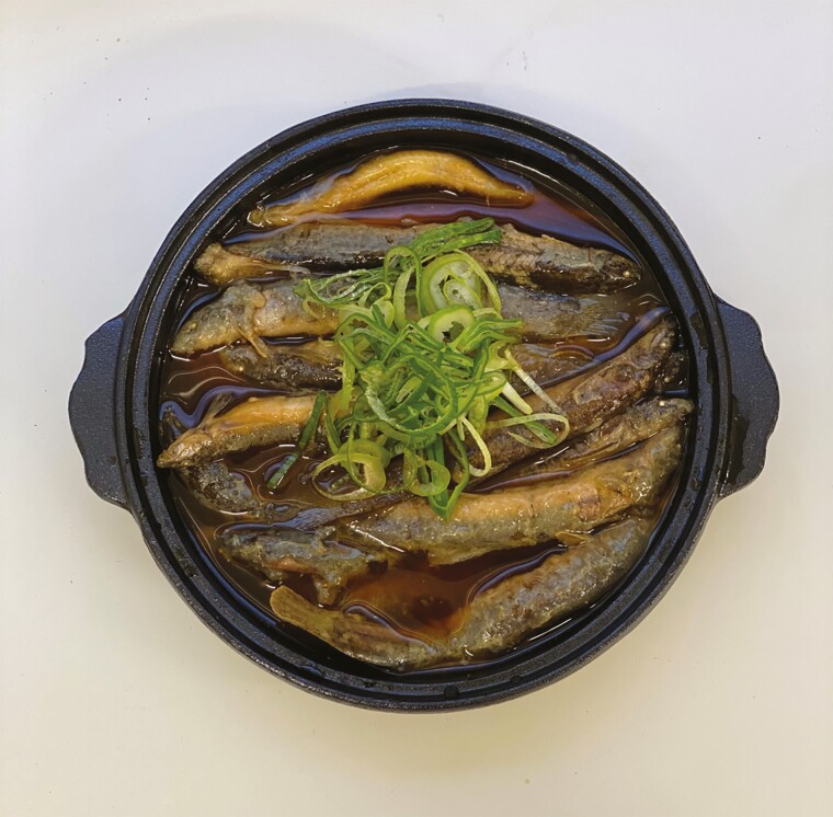
A 49-year-old Japanese man presented to our hospital with a 4-hour history of vomiting, severe watery diarrhea, intermittent abdominal pain, and generalized weakness. The patient had been diagnosed with alcoholic cirrhosis (Child-Pugh class B) 3 months previously when he developed worsening abdominal distention, shortness of breath, and fatigue. On the last evening before this admission, he had eaten Dojo nabe (Figure 1) at a local restaurant in Tokyo. He reported that there had been a dozen freshly caught loaches stewed with alcohol and soy sauce in a hot pot, some of which appeared undercooked. Twenty people, all of whom had no medical problems, had eaten the hotpot with him and none of them developed any symptoms. His medical history was otherwise unremarkable. He was taking furosemide and spironolactone, and had no known drug allergies. He reported drinking 1–2 L per day of beer for 23 years and smoking 1 pack of cigarettes per day for 30 years.
Figure 1.
Dojo nabe is loach hotpot, where loaches are put into alcohol alive and then cooked with soy sauce, broth, and green onions.
On the initial examination, he appeared pale and disoriented. He had a blood pressure of 70/40 mm Hg, heart rate of 160 beats per minute, temperature of 39.0°C, respiratory rate of 30 breaths per minute, and oxygen saturation of 80% while breathing 6 L of oxygen per minute. Physical examination revealed slightly icteric sclera, severe tenderness in the right lower quadrant without rigidity or rebound tenderness, hyperactive bowel sounds, and edema of the legs; his limbs were cool to the touch. Laboratory tests revealed a white blood cell count of 1100 cells/µL with 46.3% neutrophils; hemoglobin, 11.6 g/dL; platelets, 114 000/µL; aspartate aminotransferase, 112 U/L (reference range, 13–30 U/L); γ-glutamyl transpeptidase, 620 U/L (reference range, 13–64 U/L); alkaline phosphatase, 154 U/L (reference range, 38–113 U/L); total bilirubin, 1.8 mg/dL (reference, 0.3–1.2 mg/dL); direct bilirubin, 1.0 mg/dL (reference range, 0–0.4 mg/dL); lactate dehydrogenase, 400 U/L (reference range, 124–222 U/L); creatinine, 2.64 mg/dL (reference range, 0.65–1.07 mg/dL); and C-reactive protein, 1.34 mg/dL (reference range, 0‒0.30 mg/dL). Contrast-enhanced abdominal computed tomography revealed edematous mucosa from the ascending to transverse colon with mucosal enhancement and pericolonic fat strands, indicative of infective enterocolitis (Figure 2).
Figure 2.
Contrast-enhanced abdominal computed tomography revealed edematous mucosa from the ascending to transverse colon with mucosal enhancement and pericolonic fat strands, indicative of infective enterocolitis.
After admission, fluid resuscitation, vasopressors, and empiric antimicrobial therapy with meropenem were initiated. He was intubated and placed on mechanical ventilation and continuous hemodiafiltration. On day 2 of hospitalization, blood cultures obtained on admission grew gram-negative bacilli, which were later identified as P shigelloides using matrix-assisted laser desorption/ionization time-of-flight mass spectrometry (BioTyper version 9.0; Bruker, Billerica, Massachusetts) with a score of 2.430. Plesiomonas shigelloides was also isolated from his stool. Pending the results of final identification and susceptibility testing, levofloxacin was added to his treatment. Susceptibility testing performed using the MicroScan WalkAway 96 Plus system (Beckman Coulter, Brea, California) determined that the isolate was susceptible to ampicillin-sulbactam, piperacillin-tazobactam, ceftriaxone, cefotaxime, ceftazidime, cefepime, aztreonam, meropenem, amikacin, and levofloxacin, and resistant to ampicillin according to Clinical and Laboratory Standards Institute guidelines [3]. Meropenem was switched to cefotaxime based on previous reports of successful treatment [1, 2] and levofloxacin was continued for a week. Sequencing of the 16S ribosomal RNA gene (800 bp) of the strain showed a complete match with that of the type strain of P shigelloides (NCTC10360, accession number LT575468).
His multiorgan dysfunction gradually improved over the following week. On day 9 of hospitalization, he was extubated. After completing a 14-day course of antibiotic therapy, he was transferred to a rehabilitation hospital in stable condition. At follow-up 3 months later, he had fully recovered and his health was back to baseline.
DISCUSSION
Plesiomonas shigelloides is an anaerobic gram-negative bacillus of the order Enterobacterales, which is normally distributed in soil and fresh water [4]. It grows best in salinities of 0–4% and temperatures of 8°C–45°C [1]. Plesiomonas shigelloides has been isolated from a wide range of species including marine mammals, fish, shellfish, crustaceans, reptiles, amphibians, and birds [1].
Human infection is acquired through ingestion of raw fish and water-contaminated foods [5]. The incidence of P shigelloides enteritis shows considerable regional variation, with a higher incidence in Southeast Asia and Africa, where people consume raw fish, and is rare in North America and Europe [1]. In Japan, P shigelloides is a rare pathogen of mass food poisoning and a common cause of diarrhea in travelers from Southeast Asian countries [6]. It generally causes self-limited gastroenteritis, which resolves in 2–3 days; however, it can cause extraintestinal infections especially in neonates or adults with immunocompromising conditions, such as hematological disorders (thalassemia, sickle cell disease, leukemia), biliary tract disease, cirrhosis, asplenia, and iron overload conditions including hemochromatosis [2].
Bacteremia is a rare presentation of P shigelloides infection. Woo et al reviewed 38 cases from 1978 to 2003 [2]; however, only 4 cases have been reported worldwide in the last 15 years [7–10]. In all 43 cases (Table 1), including the present case, the patient’s median age was 38 years (range, 1 day to 94 years). The majority of patients (91%) had underlying predisposing factors: neonates (n = 11), splenectomy/functional asplenia and/or iron overload (n = 9), malignancy (n = 9), biliary tract disease (n = 6), and chronic liver disease (n = 3). Of the cases, 35 (81%) were monomicrobial infections. Thirty-four patients (79%) had documented primary site of infection: meningitis (n = 10, all neonates), cholangitis (n = 7), cellulitis (n = 5), gastroenteritis (n = 4), and abscesses (n = 3). Beta-lactams were the most commonly used antibiotics. The case fatality rate was 40% (17/43).
Table 1.
Characteristics of Patients With Plesiomonas shigelloides Bacteremia
| Characteristics | No. | (%) |
|---|---|---|
| Sex | ||
| Female | 16 | (37.2) |
| Male | 26 | (60.5) |
| Unknown | 1 | (2.3) |
| Age, y, median (range) | 38 | (0–94) |
| Predisposing factor | ||
| Neonates | 11 | (25.6) |
| Splenectomy/functional asplenia and/or iron overload | 9 | (20.9) |
| Malignancy | 9 | (20.9) |
| Biliary tract disease | 6 | (14.0) |
| None | 4 | (9.3) |
| Chronic liver disease | 3 | (7.0) |
| HIV infection | 1 | (2.3) |
| Monomicrobial infection | 35 | (81.4) |
| Diagnosis | ||
| Neonatal meningitis | 10 | (23.3) |
| Cholangitis | 7 | (16.3) |
| Primary bacteremia | 7 | (16.3) |
| Cellulitis | 5 | (11.6) |
| Gastroenteritis | 4 | (9.3) |
| Abscess | 3 | (7.0) |
| Disseminated infections | 3 | (7.0) |
| Arthritis | 1 | (2.3) |
| Empyema thoracis | 1 | (2.3) |
| Proctitis | 1 | (2.3) |
| Acute gangrenous cholecystitis | 1 | (2.3) |
| Epididymo-orchitis | 1 | (2.3) |
| Outcome | ||
| Survived | 26 | (60.5) |
| Died | 17 | (39.5) |
Abbreviation: HIV, human immunodeficiency virus.
People with cirrhosis are thought to be susceptible to P shigelloides infection through chronic iron overload, in addition to increased intestinal permeability and disturbed expression of intestinal antimicrobial peptides due to portal hypertension [11]. While isolated iron overload can cause liver injury, it is present in up to 80% of patients with cirrhosis, regardless of the etiology [12]. Plesiomonas shigelloides is siderophilic and uses heme as an iron source [13], and virulence is enhanced in iron-rich environments in a similar fashion as Vibrio vulnificus and Yersinia enterocolitica [14].
While P shigelloides enteritis is often self-limiting, antimicrobial therapy is needed in cases of extraintestinal disease. Plesiomonas shigelloides expresses chromosomally encoded noninducible β-lactamases and is resistant to ampicillin and carbenicillin, but is susceptible to aminopenicillins in combination with β-lactamase inhibitors, cephalosporins, carbapenems, aztreonam, tetracyclines, aminoglycosides, quinolones, trimethoprim, sulfamethoxazole, azithromycin, chloramphenicol, nitrofurantoin, and fosfomycin [15]. Antimicrobial susceptibility may be inoculum dependent [15]. In addition, some P shigelloides possess AmpC β-lactamase [16] and may develop resistance to third-generation cephalosporins while on treatment, which can lead to treatment failure as in other Enterobacterales [17]. The optimal antibiotic therapy against P shigelloides bacteremia remains uncertain and further studies are awaited.
Loaches are a freshwater fish that live in deep muddy ponds and rivers. Japanese people have been eating loaches since the 17th century and believe that they are very nutritious and effective for recovering stamina. Besides broiled loach fillet, Dojo nabe (loach hotpot) has been among those, where loaches are put into alcohol alive and then cooked with soy sauce, broth, and green onions (Figure 1). Nowadays local Dojo nabe restaurants are popular among foreign tourists to Tokyo and appear in many guidebooks and websites for tourists. Despite its popularity, P shigelloides infection following loach hotpot ingestion has not been reported previously. There is a potential for acquiring infections from eating loach hotpot because it is often presented undercooked so that diners can boil the hotpot by themselves before eating, which can be imperfect. Plesiomonas shigelloides exists in intestinal microbiota of loaches in paddy fields and ponds [18]. It is plausible that P shigelloides in loaches’ intestines caused a severe infection in our patient. Besides P shigelloides, Aeromonas species, Stenotrophomonas species, Shewanella species, and Lactococcus lactis also reside in loach intestines and may cause invasive infections, especially in immunocompromised patients, albeit rarely [18, 19].
As sushi has become a worldwide poplar cuisine, sushi-associated fishborne and foodborne parasitic zoonosis, such as cutaneous larva migrans with Gnathostoma species, has been well recognized [20, 21]. Our case adds another potential price for the enjoyment of exotic cuisine to the list and serves as a warning for people or travelers with immunocompromising conditions. It is thus important to ensure that loaches are well cooked before consumption.
Notes
Author contributions. All authors cared for the patient, performed the literature search, and critically reviewed the draft of the manuscript.
Patient consent statement. We obtained written informed consent from the patient.
Potential conflicts of interest. All authors: No reported conflicts of interest.
All authors have submitted the ICMJE Form for Disclosure of Potential Conflicts of Interest. Conflicts that the editors consider relevant to the content of the manuscript have been disclosed.
References
- 1.Janda JM, Abbott SL, McIver CJ. Plesiomonas shigelloides revisited. Clin Microbiol Rev 2016; 29:349–74. [DOI] [PMC free article] [PubMed] [Google Scholar]
- 2.Woo PCY, Lau SKP, Yuen KY. Biliary tract disease as a risk factor for Plesiomonas shigelloides bacteraemia: a nine-year experience in a Hong Kong hospital and review of the literature. New Microbiol 2005; 28:45–55. [PubMed] [Google Scholar]
- 3.Clinical and Laboratory Standards Institute. Performance Standards for Antimicrobial Susceptibility Testing. M45, 3rd ed. Wayne, PA: CLSI; 2016. [Google Scholar]
- 4.Miller ML, Koburger JA. Plesiomonas shigelloides: an opportunistic food and waterborne pathogen 1. J Food Prot 1985; 48:449–57. [DOI] [PubMed] [Google Scholar]
- 5.Marshman WE, Lyons CJ. Congenital endophthalmitis following maternal shellfish ingestion. Aust N Z J Ophthalmol 1998; 26:161–3. [DOI] [PubMed] [Google Scholar]
- 6.Ueda Y, Suzuki N, Furukawa T, et al. Bacteriological studies of traveller’s diarrhoea (6). Analysis of enteropathogenic bacteria at Kansai Airport quarantine station from September 4th, 1994 through December 1996. Kansenshogaku Zasshi 1999; 73:110–21. [DOI] [PubMed] [Google Scholar]
- 7.Ozdemir O, Sari S, Terzioglu S, Zenciroglu A. Plesiomonas shigelloides sepsis and meningoencephalitis in a surviving neonate. J Microbiol Immunol Infect 2010; 43:344–6. [DOI] [PubMed] [Google Scholar]
- 8.Auxiliadora-Martins M, Bellissimo-Rodrigues F, Viana JM, et al. Septic shock caused by Plesiomonas shigelloides in a patient with sickle beta-zero thalassemia. Heart Lung 2010; 39:335–9. [DOI] [PubMed] [Google Scholar]
- 9.Samannodi M, Zhao A, Nemshah Y, Shiley K. Plesiomonas shigelloides septic shock leading to death of postsplenectomy patient with pyruvate kinase deficiency and hemochromatosis. Case Rep Infect Dis 2016; 2016:1538501. [DOI] [PMC free article] [PubMed] [Google Scholar]
- 10.Pennycook KM, Pennycook KB, McCready TA, Kazanowski D. Severe cellulitis and bacteremia caused by Plesiomonas shigelloides following a traumatic freshwater injury. IDCases 2020; 19:e00637. [DOI] [PMC free article] [PubMed] [Google Scholar]
- 11.Bruns T, Zimmermann HW, Stallmach A. Risk factors and outcome of bacterial infections in cirrhosis. World J Gastroenterol 2014; 20:2542–54. [DOI] [PMC free article] [PubMed] [Google Scholar]
- 12.Deugnier Y, Turlin B. Pathology of hepatic iron overload. Semin Liver Dis 2011; 31:260–71. [DOI] [PubMed] [Google Scholar]
- 13.Oldham AL, Wood TA, Henderson DP. Plesiomonas shigelloides hugZ encodes an iron-regulated heme binding protein required for heme iron utilization. Can J Microbiol 2008; 54:97–102. [DOI] [PubMed] [Google Scholar]
- 14.Weinberg ED. Iron loading and disease surveillance. Emerg Infect Dis 1999; 5:346–52. [DOI] [PMC free article] [PubMed] [Google Scholar]
- 15.Stock I, Wiedemann B. Natural antimicrobial susceptibilities of Plesiomonas shigelloides strains. J Antimicrob Chemother 2001; 48:803–11. [DOI] [PubMed] [Google Scholar]
- 16.Adesiyan IM, Bisi-Johnson MA, Ogunfowokan AO, Okoh AI. Incidence and antimicrobial susceptibility fingerprints of Plesiomonas shigelloides isolates in water samples collected from some freshwater resources in southwest Nigeria. Sci Total Environ 2019; 665:632–40. [DOI] [PubMed] [Google Scholar]
- 17.Tamma PD, Doi Y, Bonomo RA, et al. ; Antibacterial Resistance Leadership Group. A primer on AmpC β-lactamases: necessary knowledge for an increasingly multidrug-resistant world. Clin Infect Dis 2019; 69:1446–55. [DOI] [PMC free article] [PubMed] [Google Scholar]
- 18.Yang S, Duan Y, Zhang J, et al. Observational comparisons of intestinal microbiota characterizations, immune enzyme activities, and muscle amino acid compositions of loach in paddy fields and ponds in Sichuan Province. Appl Microbiol Biotechnol 2017; 101:4775–89. [DOI] [PubMed] [Google Scholar]
- 19.Shimizu A, Hase R, Suzuki D, et al. Lactococcus lactis cholangitis and bacteremia identified by MALDI-TOF mass spectrometry: a case report and review of the literature on Lactococcus lactis infection. J Infect Chemother 2019; 25:141–6. [DOI] [PubMed] [Google Scholar]
- 20.Herman JS, Chiodini PL. Gnathostomiasis, another emerging imported disease. Clin Microbiol Rev 2009; 22:484–92. [DOI] [PMC free article] [PubMed] [Google Scholar]
- 21.Nawa Y, Hatz C, Blum J. Sushi delights and parasites: the risk of fishborne and foodborne parasitic zoonoses in Asia. Clin Infect Dis 2005; 41:1297–303. [DOI] [PubMed] [Google Scholar]


