Abstract
Objective:
To evaluate the effectiveness of low-level laser therapy (LLLT) in accelerating orthodontic tooth movement of crowded maxillary incisors.
Materials and Methods:
This two-arm, parallel-group, randomized controlled trial involved 26 patients with severe to extreme maxillary incisors irregularity according to Little's irregularity index, indicating two first premolars extraction. Patients were randomly assigned to either the laser group or the control group (13 each). Following premolars extraction, orthodontic treatment with fixed appliances was initiated for both groups. Immediately after insertion of the first archwire, patients in the laser group received a LLL dose from an 830-nm wavelength Ga-Al-As semiconductor laser device with energy of 2 J/point. The laser was applied to each maxillary incisor's root at four points (two buccal, two palatal). Application was repeated on days 3, 7, 14, and then every 15 days starting from the second month until the end of the leveling and alignment stage. Alignment progress was evaluated on the study casts taken before inserting the first archwire (T0), after 1 month of treatment commencement (T1), after 2 months (T2), and at the end of the leveling and alignment stage (T3). The outcome measures were the overall time needed for leveling and alignment and the leveling and alignment improvement percentage.
Results:
A statistically significant difference was found between the two groups in the overall treatment time (P < .001) and the leveling and alignment improvement percentage at T1 (P = .004) and T2; (P = .001).
Conclusion:
LLLT is an effective method for accelerating orthodontic tooth movement.
Keywords: Low-level laser therapy, Orthodontic tooth movement acceleration, Dental crowding, Leveling and alignment
INTRODUCTION
Dental crowding is considered the most common type of malocclusion. A survey stated that 78% of the American population have degrees of incisors irregularity, 15% of which is classified as severe to extreme.1 Leveling and alignment of such cases may take up to 8 months.2 In general, long orthodontic treatment time is one of the main reasons patients refuse to undergo treatment.3 It also has other disadvantages such as increased caries rates and root resorption.4 For these reasons, accelerating orthodontic tooth movement is desirable to prevent those effects and encourage patients to undergo treatment. Several approaches have been studied in an attempt to accelerate orthodontic tooth movement, including local injection of biological substances and surgical, mechanical, and physical methods.5
Recently, one of the physical methods, low-level laser therapy (LLLT), has proven to be effective in inducing remodeling processes in the alveolar bone by increasing osteoblast and osteoclast numbers, which leads to acceleration of orthodontic tooth movement.3,6 The application of LLLT in orthodontics has shown to be effective in reducing orthodontic pain and in the photobiomodulation that might accelerate orthodontic tooth movement.7,8 Several investigators have studied the use of LLLT in accelerating orthodontic tooth movement, most of them dealt with canine retraction cases.8,9 Some studies found laser effective while others concluded the opposite.10,11 These conflicting results may be explained by the difference in laser parameters used in each study regarding its type, application method, wavelength, dose of irradiation, and exposure time as these parameters relate directly to laser clinical results.6 Only three studies have evaluated the LLL effect during leveling and alignment of crowding cases.5,7,12 However, none of them was a randomized controlled trial (RCT), and they did not involve crowding cases with severe incisor irregularity. Recent systematic reviews stated that there is a lack of evidence regarding LLLT's effectiveness in accelerating orthodontic tooth movement, so there is a need for well-designed RCTs to determine the best protocols of laser parameters and present clear recommendations about its effects.10,11
To the best of our knowledge, this is the first published RCT having the objective of evaluating LLLT effectiveness in accelerating leveling and alignment in dental crowding cases.
MATERIALS AND METHODS
Trial Design
This study is a two-arm, parallel-group, RCT studying the effect of LLLT in accelerating tooth movement in dental crowding cases. The CONSORT statement was used as a guide for this study.13 The study was conducted in the Department of Orthodontics and Dentofacial Orthopaedics and Laser Research Unit at Damascus University between July 2015 and March 2016. Ethical approval was obtained from the Ethics Committee at the Ministry of Higher Education in Syria (26106/SM). This RCT is registered in the Clinical Trials database (NCT02568436). There is no funding to be declared.
Sample Size Calculation
Sample size was calculated using the G*power 3.1.3 program according to the following assumptions: depending on the results of a previous study2, The smallest clinically significant difference in time needed for leveling and alignment of severely crowded incisors—assuming a 40% reduction in treatment time using LLLT—would be 97.2 days. The standard deviation in the same study was 82.5 days. The statistical test to be used is a two-sample t-test with a statistical power of 80% and a significance level of 0.05. The given sample size was 26 patients (13 per group).
Participants
Participants were recruited from patients attending the Department of Orthodontics and Dentofacial Orthopaedics at Damascus University. Clinical examination was done on 94 patients. Patients were considered eligible for the study if they met the following inclusion criteria: aged between 16 and 24 years, presence of all maxillary permanent teeth except third molars, moderate crowding (tooth-size–arch-length discrepancy of 3–5 mm) in the anterior maxilla with Little's irregularity index (LII) of 7 mm or more—indicating extraction of two first premolars, the feasibility of bonding brackets on all maxillary teeth, no previous orthodontic treatment, no systemic diseases, and good oral hygiene.
Exclusion criteria were patients with severe tooth displacement (eg, ectopic canine) and those reporting the use of medications throughout the study. Twenty-six patients were selected to participate. The rights of patients were protected, and the purpose and methods of the study were completely explained to the patients and parents; an informed consent was obtained from each.
Randomization
Patients were assigned to a laser group or a control group with an allocation ratio of 1:1 using a simple randomization technique. Each patient was asked to select a folded piece of paper from a box containing 26 pieces of paper on 13 of which the word “laser” was written; on the other 13, the word “control” was written. According to which piece was selected, the patient was assigned to one of the two groups. The random allocation sequence, participants' enrollment, and assignment to intervention were done by the corresponding author.
Interventions
All 26 patients underwent conventional orthodontic treatment with fixed appliances. Patients in the laser group additionally underwent a LLL regimen throughout the leveling and alignment stages.
Five to 7 days after first premolar extraction, fixed orthodontic appliances of the MBT prescription and 0.022-inch slot height (American Orthodontics, Sheboygan, Wisc) were bonded. Then a 0.014-inch NiTi archwire (American Orthodontics) was inserted and tied to each bracket in the maxillary arch using ligature wires. Immediately after inserting the first archwire, a LLL dose was applied for the laser group patients using an 830-nm wavelength laser device (CMS Dental ApS, 55 Wildersgade, 1408 Copenhagen K, Denmark) with a 2.25-J/cm2 irradiation dose. Laser device parameters are listed in Table 1. The laser beam was applied to each root of the six maxillary incisors roots. Each root was divided into two halves: cervical and apical. The laser device tip was applied to the center of each half, perpendicular to the root and in direct contact with the mucosa from both the buccal and palatal sides so that there were four application points for each tooth with an exposure time of 1 minute/tooth. The laser application was repeated on days 3, 7, and 14 after the first application and every 15 days starting from the second month until the leveling and the alignment stage was complete. Irradiation was done by the corresponding author.
Table 1.
Laser Parameters Used in the Study
| Active Medium | Ga-Al-As |
| Emission type | Continuous |
| Wavelength | 830 nm |
| Dose of irradiation | 2.25 J/cm2 |
| Energy/point | 2 J |
| Output | 150 mW |
| Exposure time/point | 15 s |
| Application technique | Direct contact |
| Laser sessions | First mo: 4 (d 0, 3, 7, 14); starting from the second mo: every 15 d |
| Laser classification | 3B |
Clinical Procedures
For both groups, the archwire sequence used was 0.014-inch NiTi followed by 0.016 × 0.016-inch and 0.017 × 0.025-inch NiTi, and finally 0.019 × 0.025-inch stainless steel.
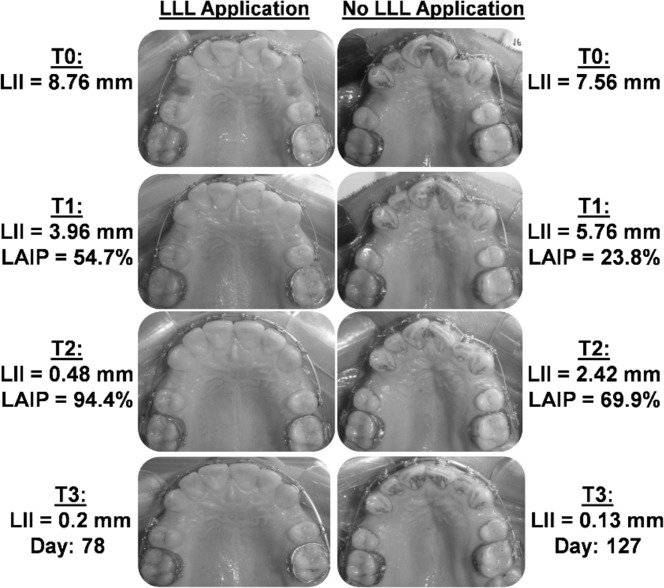
Patients were evaluated every week starting from the second month. Wire progression was achieved only if there was less than a 0.5-mm change in tooth movement within 2 weeks and the possibility of inserting the next archwire with full engagement into all brackets. Treatment was considered finished when LII was less than 1 mm, indicating complete alignment of the teeth and the feasibility of inserting the final archwire passively into all brackets, indicating complete leveling of the teeth (Figure 1).
Figure 1.
Two illustrative cases representing treatment progress in the laser group (left panel) and the control group (right panel). LII: Little's irregularity index; LAIP: Leveling and alignment improvement percentage.
Outcomes
The primary outcome measure was the overall time needed to complete leveling and aligning (OLAT) the maxillary dental arch. The secondary outcome measure was leveling and alignment improvement percentage (LAIP) of the maxillary teeth throughout the leveling and alignment stage.
To calculate outcome measures, a maxillary alginate impression was taken to make study casts at four time points: before insertion of the first archwire (T0), after 1 month of treatment commencement (T1), after 2 months (T2), and at the end of the leveling and alignment stage (T3), represented by final archwire insertion. LII was used to measure the change in tooth alignment on the casts. It involved measuring the horizontal linear distance among adjacent contact points of the six anterior teeth. The sum of these five measurements gave the value of the index.14 LII was measured using a digital caliper (Insize, Insize Co, Suzhou New District, China) to the nearest 0.01 mm by the corresponding author.
OLAT was calculated by the number of days between T0 and T3. LAIP was calculated by dividing the amount of change in the LII value at a specific time point (T1, T2, or T3; calculated by subtracting the LII value at T1, T2, or T3 from the LII value at T0) by Lll value at T0.
Error of the Method
To assess measurement reliability, 10 dental casts (of the T1 casts) were randomly chosen, and LII was remeasured 1 month after the first measurement. Reliability was evaluated using intraclass correlation (ICC), which gave a strong intraexaminer reliability (ICC = 0.998), and the Dahlberg formula, which showed minimal error that does not affect the reliability of the LII measurements.
Statistical Analysis
Statistical Analysis was performed using the SPSS program version 20 (SPSS Inc, Chicago, Ill). The Kolmogorov-Smirnov test was used to test normality of data distribution, which revealed normal distribution; therefore, parametric tests were used. A two-sample t-test was applied to evaluate the differences in OLAT and LAIP in each studied time point between the two groups. Significance level was set at 0.05.
RESULTS
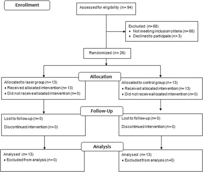
Patient flow through the study is illustrated in the CONSORT flow diagram shown in Figure 2. Twenty-six patients were recruited and allocated randomly to either the laser group or the control group. No dropout occurred, and complete follow-up and analysis were achieved for all patients. Table 2 shows the descriptive statistics of the sample regarding gender, age and initial LII (at T0).
Figure 2.
CONSORT flow diagram.
Table 2.
Sample Descriptive Statistics
|
|
N |
Sex |
Initial LII* (mm) |
Age (y) |
|||
| Male |
Female |
Mean |
SD |
Mean |
SD |
||
| Laser group | 13 | 2 | 11 | 8.91 | 1.57 | 18.53 | 2.9 |
| Control group | 13 | 4 | 9 | 10.8 | 2.29 | 21.61 | 2.63 |
| Total sample | 26 | 6 | 20 | 9.86 | 2.15 | 20.07 | 3.13 |
LII indicates Little's irregularity index.
Table 3 represents mean OLAT. A statistical significance was found between the two groups. The laser group needed less mean time (81.23 ± 15.29 days) to complete leveling and alignment than did the control group (109.23 ± 14.18 days); (P < .001), which means a 26% decrease in overall treatment time.
Table 3.
Overall Leveling and Alignment Time (D)
|
|
Min |
Max |
Mean |
SD |
P Value |
| Laser group | 57 | 106 | 81.23 | 15.29 | |
| Control group | 85 | 141 | 109.23 | 14.18 | <.001* |
Indicates significant.
Mean LAIP (Table 4) was significantly higher in the laser group than in the control group at T1 and T2. At T1, the percentage was 69.41 ± 15.45% for the laser group compared with 48.85 ± 17.04% for the control group (P = .004). At T2, the laser group LAIP was 89.42 ± 7.16% compared with 71.71 ± 16.18% for the control group (P = .001). No statistical significant difference was found between the two groups at T3 (P = .973).
Table 4.
Leveling and Alignment Improvement Percentage (%)
|
|
At T1 |
At T2 |
At T3 |
|||
| Mean |
SD |
Mean |
SD |
Mean |
SD |
|
| Laser group | 69.41 | 15.45 | 89.42 | 7.16 | 94.24 | 3.65 |
| Control group | 48.85 | 17.04 | 71.7 | 16.18 | 94.2 | 2.81 |
| P value | .004* | .001* | .973** | |||
Indicates significant; **Nonsignificant.
DISCUSSION
This study aimed to evaluate the effectiveness of LLLT in accelerating orthodontic tooth movement for leveling and alignment of dental crowding cases. We found that LLL accelerated leveling and alignment and reduced the overall time needed to achieve it by 26%.
An 830-nm wavelength laser device was used in this study. This wavelength falls in the optimal range (600–1000 nm),7 providing a proper photobiomodulation effect because it has a low absorbance coefficient in chromophores (ie, hemoglobin) and water that allows for proper penetration of the laser beam into the tissues.15 Furthermore, previous studies with a similar LLL wavelength found positive effects on orthodontic movement acceleration.8,12
The dose of irradiation has been reported as an important laser parameter that affects orthodontic tooth movement acceleration. No precise value has yet been defined. However, Goulart stated that a lower irradiation dose value has a more positive effect.16 In this study, a low dose of irradiation (2.25 J/cm2) was applied, which was effective in accelerating in accordance with the Sousa et al. study9 but contrary to the Altan et al. study,17 which also used low irradiation doses. The other important parameter of LLLT is the energy of the laser beam. Sousa et al. recommended that for tooth movement acceleration it should range between 0.2 J and 2.2 J to be clinically effective.11 For that, an energy of 2 J/point was used in this study.
Treatment progress was assessed every week starting from the second month of treatment to ensure precise evaluation to avoid missing important changes in treatment, which could affect interpretation of the results. LII was used to assess treatment outcome measure regarding LAIP. It is simple, reproducible, and considered to be an accurate and valid method of measuring anterior arch-length discrepancy.18 Patients with rotations or vertical discrepancies affecting LII accuracy were excluded from participating in the study.19
Our results showed that overall treatment time was 81.23 ± 15.29 days for the laser group and 109.23 ± 14.18 days for the control group, which means that laser application reduced leveling and alignment time by about 26%. Camacho and Cujar found similar results with a 30% treatment time reduction.12 However, they studied the whole treatment time until debonding of the fixed appliances, which may vary between patients according to specific treatment needs. Also, the study involved cases with little crowding amount and pretreatment parameters between the two groups were not adequately addressed.20
LAIP at T1 was 69.41 ± 14.45% for the laser group and 48.85 ± 17.04% for the control group. These results indicate a 30% higher leveling rate for the laser group. This percentage decreased to 20% at T2, with LAIP of 89.42 ± 7.16% for the laser group and 71.71 ± 16.18% for the control group. Results of this study agree with those of previous studies,8,9 which found a decrease in the improvement rate throughout treatment. This decrease could be explained by the gradual decrease in the targeted tissues response to the laser over time and because most of the LAIP in the laser group occurred during the first month, meaning that no important development occurred toward the end of this stage. No significant difference was found at T3 in LAIP, which seems to be normal given that the main factor to consider this stage finished was that LII was less than 1 mm.
Two previous studies found that the movement acceleration rate increased by 54%7 and more than 100%5 between the laser group and the control group. Those higher acceleration rates compared with this study's findings might be explained by the fact that those studies applied LLL on a daily basis for a long application time each day utilizing complicated extraoral devices. However, this is not considered practical for routine use in orthodontics compared with our protocol which applied the laser two or four times monthly with a LED-like portable device and an application time of 6 minutes/session.
Doshi-Mehta and Bhad-Patil applied a similar protocol to evaluate the LLLT effect on accelerating canine retraction and found a 30% higher acceleration rate for the laser group.8 Comparing the results of our study with theirs, considering two different phases of treatment, and applying similar protocols with similar results, we conclude that the laser regimen and parameters used in this study are effective; we recommend them as proper parameters for laser application in accelerating orthodontic tooth movement.
This study has some limitations: it was almost impossible to obtain the same values of LII for the 26 patients at treatment commencement. However, we tried to eliminate the effect of this factor by using the improvement percentage for each patient (instead of the LII value at each time point) as a criterion for evaluating the development of each case. Besides, it is difficult to control all the variables in the leveling and alignment stage as in other treatment stages (as canine retraction) because all teeth are involved in the movement. We tried to control that by recruiting cases with a close amount of initial crowding, standardizing wire sequence, and assessing patients weekly to avoid missing important changes in treatment. Also, no blinding was applied to either operator or patients, which sometimes risks bias. However, the risk of bias was eliminated by randomizing patient allocation to the respective groups, besides the fact that patient blinding does not affect treatment results because no patients' self-assessed outcomes were studied.
Despite these limitations, this study, which we consider the first RCT of its kind, used the best possible criteria, laser protocol, and parameters to achieve the most reliable results and recommendations for applying LLLT to accelerate orthodontic tooth movement.
CONCLUSION
-
•
Low-level laser therapy, used with the described parameters, is an effective method for accelerating orthodontic tooth movement in dental crowding cases.
REFERENCES
- 1.Proffit WR, Fields HW, Jr, Moray LJ. Prevalence of malocclusion and orthodontic treatment need in the United States: estimates from the NHANES III survey. Int J Adult Orthod Orthognath Surg. 1998;13:97–106. [PubMed] [Google Scholar]
- 2.Scott P, DiBiase AT, Sherriff M, Cobourne MT. Alignment efficiency of Damon 3 self-ligating and conventional orthodontic bracket systems: a randomized clinical trial. Am J Orthod Dentofacial Orthop. 2008. 134:470 e1–e8. [DOI] [PubMed]
- 3.Yoshida T, Yamaguchi M, Utsunomiya T, et al. Low-energy laser irradiation accelerates the velocity of tooth movement via stimulation of the alveolar bone remodeling. Orthod Craniofac Res. 2009;12:289–298. doi: 10.1111/j.1601-6343.2009.01464.x. [DOI] [PubMed] [Google Scholar]
- 4.Segal GR, Schiffman PH, Tuncay OC. Meta analysis of the treatment-related factors of external apical root resorption. Orthod Craniofac Res. 2004;7:71–78. doi: 10.1111/j.1601-6343.2004.00286.x. [DOI] [PubMed] [Google Scholar]
- 5.Kau CH, Kantarci A, Shaughnessy T, et al. Photobiomodulation accelerates orthodontic alignment in the early phase of treatment. Prog Orthod. 2013;14:30. doi: 10.1186/2196-1042-14-30. [DOI] [PMC free article] [PubMed] [Google Scholar]
- 6.Huang H, Williams RC, Kyrkanides S. Accelerated orthodontic tooth movement: molecular mechanisms. Am J Orthod Dentofacial Orthop. 2014;146:620–632. doi: 10.1016/j.ajodo.2014.07.007. [DOI] [PubMed] [Google Scholar]
- 7.Shaughnessy T, Kantarci A, Kau CH, Skrenes D, Skrenes S, Ma D. Intraoral photobiomodulation-induced orthodontic tooth alignment: a preliminary study. BMC Oral Health. 2016;16:3. doi: 10.1186/s12903-015-0159-7. [DOI] [PMC free article] [PubMed] [Google Scholar]
- 8.Doshi-Mehta G, Bhad-Patil WA. Efficacy of low-intensity laser therapy in reducing treatment time and orthodontic pain: a clinical investigation. Am J Orthod Dentofacial Orthop. 2012;141:289–297. doi: 10.1016/j.ajodo.2011.09.009. [DOI] [PubMed] [Google Scholar]
- 9.Sousa MV, Scanavini MA, Sannomiya EK, Velasco LG, Angelieri F. Influence of low-level laser on the speed of orthodontic movement. Photomed Laser Surg. 2011;29:191–196. doi: 10.1089/pho.2009.2652. [DOI] [PubMed] [Google Scholar]
- 10.de Almeida VL. de AndradeGois VL, Andrade RN, et al., editors. Efficiency of low-level laser therapy within induced dental movement: a systematic review and meta-analysis. J Photochem Photobiol B. 2016;158:258–266. doi: 10.1016/j.jphotobiol.2016.02.037. [DOI] [PubMed] [Google Scholar]
- 11.Sousa MV, Pinzan A, Consolaro A, Henriques JF, de Freitas MR. Systematic literature review: influence of low-level laser on orthodontic movement and pain control in humans. Photomed Laser Surg. 2014;32:592–599. doi: 10.1089/pho.2014.3789. [DOI] [PubMed] [Google Scholar]
- 12.Camacho AD, Velasquez Cujar SA. Acceleration effect of orthodontic movement by application of low-intensity laser. J Oral Laser Applications. 2010;10:99–105. [Google Scholar]
- 13.CONSORT http://www.consort-statement.org/consort-statement/
- 14.Little RM. The irregularity index: a quantitative score of mandibular anterior alignment. Am J Orthod. 1975;68:554–563. doi: 10.1016/0002-9416(75)90086-x. [DOI] [PubMed] [Google Scholar]
- 15.Heravi F, Moradi A, Ahrari F. The effect of low level laser therapy on the rate of tooth movement and pain perception during canine retraction. Oral Health Dent Manag. 2014;13:183–188. [PubMed] [Google Scholar]
- 16.Goulart CS, Nouer PR, Mouramartins L, Garbin IU, de Fatima Zanirato , Lizarelli R. Photoradiation and orthodontic movement: experimental study with canines. Photomed Laser Surg. 2006;24:192–196. doi: 10.1089/pho.2006.24.192. [DOI] [PubMed] [Google Scholar]
- 17.Altan AB, Sokucu O, Toker H, Sumer Z. The effects of low-level laser therapy on orthodontic tooth movement: metrical and immunological investigation. JSM Dentistry. 2014;2:1040. [Google Scholar]
- 18.Bernabe E, Flores-Mir C. Estimating arch length discrepancy through Little's irregularity index for epidemiological use. Eur J Orthod. 2006;28:269–273. doi: 10.1093/ejo/cji112. [DOI] [PubMed] [Google Scholar]
- 19.Sebastian B. Alignment efficiency of superelastic coaxial nickel-titanium vs superelastic single-stranded nickel-titanium in relieving mandibular anterior crowding: a randomized controlled prospective study. Angle Orthod. 2012;82:703–708. doi: 10.2319/072111-460.1. [DOI] [PMC free article] [PubMed] [Google Scholar]
- 20.Gkantidis N, Mistakidis I, Kouskoura T, Pandis N. Effectiveness of non-conventional methods for accelerated orthodontic tooth movement: a systematic review and meta-analysis. J Dent. 2014;42:1300–1319. doi: 10.1016/j.jdent.2014.07.013. [DOI] [PubMed] [Google Scholar]


