Abstract
Coenzyme A is synthesised from pantothenate via five enzyme-mediated steps. The first step is catalysed by pantothenate kinase (PanK). All PanKs characterised to date form homodimers. Many organisms express multiple PanKs. In some cases, these PanKs are not functionally redundant, and some appear to be non-functional. Here, we investigate the PanKs in two pathogenic apicomplexan parasites, Plasmodium falciparum and Toxoplasma gondii. Each of these organisms express two PanK homologues (PanK1 and PanK2). We demonstrate that PfPanK1 and PfPanK2 associate, forming a single, functional PanK complex that includes the multi-functional protein, Pf14-3-3I. Similarly, we demonstrate that TgPanK1 and TgPanK2 form a single complex that possesses PanK activity. Both TgPanK1 and TgPanK2 are essential for T. gondii proliferation, specifically due to their PanK activity. Our study constitutes the first examples of heteromeric PanK complexes in nature and provides an explanation for the presence of multiple PanKs within certain organisms.
Author summary
Apicomplexans are a phylum of obligate intracellular parasites that cause diseases in humans and other animals, inflicting considerable burdens on human societies. During their intracellular stage, these parasites must scavenge vitamins from their host organisms in order to survive and proliferate. One such vitamin is pantothenate (vitamin B5), which parasites convert in a universal five-step pathway to the essential cofactor coenzyme A (CoA). The first reaction in the CoA biosynthesis pathway is catalysed by the enzyme pantothenate kinase (PanK). The genomes of humans and many other organisms, including apicomplexans, encode multiple PanK homologues, although in all studied examples, the functional PanK enzyme exists as a homodimer. In this study, we demonstrate that the two PanK homologues encoded in the genomes of the apicomplexans Plasmodium falciparum and Toxoplasma gondii, PanK1 and PanK2, exist as functional heteromeric complexes. We provide evidence that both PanK homologues contribute to the PanK activity in these parasites, and that both PanK1 and PanK2 are essential for the proliferation of T. gondii parasites specifically for their PanK activity. Our data describe the first known instances of heteromeric PanK complexes in nature and may explain why some organisms that express multiple PanKs harbour seemingly non-functional isoforms.
Introduction
Coenzyme A (CoA) is an essential enzyme cofactor in all living organisms [1]. CoA itself, and the CoA-derived phosphopantetheine prosthetic group required by various carrier proteins, function as acyl group carriers and activators in key cellular processes such as fatty acid biosynthesis, β-oxidation and the citric acid cycle. Pantothenate kinase (PanK) catalyses the first step in the conversion of pantothenate (vitamin B5) to CoA [2]. PanKs are categorised into three distinct types—type I, II and III—based on their primary sequences, structural fold, enzyme kinetics and inhibitor sensitivity. PanKs from all three types have been shown to exist as homodimers based on their solved protein structures [3–10]. All eukaryotic PanKs that have been characterised so far are type II PanKs. Interestingly, many eukaryotes (such as Arabidopsis thaliana [11,12], Mus musculus [13–16] and Homo sapiens [17–21]) express multiple PanKs and in some cases it is clear that these PanKs are not functionally redundant [15,22]. For example, mutations in only one of four type II PanKs in humans causes a neurodegenerative disorder known as PanK-associated neurodegeneration [17]. Some bacteria also express multiple PanKs. For example, some Mycobacterium [23], Streptomyces [7] and Bacillus [7,24,25] species have both type I and type III PanKs, while a select few bacilli (including the category A biodefense pathogen Bacillus anthracis) carry both a type II and type III PanK [7]. In some organisms harbouring multiple PanKs, it has not been possible to demonstrate functional activity for all enzymes. One of the four human type II PanKs was shown to be catalytically inactive [21,26], as is a type III PanK from Mycobacterium tuberculosis [23], and a type II PanK from B. anthracis [7]. The reason for the presence of multiple PanKs within certain cells, and the apparent inactivity of certain PanKs, is unclear.
Two putative genes coding for PanK enzymes have been identified in each of the genomes of the pathogenic apicomplexan parasites Plasmodium falciparum (PF3D7_1420600 (Pfpank1) and PF3D7_1437400 (Pfpank2)) and Toxoplasma gondii (TGME49_307770 (Tgpank1) and TGME49_235478 (Tgpank2)). We have recently shown that mutations in PfPanK1 alter PanK activity in P. falciparum, providing evidence that PfPanK1 is an active PanK, at least in the disease-causing stage of the parasite’s lifecycle [27]. The function of PfPanK2 and its contribution to PanK activity in P. falciparum is unknown. PfPanK2 contains a unique, large insert in a loop associated with the dimerisation of PanKs in their native conformation [8] and this may affect its ability to form a dimer, rendering it inactive [28]. No functional information is available on the putative T. gondii PanKs, but a genome-wide CRISPR-Cas9 screen of the T. gondii genome predicted that both PanK genes are important for parasite proliferation in vitro [29]. Similarly, a recent genome-wide insertional mutagenesis study of P. falciparum has predicted that mutations in either PfPanK1 or PfPanK2 result in significant fitness costs to the parasite [30]. These results suggest that the PanK2 proteins of these parasites play important role(s), although their exact function remains unclear.
In this study, we demonstrate that PanK1 and PanK2 from both P. falciparum and T. gondii are part of the same, multimeric protein complex in these parasites. This constitutes the first identification of heteromeric PanK complexes in nature. Furthermore, our data provide the first evidence that PanK2 contributes to, and is essential for, PanK function in apicomplexans.
Results
PfPanK1 and PfPanK2 are part of the same protein complex
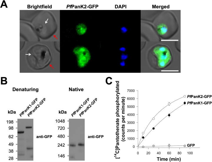
The importance and role of PanK2 in apicomplexan parasites have not previously been established. To characterise the P. falciparum PanK2 homologue (PfPanK2), we first determined where in the parasite the protein localises. We episomally expressed PfPanK2-GFP in asexual blood stage P. falciparum parasites and found that PfPanK2-GFP is localised throughout the parasite cytosol and is not excluded from the nucleus (Fig 1A). This is a similar localisation to what we observed for PfPanK1-GFP previously [27]. Western blotting (S1 Fig, red arrow) of proteins separated by SDS-PAGE revealed that PfPanK2-GFP has a molecular mass consistent with the predicted mass of the fusion protein (~118 kDa; Fig 1B), which is slightly larger than the predicted mass of PfPanK1-GFP (~87 kDa; Fig 1B). As PanKs from other organisms exist as homodimers, we undertook blue native-PAGE to determine whether PfPanK1-GFP and PfPanK2-GFP exist in protein complexes. Interestingly, under native conditions, both PfPanK1-GFP and PfPanK2-GFP were found to be part of complexes that are ~240 kDa in mass (Fig 1B).
Fig 1. PfPanK1 and PfPanK2 are part of similar-sized protein complexes that possess PanK activity.
(A) Confocal micrographs showing the subcellular location of PfPanK2-GFP within trophozoite/schizont-stage P. falciparum-infected erythrocytes. The nuclei of the parasites are stained with DAPI. From left to right: Brightfield, GFP-fluorescence, DAPI-fluorescence, and merged images. Arrows indicate the plasma membranes of the erythrocyte (red) or the parasite (white). Scale bars represent 5 μm. (B) Denaturing and native western blot analyses of the GFP-tagged proteins in the PfPanK1-GFP and PfPanK2-GFP parasite lines. The expected sizes of the proteins are ~87 kDa for PfPanK1-GFP and ~118 kDa for PfPanK2-GFP. For reference, the molecular mass of the GFP tag is ~27 kDa. Western blots were performed with anti-GFP antibodies and each of the blots shown is representative of three independent experiments, each performed with a different batch of parasites. (C) The phosphorylation of [14C]pantothenate (initial concentration of 2 μM, ~10,000 counts per minute) over time by the immunopurified complex from lysates of parasites expressing PfPanK1-GFP (black circles), PfPanK2-GFP (white circles) and untagged GFP (grey circles). Data shown are representative of two independent experiments, each performed with a different batch of parasites and carried out in duplicate. Error bars represent range/2 and are not shown if smaller than the symbols.
To determine the activity and protein composition of these complexes, we set out to purify the PfPanK1-GFP and PfPanK2-GFP complexes by immunoprecipitation. As a control, we also purified untagged GFP. We verified that most of the GFP-tagged proteins were captured from the total lysates prepared from cell lines expressing the different proteins, with bands corresponding to PfPanK1-GFP, PfPanK2-GFP and the untagged GFP epitope tag detected in the bound fraction of the respective cell lines (S2 Fig). To determine whether the purified PfPanK1 and PfPanK2 complexes possess PanK activity, we performed a [14C]pantothenate phosphorylation assay (S1 Fig, orange arrow). We found that 50 − 60% of the [14C]pantothenate initially present in the reaction was phosphorylated within 90 min by the immunopurified complex from both the PfPanK1-GFP and PfPanK2-GFP lines (Fig 1C). Conversely, the immunopurified untagged GFP did not display PanK activity (Fig 1C). These experiments provide the first indication that PfPanK1 and PfPanK2 exist as part of an active PanK enzyme complex of similar mass in P. falciparum parasites. They also provide the first indication that PfPanK2 contributes to PanK activity in these parasites.
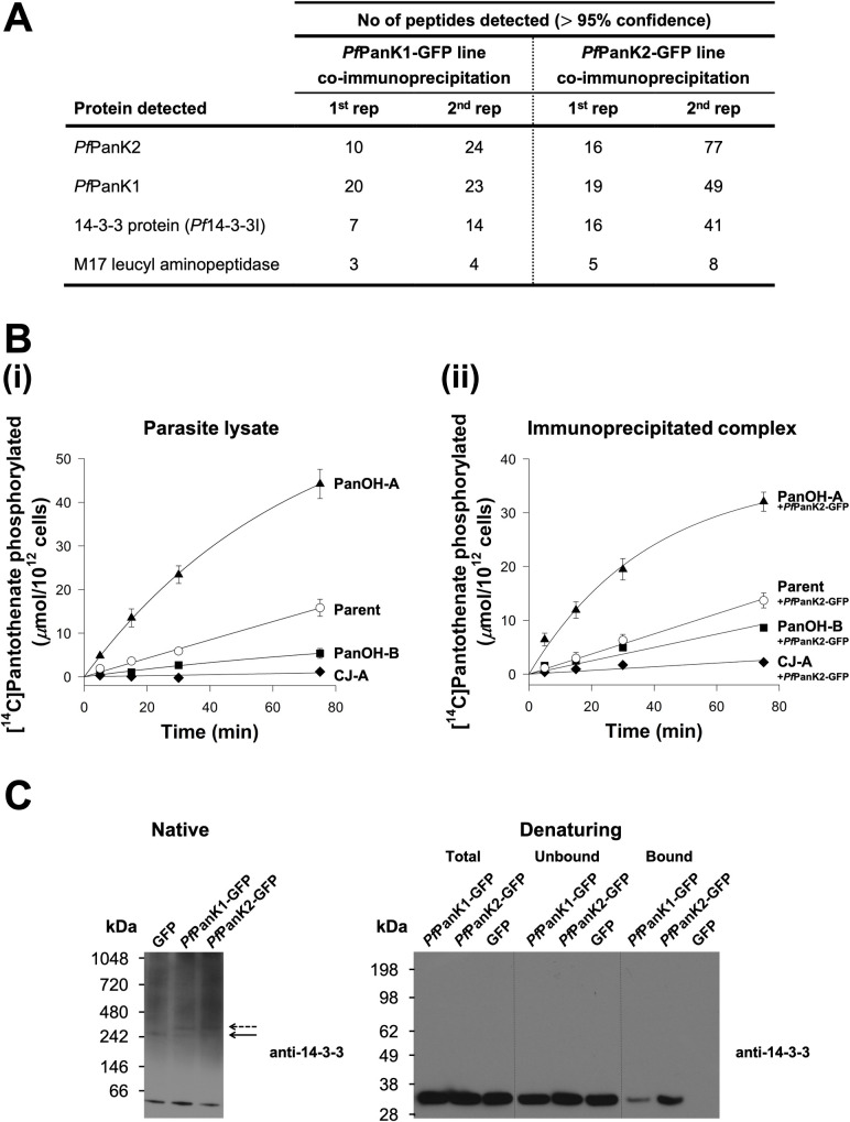
To elucidate the protein composition of the PfPanK1-GFP and PfPanK2-GFP complexes, the immunoprecipitated samples were subjected to mass spectrometry (MS)-based proteomic analysis (S1 Fig, green arrow; bound fractions of untagged GFP-expressing and 3D7 wild-type parasites were included as negative controls). Both PfPanK1 (36–50% coverage, S3 Fig) and PfPanK2 (29–49% coverage, S4 Fig) were unequivocally detected as the two most abundant proteins in the immunopurified complex from both the PfPanK1-GFP and PfPanK2-GFP lines (Fig 2A). Interestingly the next most abundant protein detected in both complexes was Pf14-3-3I (43–67% coverage, Figs 2A and S5). These results are consistent with PfPanK1, PfPanK2 and Pf14-3-3I being part of the same protein complex. Other proteins, such as M17 leucyl aminopeptidase (fourth most abundant), were also detected in the MS analysis, albeit with a comparatively fewer number of peptides (Fig 2A and S1 Table).
Fig 2. PfPanK1 and PfPanK2 are part of a single PanK complex that includes Pf14-3-3I.
(A) The four most abundant proteins identified in the MS analysis of proteins immunoprecipitated with GFP-Trap from the PfPanK1-GFP and PfPanK2-GFP lines. Data shown are representative of two independent analyses (1st and 2nd rep), each performed with a different batch of parasites. Proteins detected in the untagged GFP line or wild-type 3D7 parasite immunoprecipitations (negative controls) were removed. Only proteins with three or more peptides detected in both replicate co-immunoprecipitation experiments are shown. Proteins identified but which did not meet these criteria are shown in S1 Table. Proteins are listed in descending order according to the total number of peptides detected across all replicates (total peptides in all four columns). (B) The phosphorylation of [14C]pantothenate (initial concentration of 2 μM) over time by (i) lysates generated from Parent (white circles), PanOH-A (black triangles), PanOH-B (black squares) and CJ-A (black diamonds) parasite lines (reproduced from [27]) and (ii) proteins immunoprecipitated with GFP-Trap from Parent+PfPanK2-GFP (white circles), PanOH-A+PfPanK2-GFP (black triangles), PanOH-B+PfPanK2-GFP (black squares) and CJ-A+PfPanK2-GFP (black diamonds) parasite lysates. Values in (ii) are averaged from three independent experiments, each performed with a different batch of parasites and carried out in duplicate. Error bars represent SEM and are not shown if smaller than the symbols. (C) Native western blot analysis of the lysates and denaturing western blot analyses of the different GFP-Trap immunoprecipitation fractions generated from PfPanK1-GFP and PfPanK2-GFP parasite lines, with the untagged GFP line as a control. Protein samples used in the denaturing western blot were derived from the same immunoprecipitation depicted in S2 Fig. Western blots were performed with pan-specific anti-14-3-3 antibodies (previously shown to detect Plasmodium 14-3-3 [32]). Arrows indicate the position of 14-3-3-containing complexes of comparable masses in all three lines (solid arrow) and the complexes found only in the PfPanK1-GFP and PfPanK2-GFP lines (dashed arrow). The native blot shown is a representative of three independent experiments, while the denaturing blot is a representative of two independent experiments, each performed with a different batch of parasites.
Our group had previously generated three mutant strains, termed PanOH-A, PanOH-B and CJ-A, by drug-pressuring P. falciparum parasites with antiplasmodial pantothenate analogues [27]. These strains harbour mutations in PfPanK1 (D507N, ΔG95 and G95A, respectively) that affect PfPanK catalytic activity [27]. To test further whether PfPanK1 and PfPanK2 are part of the same protein complex, we introduced episomally-expressed PfPanK2-GFP into these parasite strains (the newly generated lines are designated with “+PfPanK2-GFP” superscript). We then immunopurified the PfPanK2-GFP complex from the PanOH-A+PfPanK2-GFP, PanOH-B+PfPanK2-GFP and CJ-A+PfPanK2-GFP lines, as well as from the wild type control (Parent+PfPanK2-GFP), and performed [14C]pantothenate phosphorylation assays with the complex derived from immunopurified PfPanK2-GFP (S1 Fig, orange arrow). As we reported previously, the PfPanK1 mutations alter the PfPanK activity of PanOH-A, PanOH-B and CJ-A parasites (using lysate, S1 Fig, blue arrow) such that the following rank order of enzyme activity relative to the Parent line is observed: PanOH-A > Parent > PanOH-B > CJ-A (Fig 2B(i), [27]). Notably, PanK activity of the complex immunopurified from the various PfPanK2-GFP-expressing mutant lines followed the same rank order: PanOH-A+PfPanK2-GFP > Parent+PfPanK2-GFP > PanOH-B+PfPanK2-GFP > CJ-A+PfPanK2-GFP (Fig 2B(ii)). This difference in pantothenate phosphorylation rates was not due to variations in the amount of PfPanK2-GFP protein in the immunopurified complexes used for the assays (S6 Fig). Further, the initial rate of pantothenate phosphorylation by the Parent lysate was 0.211 ± 0.026 (mean ± SEM) μmol/1012 cells/min (Fig 2B(i)) while that of the immunopurified complex from the Parent+PfPanK2-GFP line was 0.177 ± 0.016 (mean ± SEM) μmol/1012 cells/min (Fig 2B(ii)), demonstrating that the PanK activity in the immunopurified complexes is indistinguishable (95% CI = -0.119 to 0.051; include 0) from that observed in parasite lysates. Overall, the data shown in Fig 2B are consistent with PfPanK2-GFP associating with the mutant PfPanK1 from each cell line and indicate that both proteins are part of the same PanK complex in P. falciparum.
Our proteomic analysis identified Pf14-3-3I as being co-immunoprecipitated with both PfPanK1 and PfPanK2 (Fig 2A). To test whether Pf14-3-3I is a bona fide component of the PanK complex of P. falciparum, we performed western blotting with a pan-specific anti-14-3-3 antibody. Under native conditions (S1 Fig, red arrow), the 14-3-3 antibody detected a major protein band at <66 kDa (Fig 2C), which likely represents dimeric Pf14-3-3I of the parasite [31]. We also observed a protein complex of ~240 kDa in the PfPanK1-GFP, PfPanK2-GFP and untagged GFP lines (solid arrow, Fig 2C). In addition, a protein complex of slightly higher molecular mass, likely corresponding to the PanK complex that includes the GFP epitope tag, was also observed in the PfPanK1-GFP and PfPanK2-GFP lines but not the untagged GFP line (dashed arrow, Fig 2C). As a direct test for whether Pf14-3-3I exists in the same complex as PfPanK1 and PfPanK2, we performed western blotting on proteins immunopurified with anti-GFP antibodies from the PfPanK1-GFP, PfPanK2-GFP and untagged GFP parasite lines (S1 Fig, purple arrow). We found that Pf14-3-3I protein was detected in the immunopurified complex from both the PfPanK1-GFP and PfPanK2-GFP lines, but not in that purified from parasites expressing untagged GFP (Fig 2C). Together with the native western blot (Fig 1B) and proteomic (Fig 2A) analyses, these results are consistent with PfPanK1 and PfPanK2 being part of the same complex that also contains Pf14-3-3I, and that this complex is responsible for the PanK activity observed in the intraerythrocytic stage of P. falciparum.
TgPanK1 and TgPanK2 also constitute a single complex with PanK activity that is essential for parasite proliferation
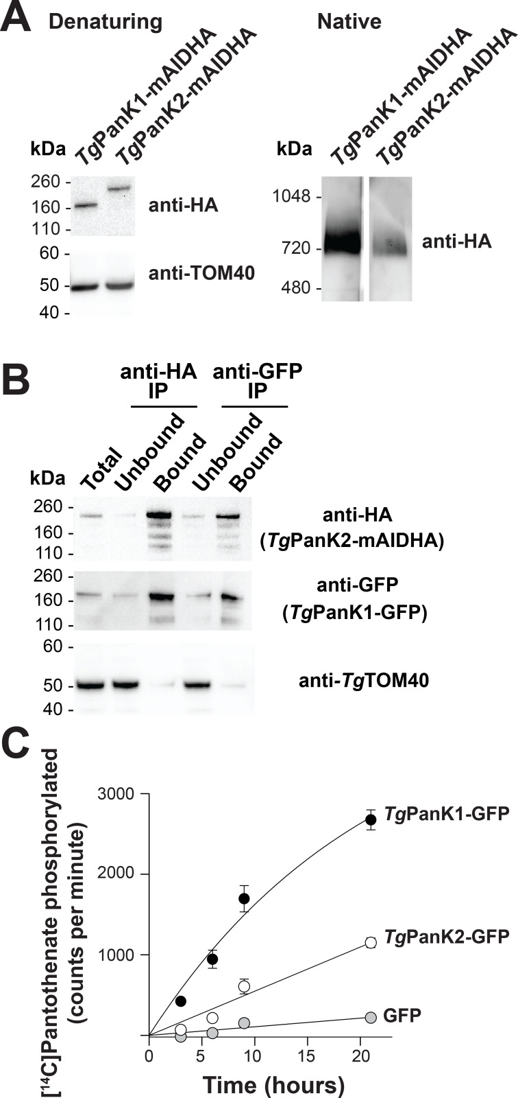
Based on sequence similarity, TgPanK1 and TgPanK2 are homologous to their P. falciparum counterparts (S7 Fig). To begin characterising TgPanK1 and TgPanK2, we introduced the coding sequence for a mini-Auxin-Inducible Degron (mAID)-haemagglutinin (HA) tag into the 3’ region of the open reading frames of TgPanK1 or TgPanK2 in RH TATiΔKu80:TIR1 strain T. gondii parasites [33] also expressing a ‘tdTomato’ red fluorescent protein (RFP) (S8A Fig). Successful integration of the mAIDHA tag was verified by PCR (S8B Fig). Western blotting (S1 Fig, red arrow) revealed that the TgPanK1-mAIDHA and TgPanK2-mAIDHA proteins have molecular masses of ~160 and ~200 kDa, respectively (Fig 3A), corresponding to the predicted sizes of TgPanK1-mAIDHA (143 kDa) and TgPanK2-mAIDHA (189 kDa). When analysed under native conditions, TgPanK1-mAIDHA and TgPanK2-mAIDHA both exist in protein complexes of ~720 kDa in mass (Fig 3A).
Fig 3. TgPanK1 and TgPanK2 are part of a single protein complex with PanK activity.
(A) Denaturing and native western blot analyses of the HA-tagged proteins in TgPanK1-mAIDHA and TgPanK2-mAIDHA parasite lines. The expected sizes of TgPanK1-mAIDHA and TgPanK2-mAIDHA are ~143 kDa and ~189 kDa, respectively. Western blots were performed with an anti-HA antibody and each blot shown is a representative of three independent experiments, each performed with different batches of parasites. Denaturing western blots were also probed with anti-TgTOM40, which served as a loading control. (B) Western blot analysis of proteins from TgPanK1-GFP/TgPanK2-mAIDHA parasite lysates immunoprecipitated with GFP-Trap and anti-HA beads (TgPanK1-GFP is 160 kDa). Protein samples were collected before immunoprecipitation (Total), from the fraction not bound to the GFP-Trap/anti-HA beads (Unbound), and from the fraction bound to the GFP-Trap/anti-HA beads (Bound). Membranes were probed with anti-GFP and anti-HA antibodies, and the blot shown is representative of three independent experiments, each performed with different batches of parasites. TgTOM40 served as a control protein that is part of an unrelated protein complex. Bound fractions contain protein from 4 × as many cells as the total and unbound lanes. (C) The phosphorylation of [14C]pantothenate (initial concentration 2 μM) over time by protein samples immunoprecipitated with GFP-Trap from TgPanK1-GFP/TgPanK2-mAIDHA (black circles),TgPanK1-HA/TgPanK2-GFP (white circles) and untagged GFP (grey circles) lines. Data shown are representative of two independent experiments, each performed with a different batch of parasites and carried out in duplicate. Error bars represent the range/2 and are not shown if smaller than the symbols.
To investigate if TgPanK1 and TgPanK2 are part of the same ~720 kDa complex, we introduced a sequence encoding a GFP tag into the genomic locus of Tgpank1 in the TgPanK2-mAIDHA strain (S8A and S8C Fig). Co-immunoprecipitation experiments (S1 Fig, purple arrow) revealed that TgPanK1-GFP (160 kDa) co-purified with TgPanK2-mAIDHA (Fig 3B). Analogous experiments with a TgPanK1-HA/TgPanK2-GFP line, wherein we integrated a sequence encoding a GFP tag into the Tgpank2 locus and a sequence encoding a HA tag into the Tgpank1 locus (S8A and S8D Fig), yielded similar results (S9 Fig). We therefore conclude that, like PfPanK1 and PfPanK2 in P. falciparum (Figs 1 and 2), TgPanK1 and TgPanK2 are components of the same protein complex. We also tested whether the PanK complex in T. gondii contains an orthologue of 14-3-3I. Western blot analysis (S1 Fig, purple arrow) of the fractions from TgPanK1-GFP/TgPanK2-mAIDHA co-immunoprecipitation using a pan-14-3-3 antibody showed that Tg14-3-3 could be detected in T. gondii parasite lysates but not in the TgPanK complex (S10 Fig).
To determine whether the TgPanK1/TgPanK2 complex has pantothenate kinase activity, we immunopurified proteins from TgPanK1-GFP/TgPanK2-mAIDHA, TgPanK1-HA/TgPanK2-GFP and control (expressing untagged GFP) cell lines using GFP-Trap, and measured the ability of the purified proteins to phosphorylate pantothenate (S1 Fig, orange arrow). The samples purified from the TgPanK1-GFP/TgPanK2-mAIDHA and TgPanK1-HA/TgPanK2-GFP lines exhibited higher pantothenate phosphorylation activity than that from the control parasites expressing untagged GFP (Fig 3C). These findings indicate that, like the P. falciparum PanK complex (Figs 1 and 2), the TgPanK1/TgPanK2 complex possesses PanK activity.
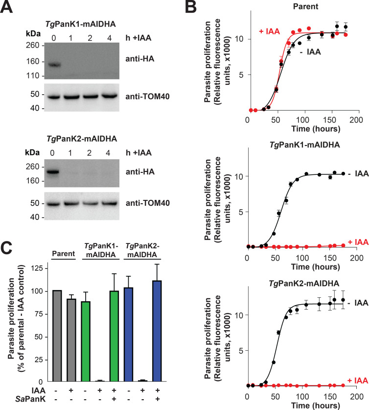
Active PanK proteins from other organisms contain conserved nucleotide binding motifs [34,35]. As is the case for PfPanK2, the nucleotide-binding motifs of TgPanK2 deviate substantially from those of other eukaryotic PanKs (S7 Fig). It is therefore unclear whether pantothenate phosphorylation is catalysed solely by TgPanK1 or if TgPanK2 also contributes to PanK activity. To answer this, we first investigated whether TgPanK1 and TgPanK2 are important for parasite proliferation (S1 Fig, pink arrows). TgPanK1 and TgPanK2 were individually knocked down by exposing the mAID-regulated lines to 100 μM indole-3-acetic acid (IAA–a plant hormone of the auxin class), a concentration that we determined was not detrimental to wild-type parasite proliferation. TgPanK1-mAIDHA and TgPanK2-mAIDHA were degraded within an hour of exposing the parasites to IAA (Fig 4A). Both the TgPanK1-mAIDHA and TgPanK2-mAIDHA lines express RFP, which enabled us to monitor parasite proliferation using fluorescence growth assays, as described previously [36]. We measured proliferation of the TgPanK1-mAIDHA, TgPanK2-mAIDHA and parental lines cultured in the presence or absence of 100 μM IAA over seven days. In the absence of IAA, we observed a normal sigmoidal growth curve for all three lines (Fig 4B). By contrast, we observed a complete cessation of proliferation of the parasite lines expressing TgPanK1-mAIDHA and TgPanK2-mAIDHA, but not the parental strain, in the presence of 100 μM IAA (Fig 4B). These data indicate that both TgPanK1 and TgPanK2 are crucial for T. gondii proliferation and, notably, that neither can substitute for the other. To establish whether TgPanK1 and TgPanK2 are essential due to the PanK activity of the complex, we set out to complement the knocked down TgPanKs with a well-characterised PanK. The Staphylococcus aureus PanK (SaPanK) was selected for this purpose because (i) it is a type II PanK (like the TgPanKs), (ii) it has been shown to function as a homodimer (so only the one gene needs to be expressed) and (iii) it is refractory to negative feedback inhibition by CoA (allowing the enzyme to function even if the parasite maintains high levels of CoA). We therefore constitutively expressed Sapank fused with the coding sequence for a Ty1 epitope tag in both the TgPanK1-mAIDHA and TgPanK2-mAIDHA parasite lines, generating lines that we termed TgPanK1-mAIDHA+SaPanK-Ty1 and TgPanK2-mAIDHA+SaPanK-Ty1. The expression of the SaPanK protein in these strains was verified by immunofluorescence microscopy and western blot (S11 Fig). We measured the proliferation of the TgPanK1-mAIDHA+SaPanK-Ty1 and TgPanK2-mAIDHA+SaPanK-Ty1 lines in the presence and absence of 100 μM IAA, and compared this with the proliferation of the TgPanK1-mAIDHA, TgPanK2-mAIDHA and Parent lines. We obtained fluorescence measurements over a 7 day period and compared the proliferation of each strain when the Parent strain cultured in the absence of IAA reached mid-log phase. We found that both the TgPanK1-mAIDHA+SaPanK-Ty1 and TgPanK2-mAIDHA+SaPanK-Ty1 lines proliferated at a similar rate to the Parent control line when cultured in the presence of IAA, in contrast to the TgPanK1-mAIDHA and TgPanK2-mAIDHA lines, where minimal proliferation was observed (Fig 4C).
Fig 4. Expression of both TgPanK1 and TgPanK2 is necessary for PanK activity and for T. gondii tachyzoite proliferation.
(A) IAA-induced knockdown of TgPanK1-mAIDHA or TgPanK2-mAIDHA protein over time. Western blot analysis of TgPanK1-mAIDHA and TgPanK2-mAIDHA lines incubated with either 100 μM IAA (+IAA) for 1, 2 and 4 h or an ethanol vehicle control (0 h). Membranes were probed with anti-HA antibody to detect the TgPanK1-mAIDHA and TgPanK2-mAIDHA proteins, and with anti-TgTom40 as a loading control. Western blots shown are representative of three independent experiments, each performed with a different batch of parasites. (B) The effect of TgPanK1-mAIDHA or TgPanK2-mAIDHA knockdown on T. gondii tachyzoite proliferation. Parent, TgPanK1-mAIDHA and TgPanK2-mAIDHA lines (all expressing tdTomato RFP) were cultured over 7 days in the presence (red circles) or absence (black circles) of 100 μM IAA. Parasite proliferation was measured over time by assessing the RFP expression using a fluorescence reader. Graphs shown are representative of three independent experiments carried out in triplicate, each performed with a different batch of parasites. Error bars represent SD and are not shown if smaller than the symbols. (C) Complementation of TgPanK1 and TgPanK2 knockdown with SaPanK. SaPanK was constitutively expressed (+) in TgPanK1-mAIDHA+SaPanK-Ty1 (green bars) and TgPanK2-mAIDHA+SaPanK-Ty1 (blue bars) parasites. These lines were cultured alongside the non-complemented (-) TgPanK1-mAIDHA (green bars), TgPanK2-mAIDHA (blue bars) and parent lines (grey bars). All parasite lines were cultured either in the presence (+) or absence (-) of 100 μM IAA. Parasite proliferation was monitored 1–2 times daily for 7 days. Proliferation was compared when the Parent strain cultured in the absence of IAA was at the mid-log phase of parasite proliferation. Values are averaged from three independent experiments, each performed with a different batch of parasites and carried out in triplicate. Error bars represent SEM.
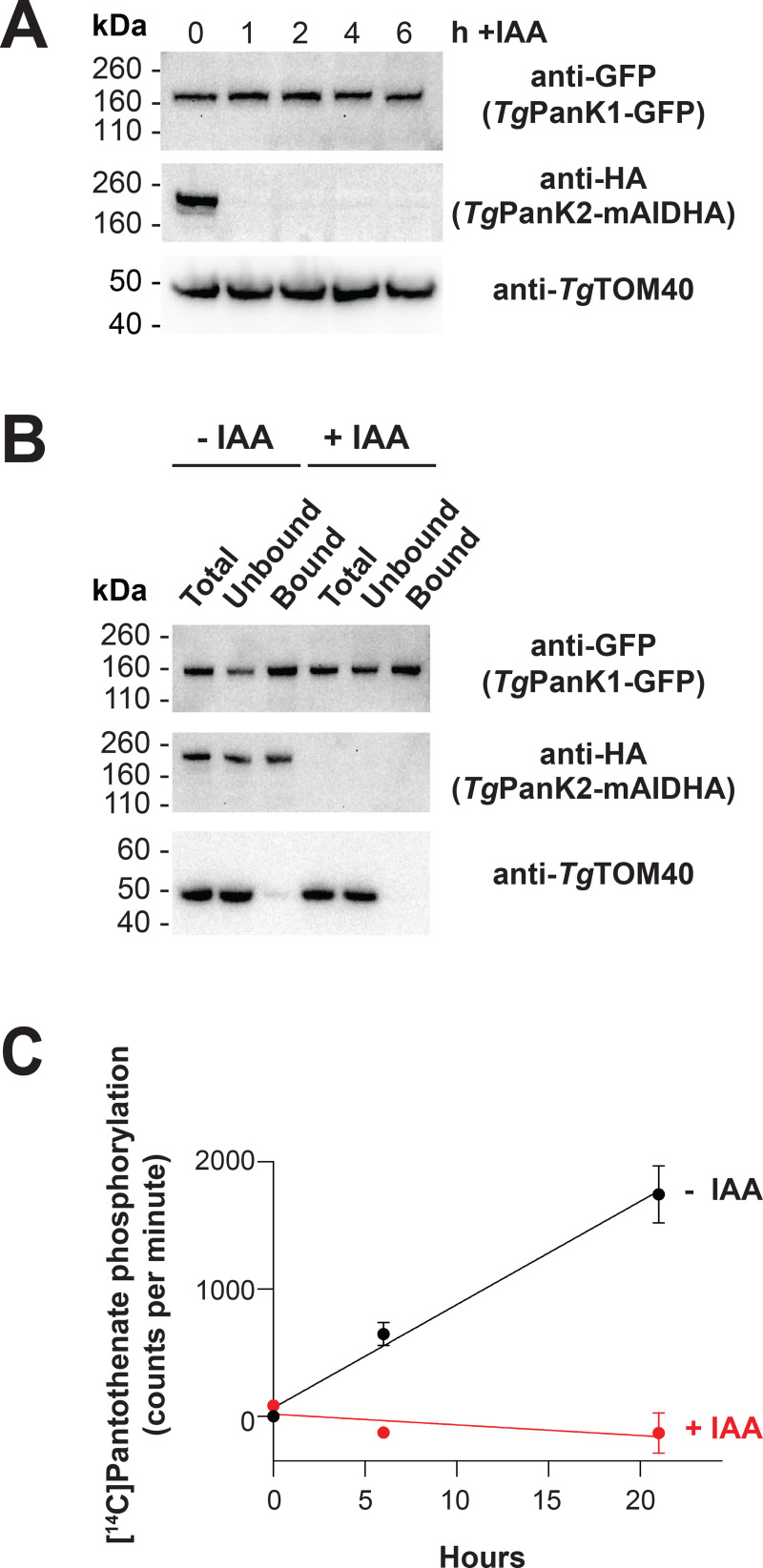
To test whether TgPanK2 is required for TgPanK1-dependent PanK activity in the parasites, we first investigated the stability of TgPanK1 when TgPanK2 was degraded by exposing the TgPanK1-GFP/TgPanK2-mAIDHA parasite line to 100 μM IAA over 6 hours. TgPanK1 abundance remained unchanged following TgPanK2 knockdown (Fig 5A), indicating that TgPanK1 stability or turnover is not dependent on TgPanK2. We next immunopurified TgPanK1-GFP from TgPanK1-GFP/TgPanK2-mAIDHA parasites that were incubated in the presence (+IAA) or absence (-IAA) of 100 μM IAA for 2–3 h, and tested the purified protein for PanK activity (S1 Fig, orange arrow). Although we observed similar abundance of purified TgPanK1-GFP between the +IAA and -IAA samples (Fig 5B), the samples immunoprecipitated from parasites exposed to IAA were devoid of PanK activity, whereas samples not exposed to IAA were able to phosphorylate pantothenate (Fig 5C). These data demonstrate that TgPanK1 is inactive in the absence of TgPanK2. Collectively, our studies on TgPanK1 and TgPanK2 reveal that (i) TgPanK1 and TgPanK2 are part of the same protein complex, (ii) expression of both proteins is required for PanK activity, and (iii) PanK activity of the complex is important for T. gondii proliferation during the disease-causing tachyzoite stage.
Fig 5. TgPanK2 is required for PanK activity in T. gondii parasites.
(A) Denaturing western blot analysis of the GFP- and HA-tagged proteins in the TgPanK1-GFP/TgPanK2-mAIDHA parasite line cultured in the absence or presence of IAA. TgPanK1-GFP/TgPanK2-mAIDHA parasites were incubated with either 100 μM IAA (+IAA) for 1, 2, 4 and 6 h, or an ethanol vehicle control (0 h). TgTOM40 served as a loading control. (B) Denaturing western blot analysis of the GFP- and HA-tagged proteins in the TgPanK1-GFP/TgPanK2-mAIDHA parasite line before and after immunopurification. TgPanK1-GFP/TgPanK2-mAIDHA parasites were incubated with either 100 μM IAA (+IAA), or an ethanol vehicle control (-IAA) for 2–3 h. The cells were lysed and a sample of total lysate (Total) was incubated with GFP-Trap. The GFP-Trap-immunoprecipitated protein (Bound) samples were then separated from the supernatant (Unbound) and all three fractions analysed by western blot. TgTOM40 served as a control protein that is not expected to be in the TgPanK complex. The western blot shown is a representative of three independent experiments, each performed with a different batch of parasites. (C) The phosphorylation of [14C]pantothenate (initial concentration of 2 μM) over time by GFP-Trap-immunoprecipitated protein samples from TgPanK1-GFP/TgPanK2-mAIDHA parasite lysates. The lysates were generated from parasites that were incubated in either the presence (+IAA) or absence (-IAA) of 100 μM IAA for 2–3 h. Data shown are averaged from three independent experiments, each performed with a different batch of parasites and carried out in duplicate. Error bars represent SEM and are not shown if smaller than the symbols.
Discussion
All PanKs characterised to date have been shown to exist as homodimers [3–10]. Here we present data consistent with PanK1 and PanK2 of the apicomplexan parasites P. falciparum and T. gondii forming a heteromeric complex (Figs 1–3), a hitherto undescribed phenomenon in nature.
There have been several attempts by us [unpublished] and others [37–41] to express a functional PfPanK1. Whilst the protein has been successfully expressed in soluble form using various heterologous expression systems (Escherichia coli, insect cells, Saccharomyces cerevisiae), until recently [41], no study had reported PanK activity from the heterologously-expressed and purified protein. Nurkanto et al. [41] have recently reported the functional expression of PfPanK1 in E. coli. The expressed protein was initially insoluble but was solubilised using high concentrations of trehalose. The authors characterised the protein’s PanK activity (using an enzyme-coupled assay) and reported a pantothenate KM of 44.6 μM. This KM is more than two orders of magnitude higher than the KM that has been reported previously for PfPanK activity in parasite lysates [27,42,43]. The physiological significance of the PfPanK1 activity reported in the Nurkanto et al. study is therefore unclear. Our observations that the PanK activity of the immunoprecipitated PfPanK complex (Fig 2B(ii)), which includes the presence of PfPanK2 (as well as, potentially, additional proteins), is indistinguishable from the PanK activity of parasite lysates (Fig 2B(i)), is consistent with the PfPanK complex described here being responsible for the malaria parasite’s PanK activity.
A comparison of the amino acid sequences of P. falciparum and T. gondii PanKs with those of other type II PanKs, such as human PanK3 (HsPanK3), provides a possible explanation for why PanKs from these apicomplexan parasites exist in heteromeric complexes (S7 Fig). Each of the two identical active sites of the homodimeric HsPanK3 are formed by parts of both of its protomers. Certain residues form hydrogen bonds with pantothenate (Glu138, Ser195, Arg207 from one protomer and Val268’ and Ala269’ from the second protomer), while others interact to stabilise the active site (Asp137 with Tyr258’, and Glu138 with Tyr254’) [34,35] (S12 Fig). Notably, the hydrogen bond between Glu138 and Tyr254’ is important for the allosteric activation of the enzyme [35]. Critically, one of the important residues involved in active site stabilisation, Asp137, is only conserved in the PanK1 of P. falciparum and T. gondii but not their PanK2, while others, such as Tyr254’ and Tyr258’ are conserved in their PanK2 but not PanK1 (S7 and S12 Figs). This raises the possibility that PanK1 and PanK2 homodimers are not functional, and that only a heteromeric PanK1/PanK2 complex, with a single complete active site, can serve as a functional PanK enzyme in these apicomplexan parasites. This is consistent with the previous observation that two of the nucleotide-binding motifs of PfPanK2 deviate from those of other eukaryotic PanKs [28]. Whether the incomplete second active site plays an additional, as yet undetermined, role(s) remains to be seen. It should be noted that the PanKs of other apicomplexan parasites (including species from the genera Babesia, Cryptosporidium and Eimeria) exhibit a similar conservation of residues as that described above for P. falciparum and T. gondii (S13 Fig), raising the possibility that heteromeric PanK complexes are ubiquitous in Apicomplexa.
The apparent molecular weight of the PfPanK heterodimer complex (as determined from native western blotting) is consistent with that of a complex that includes PfPanK1, PfPanK2 and a Pf14-3-3I dimer (Fig 1B). However, due to various limitations of native gels [44], it is difficult to obtain an accurate estimate of the molecular weight of the complex. Although we cannot rule out the inclusion of other proteins in the PfPanK complex, such as M17 leucyl aminopeptidase (Fig 2A and S1 Table), we think that this is unlikely, since peptides from these proteins were detected at lower abundance than peptides from PfPanK1, PfPanK2 and Pf14-3-3I. The role of Pf14-3-3I in the heteromeric PfPanK complex (Fig 2A and 2C) is not clear. The 14-3-3 protein family comprises highly conserved proteins that occur in a wide array of eukaryotic organisms, including apicomplexans such as P. falciparum [32,45–47]. Multiple isoforms of 14-3-3 are found to occur in every organism that expresses the protein [48]. 14-3-3 proteins bind to, and regulate, the function of proteins that are involved in a large range of cellular functions, including cell cycle regulation, signal transduction and apoptosis (reviewed in [49]). They typically bind to phosphorylated Ser/Thr residues on target proteins, and modify their target protein’s trafficking/targeting (reviewed in [50]), conformation, co-localisation, and/or activity (reviewed in [51]). The TgPanK heterodimer complex has a molecular weight that is much larger than the combined molecular weights of TgPanK1 and TgPanK2. Unfortunately, mass spectrometry analysis aimed at identifying the protein composition of the T. gondii PanK complex was unsuccessful, presumably because the native level of expression of the complex is too low. Nevertheless, we were unable to detect Tg14-3-3 in the TgPanK complex with a pan-specific 14-3-3 antibody even though we could detect it in T. gondii parasite lysates (S10 Fig). Whether this means that the presence of 14-3-3 in the P. falciparum PanK complex is not essential for enzyme activity, or if a different protein fulfils a similar role to 14-3-3 in the TgPanK complex, remains to be seen.
In this study, we have characterised, for the first time, PanK activity in T. gondii. The [14C]pantothenate phosphorylation data generated with the purified TgPanK complex (Fig 3C) provide the first biochemical evidence indicating that these putative PanKs are able to phosphorylate pantothenate. This finding, combined with the results of the knockdown and SaPanK complementation experiment in T. gondii (Fig 4B and 4C), as well as our demonstration that TgPanK1 alone is inactive (Fig 5), not only demonstrate the essentiality of TgPanK1 and TgPanK2 and their dependence on each other, but also show that the essentiality is due to their role in phosphorylating pantothenate.
T. gondii parasites inhabit metabolically active mammalian cells that contain their own CoA biosynthesis pathway. Our data indicate that T. gondii parasites are unable to scavenge sufficient downstream intermediates in the CoA biosynthesis pathway, including CoA, from their host cells, for their survival, and therefore must maintain their own active CoA biosynthesis pathway. The requirement for CoA biosynthesis in T. gondii, coupled with the intense investigation of this pathway as a drug target in P. falciparum [27,39–41,52–67], suggests that further characterisation of TgPanK, and the CoA biosynthesis pathway in T. gondii, could yield novel drug targets for chemotherapy.
It has been an open question as to why many organisms (eukaryotes [11–21] and prokaryotes [7,23–25]), including all apicomplexan parasites [68], express more than one PanK and why some PanKs appear to be non-functional [7,21,23] (either by analysis of their sequence or through failed attempts to demonstrate PanK activity experimentally). The data that we present here provides a possible answer to this question, at least in apicomplexan parasites.
Methods
Parasite and host cell culture
P. falciparum parasites were maintained in RPMI 1640 medium supplemented with 11 mM glucose (to a final concentration of 22 mM), 200 μM hypoxanthine, 24 μg/mL gentamicin and 6 g/L Albumax II as described previously [69]. T. gondii was cultured in human foreskin fibroblasts (HFF cells) as described previously [70]. T. gondii parasites were grown in flasks with a confluent HFF cell layer in either Dulbecco’s modified Eagle’s medium (DMEM) or complete RPMI 1640, with both media containing 2 g/L sodium bicarbonate and supplemented with 1% (v/v) fetal bovine serum (FBS), 50 units/mL penicillin, 50 μg/mL streptomycin, 10 μg/mL gentamicin, 0.2 mM L-glutamine, and 0.25 μg/mL amphotericin B.
Plasmid preparation and parasite transfection
The PfPanK1-GFP line was generated in a previous study [27], while the untagged GFP line was a generous gift from Professor Alex Maier (Research School of Biology, Australian National University, Canberra). A Pfpank2-pGlux-1 vector was generated for the overexpression of PfPanK2-GFP in 3D7 strain P. falciparum as detailed in S1 Text. The primers used are listed in S2 Table. The same construct was also transfected into each of the PfPanK1 mutants and their Parent line described previously by Tjhin et al. [27]. Transfections were performed with ring-stage parasites and transformants were subsequently selected and maintained using WR99210 (10 nM) as described previously [71].
Transgenic T. gondii parasite lines were generated using a CRISPR/Cas9 strategy as previously described in Shen et al. [72], which is detailed in the S1 Text. The guide RNAs, primers, and the sequences of gBlocks used are provided in S2 and S3 Tables.
The complementation lines TgPanK1-mAIDHA+SaPanK-Ty1 and TgPanK2-mAIDHA+SaPanK-Ty1 were created by expressing the S. aureus type II PanK gene (Sapank) in T. gondii under the regulation of the tubulin promoter (details in the S1 Text, S2 and S3 Tables).
Immunofluorescence assays and microscopy
Fixed PfPanK2-GFP-expressing 3D7 strain P. falciparum parasites within infected erythrocytes were observed and imaged with a Leica TCS-SP2-UV confocal microscope (Leica Microsystems) using a 63× water immersion lens as described in the S1 Text. To confirm the expression of SaPanK-Ty1 in the TgPanK1-mAIDHA+SaPanK-Ty1 line, immunofluorescence assays were performed based on the protocol described by van Dooren et al. [73]. T. gondii parasites were incubated with mouse anti-Ty1 antibodies (1:200 dilution). Secondary antibodies used were goat anti-mouse AlexaFluor 488 at a 1:250 dilution. The nucleus was stained with DAPI. Immunofluorescence images were acquired on a DeltaVision Elite system (GE Healthcare) using an inverted Olympus IX71 microscope with a 100× UPlanSApo oil immersion lens (Olympus) paired with a Photometrics CoolSNAP HQ2 camera. Images taken on the DeltaVision setup were deconvolved using SoftWoRx Suite 2.0 software. Images were adjusted linearly for contrast and brightness.
Polyacrylamide gel electrophoresis and western blotting
Parasite protein samples were analysed using either denaturing or blue native gels to determine the presence and abundance of a single protein or protein complex of interest, respectively. Briefly, mature trophozoite-stage P. falciparum parasites were isolated from infected erythrocytes by saponin lysis, as described previously [74]. Saponin-isolated parasites were then pelleted and lysed in the appropriate buffers (as detailed in the S1 Text). T. gondii protein samples were prepared as described previously, with samples for blue native-PAGE solubilised in Native PAGE sample buffer (ThermoFisher) containing 1% (v/v) Triton X-100 [73]. Protein samples generated from both P. falciparum and T. gondii parasites were separated by polyacrylamide gel electrophoresis (PAGE) in precast NuPAGE (4–12% or 12%) or NativePAGE (4–16%) gels (ThermoFisher) according to the manufacturer’s instructions with minor modifications (detailed in the S1 Text). The separated proteins were transferred to the appropriate membranes (nitrocellulose or polyvinylidene fluoride (PVDF)) and blocked (detailed in the S1 Text) before immunoblotting. Blocked membranes were exposed (45 min– 2 h) to specific primary and secondary antibodies to allow for the detection of the protein of interest. To visualise the protein band(s), membranes were incubated in Pierce enhanced chemiluminescence (ECL) Plus Substrate (ThermoFisher) according to the manufacturer’s instructions or home-made ECL substrate (0.04% w/v luminol, 0.007% w/v coumaric acid, 0.01% v/v H2O2, 100 mM Tris, pH 9.35). Protein bands were then either imaged onto X-ray films and scanned or visualised on a ChemiDoc MP Imaging System (Bio-Rad).
Flow cytometry
Saponin-isolated mature trophozoites from 3D7 wild-type, Parent+PfPanK2-GFP, PanOH-A+PfPanK2-GFP, PanOH-B+PfPanK2-GFP and CJ-A+PfPanK2-GFP cultures were subjected to flow cytometry analysis to determine the proportion of GFP-positive cells (S6 Fig). Aliquots of each isolated parasite suspension were diluted in a saline solution (125 mM NaCl, 5 mM KCl, 25 mM HEPES, 20 mM glucose and 1 mM MgCl2, pH 7.1) to a concentration of ~106–107 cells/mL in 1.2 mL Costar polypropylene cluster tubes (Corning) and sampled for flow cytometry analysis (in measurements of 100,000 cells, low sampling speed) with the following settings: forward scatter = 450 V (log scale), side scatter = 350 V (log scale) and AlexaFluor 488 = 600 V (log scale). The 3D7 wild-type cells were used to establish a gating strategy that defined a threshold below which parasites were deemed to be auto-fluorescent. This strategy was then applied in all analyses to determine the proportion of cells in each cell line that was GFP-positive (i.e. above the defined threshold).
Immunoprecipitations
In order to immunopurify GFP-tagged or HA-tagged proteins from parasite lysates, immunoprecipitation was performed using either GFP-Trap (high affinity anti-GFP alpaca nanobody bound to agarose beads; Chromotek) or anti-HA beads (Sigma-Aldrich), respectively. P. falciparum lysate was prepared from saponin-isolated trophozoites, and T. gondii lysate was prepared from tachyzoites, as described previously ([74] and [73], respectively). Immunoprecipitation was then performed (as detailed in the S1 Text). In P. falciparum experiments where the amount of immunoprecipitated proteins were to be standardised across cell lines and biological repeats, the number of GFP-positive cells to be used for lysate preparation was calculated by a combination of haemocytometer count and flow cytometry. All immunoprecipitated samples from Parent+PfPanK2-GFP, PanOH-A+PfPanK2-GFP, PanOH-B+PfPanK2-GFP and CJ-A+PfPanK2-GFP cell lines contained protein from 5 × 10⁷ GFP-positive cells. Each of these samples was subsequently divided into two equal aliquots, one used in the [14C]pantothenate phosphorylation assay and the other for denaturing western blot.
When an aliquot of the immunoprecipitation sample (beads that have bound proteins from ~106–107 GFP-positive cells for P. falciparum and ~107–108 cells for T. gondii) was required for western blot, the bead suspension was centrifuged (2,500 × g, 2 min), the supernatant removed, and the beads resuspended in 50 μL sample buffer containing 2 × NuPAGE lithium dodecyl sulfate (LDS) sample buffer (ThermoFisher) and 2 × NuPAGE sample reducing agent (ThermoFisher). In some experiments, 10 μL aliquots of the total or unbound lysate fractions were each mixed with 10 μL of the same sample buffer. These samples were then boiled (95°C, 10 min) and 10 μL of each was then used in a denaturing western blot as described above.
[14C]Pantothenate phosphorylation assays
In order to determine the PanK activity of the protein(s) isolated in the GFP-Trap immunoprecipitation assays, the immunopurified complexes were used to perform a [14C]pantothenate phosphorylation time course. The bead suspensions containing the immunoprecipitated proteins from P. falciparum and T. gondii were centrifuged (2,500 × g, 2 min), the supernatant removed, and the beads resuspended in 250–300 μL (T. gondii) or 500 μL (for P. falciparum) buffer containing 100 mM Tris-HCl (pH 7.4), 10 mM ATP and 10 mM MgCl2 (i.e. all reagents were at twice the final concentration required for the phosphorylation reaction). Each time course was then initiated by the addition of 250–300 μL (for T. gondii) or 500 μL (for P. falciparum) 4 μM (0.2 μCi/mL) [14C]pantothenate in water (pre-warmed to 37°C), to the bead suspension. Aliquots of each reaction (50 μL in duplicate) were terminated at pre-determined time points by mixing with 50 μL 150 mM barium hydroxide preloaded within the wells of a 96-well, 0.2 μm hydrophilic PVDF membrane filter bottom plate (Corning). Phosphorylated compounds in each well were then precipitated by the addition of 50 μL 150 mM zinc sulfate to generate the Somogyi reagent [75], the wells processed, and the radioactivity in the plate determined as detailed previously [43]. Total radioactivity in each phosphorylation reaction was determined by mixing 50 μL aliquots of each reaction (in duplicate) thoroughly with 150 μL Microscint-40 (PerkinElmer) by pipetting the mixture at least 5 times, in the wells of an OptiPlate-96 microplate (PerkinElmer) [43].
Mass spectrometry of immunoprecipitated samples
The identities of the proteins co-immunoprecipitated from lysates of the wild-type 3D7, PfPanK1-GFP, PfPanK2-GFP and untagged GFP lines were determined by mass spectrometry. Aliquots of bead-bound co-immunoprecipitated samples were resuspended in 2 × NuPAGE LDS sample buffer and 2 × NuPAGE sample reducing agent and sent (at ambient temperature, travel time less than 24 h) to the Australian Proteomics Analysis Facility (Sydney) for processing and mass spectrometry analysis (as detailed in the S1 Text).
Fluorescent T. gondii proliferation assay
Fluorescent T. gondii proliferation assays were performed as previously described [36]. Briefly, 2000 parasites suspended in complete RPMI were added to the wells of optical bottom black 96 well plates (ThermoFisher) containing a confluent layer of HFF cells, either in the presence of 100 μM IAA dissolved in ethanol (final ethanol concentration of 0.1%, v/v) or with ethanol (0.1%, v/v) as a vehicle control, in triplicate. Fluorescent measurements (Excitation filter, 540 nm; Emission filter, 590 nm) using a FLUOstar OPTIMA Microplate Reader (BMG LABTECH) were taken over 7 days.
Knockdown of mAID protein
Flasks containing a confluent layer of HFF cells were seeded with TgPanK1-mAIDHA, TgPanK2-mAIDHA, TgPanK1-mAIDHA+SaPanK-Ty1 or TgPanK2-mAIDHA+SaPanK-Ty1 T. gondii parasites. While the parasites were still intracellular, 100 μM of IAA dissolved in ethanol (final ethanol concentration of 0.1%, v/v) was added to induce the knockdown of TgPanK1-mAIDHA or TgPanK2-mAIDHA, with ethanol (0.1%, v/v) added to another flask as a vehicle control. Flasks with IAA added were processed at 1, 2 and 4 h time points, and the control flask was processed at the 4-h time point. Parasite concentrations were determined using a haemocytometer and 1.5 × 10⁷ parasites were resuspended in LDS sample buffer, and boiled at 95°C for 10 minutes. An aliquot from each sample was analysed by western blotting. The knockdown of TgPanK2-mAIDHA protein in the TgPanK1-GFP/TgPanK2-mAIDHA line was carried out using the same protocol, but with the addition of a 6 h time point.
Alignment of PanKs
PanK homologues from P. falciparum and T. gondii, and a selection of other type II PanKs from other eukaryotic organisms and S. aureus were aligned using PROMALS3D [76] (available at: http://prodata.swmed.edu/promals3d/promals3d.php). The default parameters were selected except for the ‘Identity threshold above which fast alignment is applied’ parameter, which was changed to “1” to allow for a more accurate alignment. The following PanK type II homologues were aligned (accession number included in brackets): Staphylococcus aureus (Q2FWC7); Saccharomyces cerevisiae (Q04430); Aspergillus nidulans (O93921); Homo sapiens PanK1 (Q8TE04), PanK2 (Q9BZ23), PanK3 (Q9H999) and PanK4 (Q9NVE7); Arabidopsis thaliana PanK1 (O80765) and PanK2 (Q8L5Y9); Plasmodium falciparum PanK1 (Q8ILP4) and PanK2 (Q8IL92) and Toxoplasma gondii PanK1 (A0A125YTW9) and PanK2 (V5B595).
Statistical analysis
Statistical analysis between the means of the pantothenate phosphorylation rate by the Parent lysate and that of the immunopurified complex from the Parent+PfPanK2-GFP line was carried out with unpaired, two-tailed, Student’s t test using GraphPad Prism 8 (GraphPad Software, Inc) from which the 95% confidence interval of the difference between the means (95% CI) was obtained. All regression analysis was done using SigmaPlot version 11.0 for Windows (Systat Software, Inc) or GraphPad Prism 8 (GraphPad Software, Inc).
Supporting information
Proteins detected in each immunoprecipitation experiment are listed in descending order according of the total number of peptides detected across the two replicates (total peptides in 1st and 2nd rep columns). Only proteins that are present in the immunoprecipitation fractions of both parasite lines and absent in the negative controls (bound fractions of untagged GFP and 3D7 parasite lysates) are shown. Proteins shown in Fig 2A are indicated in red.
(TIF)
(TIF)
(TIF)
Flow chart highlighting the cell lines generated for the study and the main experimental steps that were performed. The coloured arrows represent final experimental results, and the associated figures within which the data are presented, are indicated under each experiment.
(TIF)
Denaturing western blot analysis of the GFP-tagged proteins present in the total lysate, unbound and GFP-Trap-bound fractions of PfPanK1-GFP, PfPanK2-GFP and untagged GFP lines. Western blots were performed with anti-GFP antibody and the blot shown is representative of two independent experiments each performed with a different batch of parasites.
(TIF)
PfPanK1 peptides detected in the two independent MS analyses of the GFP-Trap immunoprecipitation from the PfPanK1-GFP and PfPanK2-GFP lines. Residues in green were detected in either analysis with >95% confidence, while residues in orange were detected in either analysis with >90% (but <95%) confidence. Percentage coverage was calculated using only the residues labelled green.
(TIF)
PfPanK2 peptides detected in the two independent MS analyses of the GFP-Trap immunoprecipitation from the PfPanK1-GFP and PfPanK2-GFP lines. Residues in green were detected in either analysis with >95% confidence, while residues in orange were detected in either analysis with >90% (but <95%) confidence. Percentage coverage was calculated using only the residues labelled green.
(TIF)
Pf14-3-3I peptides detected in the two independent MS analyses of the GFP-Trap immunoprecipitation from the PfPanK1-GFP and PfPanK2-GFP lines. Residues in green were detected in either analysis with >95% confidence, while residues in orange were detected in either analysis with >90% (but <95%) confidence. Percentage coverage was calculated using only the residues labelled green.
(TIF)
(A) The proportion of GFP-positive saponin-isolated 3D7, Parent+PfPanK2-GFP, PanOH-A+PfPanK2-GFP, PanOH-B+PfPanK2-GFP and CJ-A+PfPanK2-GFP trophozoites was determined by FACS analysis. The forward scatter (FSC) intensity on each x-axis corresponds to cell size and the y-axis corresponds to the intensity of GFP fluorescence. The proportion of GFP-positive cells in each transgenic line (percentage value in each plot) was determined by using 3D7 trophozoites to set a gating threshold below which parasites were defined to be auto-fluorescent. Data shown are representative of three independent experiments, each performed prior to the [14C]pantothenate phosphorylation assays presented in Fig 2B(ii). The flow cytometry data were used to standardise the amount of PfPanK2-GFP immunoprecipitated from each cell line used in each [14C]pantothenate phosphorylation assay. (B) Denaturing western blot analysis of PfPanK2-GFP in the GFP-Trap immunoprecipitated complexes that were used in the [14C]pantothenate phosphorylation assays performed to generate the data in Fig 2B(ii). Western blots were performed with an anti-GFP antibody and each blot shows the relative amounts of PfPanK2-GFP immunopurified from the four different cell lines used in each of the three [14C]pantothenate phosphorylation experiment. The same volume of samples (10 μL per lane) was used for all three experiments.
(TIF)
The conserved PHOSPHATE 1, PHOSPHATE 2, and ADENOSINE 1 motifs of the acetate and sugar kinases/Hsc70/actin (ASKHA) superfamily of kinases are labelled at the top of the alignment. The Glu (E) residue involved in catalysis and the Arg (R) residue involved in positioning the substrate, are shown on a black background. Residues that have been found to interact with pantothenate and acetyl-CoA in human PanK3 [34,35] are marked with a blue asterisk. Residues that were found to interact to stabilise the human PanK3 active site are marked with a red asterisk. The catalytic Glu (E) residue is marked with a red and blue asterisk as it is involved in both the interaction with pantothenate and the stabilisation of the active site through interaction with a Tyr (Y) residue of the opposite protomer. The numbers at the start and end of each sequence indicate the position of the first and last residue in the alignment, respectively. The lengths of insertions are specified within the square brackets and the total length of protein sequences are shown in round brackets. Residues within the ASKHA superfamily motifs and conserved residues are highlighted based on the consensus AA guide for the column as follows: identical = bold, hydrophobic (W,F,Y,M,L,I,V,A,C,T,H) = yellow, charged/polar/small (D,E,K,R,H/D,E,H,K,N,Q,R,S,T/A,G,C,S,V,N,D,T,P) = grey and Gly (G) = red. The two insertion regions (Ins 1 and Ins 2) common to eukaryotic type II PanKs, but absent in prokaryotic PanKs are indicated by the black horizontal bars, while the PfPank1/TgPanK1 and PfPanK2/TgPanK2 specific inserts are highlighted on a red and blue background, respectively. Conservation refers to the conservation index [77]. Values at and above the conservation index cut-off (5) are displayed above the amino acid. Consensus AA: refers to the consensus level alignment parameters for the consensus amino acid sequence. This is displayed if the weighted frequency of a certain class of residues in a position is above 0.8. Consensus symbols: conserved amino acids are in bold and uppercase letters; aliphatic (I, V, L): l; aromatic (Y, H, W, F): @; hydrophobic (W, F, Y, M, L, I, V, A, C, T, H): h; alcohol (S, T): o; polar residues (D, E, H, K, N, Q, R, S, T): p; tiny (A, G, C, S): t; small (A, G, C, S, V, N, D, T, P): s; bulky residues (E, F, I, K, L, M, Q, R, W, Y): b; positively charged (K, R, H): +; negatively charged (D, E): -; charged (D, E, K, R, H): c. Marked below the alignment, 85% consensus includes those residues that occur in either the superfamily motifs and/or conserved residues where the same residue occurs more than 85% (10 out of 13 sequences). Consensus secondary structure (ss) elements: h = alpha helix, e = beta strand. Species names are abbreviated as follows: Sa = Staphylococcus aureus, Sc = Saccharomyces cerevisiae, An = Aspergillus nidulans, Hs = Homo sapiens, At = Arabidopsis thaliana, Pf = Plasmodium falciparum and Tg = Toxoplasma gondii. The alignment was created using PROMALS3D [76].
(TIF)
(A) Schematic of theTgpank1 and Tgpank2 genomic loci, indicating the incorporation sites of the epitope tag coding sequence. The expected sizes of the PCR products when screened with each set of screening primers are shown above the corresponding epitope tag coding sequence. The screening primers are Tgpank1 screen fwd and rvs for Tgpank1 (referred to as Tgpank1 primers in panels B-D), and Tgpank2 screen fwd and rvs for Tgpank2 (referred to as Tgpank2 primers in panels B-D). Primers are detailed in S2 Table. (B) PCR analysis of the parental strain (TgParent), and singly-tagged TgPanK1-mAIDHA and TgPanK2-mAIDHA lines. Both TgPanK1-mAIDHA and TgPanK2-mAIDHA have successfully incorporated mAIDHA tags. (C) PCR analysis of the doubly-tagged TgPanK1-GFP/TgPanK2-mAIDHA line. CRISPR/Cas9 was utilised to incorporate a sequence encoding a TEV-GFP tag into the genomic locus of the Tgpank1 gene within the TgPanK2-mAIDHA line. (D) PCR analysis of the TgPanK1-HA/TgPanK2-GFP doubly-tagged line. CRISPR/Cas9 was utilised to incorporate a sequence encoding a TEV-HA tag into the genomic locus of the Tgpank1 gene and a sequence encoding a TEV-GFP tag into the genomic locus of the Tgpank2 gene.
(TIF)
Anti-HA and anti-GFP denaturing western blot analysis of fractions from GFP-Trap and anti-HA immunoprecipitations performed using lysates prepared from the parasite lines expressing TgPanK1-HA/TgPanK2-GFP. The expected molecular masses of TgPanK1-HA and TgPanK2-GFP are 136 kDa and 206 kDa, respectively. The blot shown is representative of three independent experiments, each performed with a different batch of parasites. Denaturing western blots were also probed with anti-TgTOM40, which served as a control for a protein that is not part of the PanK complex.
(TIF)
Anti-HA, anti-GFP and anti-14-3-3 denaturing western blot analysis of fractions from GFP-Trap immunoprecipitation of lysates prepared from the parasite line expressing TgPanK1-GFP/TgPanK2-mAIDHA. The expected molecular masses of TgPanK1-GFP, TgPanK2-mAIDHA and 14-3-3 are approximately 160 kDa, 189 kDa and 37 kDa, respectively. The blots shown are representative of two independent experiments, each performed with a different batch of parasites.
(TIF)
(A) Anti-HA and anti-Ty1 denaturing western blot analysis of SaPanK-Ty1-complemented and non-complemented (i) TgPanK1-mAIDHA and (ii) TgPanK2-mAIDHA lines, in the absence or presence (for 1 h) of 100 μM IAA. The expected molecular masses of TgPanK1-mAIDHA, TgPanK2-mAIDHA and SaPanK-Ty1 are ~143 kDa, ~189 kDa and ~29 kDa, respectively. Denaturing western blots were also probed with anti-TgTOM40, which served as a loading control. Each blot shown is representative of three independent experiments, each performed with a different batch of parasites. (B) Fluorescence micrographs of a HFF cell infected with four tachyzoite-stage TgPanK1-mAIDHA+SaPanK-Ty1 parasites within a vacuole, indicating the presence of SaPanK-Ty1. From left to right: Differential interference contrast (DIC), tdTomato RFP (a marker of the nucleus and cytosol; red), anti SaPanK-Ty1 (green), and merged images. Scale bar represents 2 μm.
(TIF)
(A) H. sapiens AMP-PNP-pantothenate-bound PanK3 crystal structure (PDB ID: 5KPR, Subramanian et al. [35]). The homodimeric protein is made up of two identical protomers (lilac and yellow) forming two identical active sites, each binding pantothenate (carbon atoms shown in green). The red square encompasses one of the active sites. (B) Magnification of the region outlined by the red square in (A). Residues from both protomers contribute to the stabilisation of the binding pocket (E138 forms a hydrogen bond with Y254’ and D137 with Y258’) and interact with pantothenate (E138, S195, R207, A269’ and V268’). Hydrogen bonds with and between the sidechains of these residues are shown in red. An apostrophe denotes residues from the lilac protomer. (C) List of residues annotated in the HsPanK3 model that participate in the stabilisation of the binding pocket (highlighted cyan), and a comparison to the equivalent residues in P. falciparum and T. gondii PanKs. The PanKs from P. falciparum and T. gondii do not individually contain the complete set of residues required for the stabilisation of the binding pocket, but the combination of residues (highlighted cyan) from PanK1 and PanK2 suggests that each PanK1/PanK2 heterodimer will have only one stabilised binding site.
(TIF)
The apicomplexan PanK residues corresponding to the HsPanK3 residues that are involved in the stabilisation of the binding pocket (D137, E138, Y254 and Y258) are highlighted in cyan if they are conserved and in grey if they are not conserved. The numbers before each alignment indicate the position of the first residue in the alignment. Each apicomplexan PanK is grouped into PanK1 or PanK2 based on their similarity to either PfPanK1/TgPanK1 or PfPanK2/TgPanK2, respectively. The alignment was created using PROMALS3D [76].
(TIF)
(DOCX)
Acknowledgments
Proteomics was undertaken at APAF, the infrastructure provided by the Australian Government through the National Collaborative Research Infrastructure Strategy (NCRIS). We are grateful to the Canberra Branch of the Australian Red Cross Lifeblood for the provision of red blood cells, and Professor Alex Maier (ANU) for the untagged GFP-expressing P. falciparum parasites and pGlux-1 plasmid.
Data Availability
All relevant data are within the manuscript and its Supporting Information files.
Funding Statement
ETT, VMH and CS were supported by Research Training Program scholarships from the Australian Government. CS was also funded by an NHMRC Overseas Biomedical Fellowship (1016357). This work was, in part, supported by a Project Grant (APP1129843) from the National Health and Medical Research Council to KJS and a Discovery Grant (DP150102883) from the Australian Research Council to GvD. The funders had no role in study design, data collection and analysis, decision to publish, or preparation of the manuscript.
References
- 1.Leonardi R, Zhang Y-M, Rock CO, Jackowski S. Coenzyme A: back in action. Prog Lipid Res. 2005;44: 125–153. doi: 10.1016/j.plipres.2005.04.001 [DOI] [PubMed] [Google Scholar]
- 2.Spry C, Kirk K, Saliba KJ. Coenzyme A biosynthesis: an antimicrobial drug target. FEMS Microbiol Rev. 2008;32: 56–106. doi: 10.1111/j.1574-6976.2007.00093.x [DOI] [PubMed] [Google Scholar]
- 3.Yun M, Park CG, Kim JY, Rock CO, Jackowski S, Park H-W. Structural basis for the feedback regulation of Escherichia coli pantothenate kinase by coenzyme A. J Biol Chem. 2000;275: 28093–28099. doi: 10.1074/jbc.M003190200 [DOI] [PubMed] [Google Scholar]
- 4.Das S, Kumar P, Bhor V, Surolia A, Vijayan M. Invariance and variability in bacterial PanK: a study based on the crystal structure of Mycobacterium tuberculosis PanK. Acta Crystallogr D Biol Crystallogr. 2006;62: 628–638. doi: 10.1107/S0907444906012728 [DOI] [PubMed] [Google Scholar]
- 5.Yang K, Eyobo Y, Brand LA, Martynowski D, Tomchick D, Strauss E, et al. Crystal structure of a type III pantothenate kinase: insight into the mechanism of an essential coenzyme A biosynthetic enzyme universally distributed in bacteria. J Bacteriol. 2006;188: 5532–5540. doi: 10.1128/JB.00469-06 [DOI] [PMC free article] [PubMed] [Google Scholar]
- 6.Hong BS, Yun MK, Zhang Y-M, Chohnan S, Rock CO, White SW, et al. Prokaryotic type II and type III pantothenate kinases: The same monomer fold creates dimers with distinct catalytic properties. Structure. 2006;14: 1251–1261. doi: 10.1016/j.str.2006.06.008 [DOI] [PubMed] [Google Scholar]
- 7.Nicely NI, Parsonage D, Paige C, Newton GL, Fahey RC, Leonardi R, et al. Structure of the type III pantothenate kinase from Bacillus anthracis at 2.0 Å resolution: implications for coenzyme A-dependent redox biology. Biochemistry. 2007;46: 3234–3245. doi: 10.1021/bi062299p [DOI] [PMC free article] [PubMed] [Google Scholar]
- 8.Hong BS, Senisterra G, Rabeh WM, Vedadi M, Leonardi R, Zhang Y-M, et al. Crystal structures of human pantothenate kinases. Insights into allosteric regulation and mutations linked to a neurodegeneration disorder. J Biol Chem. 2007;282: 27984–27993. doi: 10.1074/jbc.M701915200 [DOI] [PubMed] [Google Scholar]
- 9.Li B, Tempel W, Smil D, Bolshan Y, Schapira M, Park H-W. Crystal structures of Klebsiella pneumoniae pantothenate kinase in complex with N-substituted pantothenamides. Proteins. 2013;81: 1466–1472. doi: 10.1002/prot.24290 [DOI] [PubMed] [Google Scholar]
- 10.Franklin MC, Cheung J, Rudolph MJ, Burshteyn F, Cassidy M, Gary E, et al. Structural genomics for drug design against the pathogen Coxiella burnetii. Proteins. 2015;83: 2124–2136. doi: 10.1002/prot.24841 [DOI] [PubMed] [Google Scholar]
- 11.Kupke T, Hernández-Acosta P, Culiáñez-Macià FA. 4’-Phosphopantetheine and coenzyme A biosynthesis in plants. J Biol Chem. 2003;278: 38229–38237. doi: 10.1074/jbc.M306321200 [DOI] [PubMed] [Google Scholar]
- 12.Tilton GB, Wedemeyer WJ, Browse J, Ohlrogge J. Plant coenzyme A biosynthesis: characterization of two pantothenate kinases from Arabidopsis. Plant Mol Biol. 2006;61: 629–642. doi: 10.1007/s11103-006-0037-4 [DOI] [PubMed] [Google Scholar]
- 13.Rock CO, Calder RB, Karim MA, Jackowski S. Pantothenate kinase regulation of the intracellular concentration of coenzyme A. J Biol Chem. 2000;275: 1377–1383. doi: 10.1074/jbc.275.2.1377 [DOI] [PubMed] [Google Scholar]
- 14.Rock CO, Karim MA, Zhang Y-M, Jackowski S. The murine pantothenate kinase (Pank1) gene encodes two differentially regulated pantothenate kinase isozymes. Gene. 2002;291: 35–43. doi: 10.1016/s0378-1119(02)00564-4 [DOI] [PubMed] [Google Scholar]
- 15.Kuo YM, Duncan JL, Westaway SK, Yang H, Nune G, Xu EY, et al. Deficiency of pantothenate kinase 2 (Pank2) in mice leads to retinal degeneration and azoospermia. Hum Mol Genet. 2005;14: 49–57. doi: 10.1093/hmg/ddi005 [DOI] [PMC free article] [PubMed] [Google Scholar]
- 16.Zhang Y-M, Rock CO, Jackowski S. Feedback regulation of murine pantothenate kinase 3 by coenzyme A and coenzyme A thioesters. J Biol Chem. 2005;280: 32594–32601. doi: 10.1074/jbc.M506275200 [DOI] [PubMed] [Google Scholar]
- 17.Zhou B, Westaway SK, Levinson B, Johnson MA, Gitschier J, Hayflick SJ. A novel pantothenate kinase gene (PANK2) is defective in Hallervorden-Spatz syndrome. Nat Genet. 2001;28: 345–349. doi: 10.1038/ng572 [DOI] [PubMed] [Google Scholar]
- 18.Ni X, Ma Y, Cheng H, Jiang M, Ying K, Xie Y, et al. Cloning and characterization of a novel human pantothenate kinase gene. Int J Biochem Cell Biol. 2002;34: 109–115. doi: 10.1016/s1357-2725(01)00114-5 [DOI] [PubMed] [Google Scholar]
- 19.Hörtnagel K, Prokisch H, Meitinger T. An isoform of hPANK2, deficient in pantothenate kinase-associated neurodegeneration, localizes to mitochondria. Hum Mol Genet. 2003;12: 321–327. doi: 10.1093/hmg/ddg026 [DOI] [PubMed] [Google Scholar]
- 20.Ramaswamy G, Karim MA, Murti KG, Jackowski S. PPARα controls the intracellular coenzyme A concentration via regulation of PANK1α gene expression. J Lipid Res. 2004;45: 17–31. doi: 10.1194/jlr.M300279-JLR200 [DOI] [PubMed] [Google Scholar]
- 21.Zhang Y-M, Chohnan S, Virga KG, Stevens RD, Ilkayeva OR, Wenner BR, et al. Chemical knockout of pantothenate kinase reveals the metabolic and genetic program responsible for hepatic coenzyme A homeostasis. Chem Biol. 2007;14: 291–302. doi: 10.1016/j.chembiol.2007.01.013 [DOI] [PMC free article] [PubMed] [Google Scholar]
- 22.Leonardi R, Rehg JE, Rock CO, Jackowski S. Pantothenate kinase 1 is required to support the metabolic transition from the fed to the fasted state. PLoS One. 2010;5: e11107. doi: 10.1371/journal.pone.0011107 [DOI] [PMC free article] [PubMed] [Google Scholar]
- 23.Awasthy D, Ambady A, Bhat J, Sheikh G, Ravishankar S, Subbulakshmi V, et al. Essentiality and functional analysis of type I and type III pantothenate kinases of Mycobacterium tuberculosis. Microbiology. 2010;156: 2691–2701. doi: 10.1099/mic.0.040717-0 [DOI] [PubMed] [Google Scholar]
- 24.Yocum RR, Patterson TA, Inc OB. Microorganisms and assays for the identification of antibiotics. U S Patent 6,830,898. 2004. [Google Scholar]
- 25.Brand LA, Strauss E. Characterization of a new pantothenate kinase isoform from Helicobacter pylori. J Biol Chem. 2005;280: 20185–20188. doi: 10.1074/jbc.C500044200 [DOI] [PubMed] [Google Scholar]
- 26.Yao J, Subramanian C, Rock CO, Jackowski S. Human pantothenate kinase 4 is a pseudo-pantothenate kinase. Protein Sci. 2019;28: 1031–1047. doi: 10.1002/pro.3611 [DOI] [PMC free article] [PubMed] [Google Scholar]
- 27.Tjhin ET, Spry C, Sewell AL, Hoegl A, Barnard L, Sexton AE, et al. Mutations in the pantothenate kinase of Plasmodium falciparum confer diverse sensitivity profiles to antiplasmodial pantothenate analogues. PLoS Pathog. 2018;14: e1006918. doi: 10.1371/journal.ppat.1006918 [DOI] [PMC free article] [PubMed] [Google Scholar]
- 28.Spry C, van Schalkwyk DA, Strauss E, Saliba KJ. Pantothenate utilization by Plasmodium as a target for antimalarial chemotherapy. Infect Disord Drug Targets. 2010;10: 200–216. doi: 10.2174/187152610791163390 [DOI] [PubMed] [Google Scholar]
- 29.Sidik SM, Huet D, Ganesan SM, Huynh M-H, Wang T, Nasamu AS, et al. A genome-wide CRISPR screen in Toxoplasma identifies essential apicomplexan genes. Cell. 2016;166: 1423–1435.e12. doi: 10.1016/j.cell.2016.08.019 [DOI] [PMC free article] [PubMed] [Google Scholar]
- 30.Zhang M, Wang C, Otto TD, Oberstaller J, Liao X, Adapa SR, et al. Uncovering the essential genes of the human malaria parasite Plasmodium falciparum by saturation mutagenesis. Science. 2018;360. doi: 10.1126/science.aap7847 [DOI] [PMC free article] [PubMed] [Google Scholar]
- 31.Aitken A. 14-3-3 and its possible role in co-ordinating multiple signalling pathways. Trends Cell Biol. 1996;6: 341–347. doi: 10.1016/0962-8924(96)10029-5 [DOI] [PubMed] [Google Scholar]
- 32.Lalle M, Currà C, Ciccarone F, Pace T, Cecchetti S, Fantozzi L, et al. Dematin, a component of the erythrocyte membrane skeleton, is internalized by the malaria parasite and associates with Plasmodium 14-3-3. J Biol Chem. 2011;286: 1227–1236. doi: 10.1074/jbc.M110.194613 [DOI] [PMC free article] [PubMed] [Google Scholar]
- 33.Brown KM, Long S, Sibley LD. Plasma membrane association by N-acylation governs PKG function in Toxoplasma gondii. MBio. 2017;8. doi: 10.1128/mBio.00375-17 [DOI] [PMC free article] [PubMed] [Google Scholar]
- 34.Leonardi R, Zhang Y-M, Yun MK, Zhou R, Zeng F-Y, Lin W, et al. Modulation of pantothenate kinase 3 activity by small molecules that interact with the substrate/allosteric regulatory domain. Chem Biol. 2010;17: 892–902. doi: 10.1016/j.chembiol.2010.06.006 [DOI] [PMC free article] [PubMed] [Google Scholar]
- 35.Subramanian C, Yun MK, Yao J, Sharma LK, Lee RE, White SW, et al. Allosteric regulation of mammalian pantothenate kinase. J Biol Chem. 2016;291: 22302–22314. doi: 10.1074/jbc.M116.748061 [DOI] [PMC free article] [PubMed] [Google Scholar]
- 36.Rajendran E, Hapuarachchi SV, Miller CM, Fairweather SJ, Cai Y, Smith NC, et al. Cationic amino acid transporters play key roles in the survival and transmission of apicomplexan parasites. Nat Commun. 2017;8: 14455. doi: 10.1038/ncomms14455 [DOI] [PMC free article] [PubMed] [Google Scholar]
- 37.Mehlin C, Boni E, Buckner FS, Engel L, Feist T, Gelb MH, et al. Heterologous expression of proteins from Plasmodium falciparum: results from 1000 genes. Mol Biochem Parasitol. 2006;148: 144–160. doi: 10.1016/j.molbiopara.2006.03.011 [DOI] [PubMed] [Google Scholar]
- 38.Shen D, Huang Q, Sha Y, Zhou B. Cloning and confirmation of several potential pantothenate kinases and their interaction with pantothenate analogues. J Tsinghua Univ. 2008;48: 403–407. [Google Scholar]
- 39.Chiu JE, Thekkiniath J, Choi J-Y, Perrin BA, Lawres L, Plummer M, et al. The antimalarial activity of the pantothenamide α-PanAm is via inhibition of pantothenate phosphorylation. Sci Rep. 2017;7: 14234. doi: 10.1038/s41598-017-14074-9 [DOI] [PMC free article] [PubMed] [Google Scholar]
- 40.Schalkwijk J, Allman EL, Jansen PAM, de Vries LE, Verhoef JMJ, Jackowski S, et al. Antimalarial pantothenamide metabolites target acetyl-coenzyme A biosynthesis in Plasmodium falciparum. Sci Transl Med. 2019;11: eaas9917. doi: 10.1126/scitranslmed.aas9917 [DOI] [PubMed] [Google Scholar]
- 41.Nurkanto A, Jeelani G, Santos HJ, Rahmawati Y, Mori M, Nakamura Y, et al. Characterization of Plasmodium falciparum pantothenate kinase and identification of its inhibitors from natural products. Front Cell Infect Microbiol. 2021;11: 639065. doi: 10.3389/fcimb.2021.639065 [DOI] [PMC free article] [PubMed] [Google Scholar]
- 42.Saliba KJ, Kirk K. H+-coupled pantothenate transport in the intracellular malaria parasite. J Biol Chem. 2001;276: 18115–18121. doi: 10.1074/jbc.M010942200 [DOI] [PubMed] [Google Scholar]
- 43.Spry C, Saliba KJ, Strauss E. A miniaturized assay for measuring small molecule phosphorylation in the presence of complex matrices. Anal Biochem. 2014;451: 76–78. doi: 10.1016/j.ab.2013.12.010 [DOI] [PubMed] [Google Scholar]
- 44.Crichton PG, Harding M, Ruprecht JJ, Lee Y, Kunji ERS. Lipid, detergent, and Coomassie Blue G-250 affect the migration of small membrane proteins in blue native gels: mitochondrial carriers migrate as monomers not dimers. J Biol Chem. 2013;288: 22163–22173. doi: 10.1074/jbc.M113.484329 [DOI] [PMC free article] [PubMed] [Google Scholar]
- 45.Al-Khedery B, Barnwell JW, Galinski MR. Stage-specific expression of 14-3-3 in asexual blood-stage Plasmodium. Mol Biochem Parasitol. 1999;102: 117–130. doi: 10.1016/s0166-6851(99)00090-0 [DOI] [PubMed] [Google Scholar]
- 46.Di Girolamo F, Raggi C, Birago C, Pizzi E, Lalle M, Picci L, et al. Plasmodium lipid rafts contain proteins implicated in vesicular trafficking and signalling as well as members of the PIR superfamily, potentially implicated in host immune system interactions. Proteomics. 2008;8: 2500–2513. doi: 10.1002/pmic.200700763 [DOI] [PubMed] [Google Scholar]
- 47.Dastidar EG, Dzeyk K, Krijgsveld J, Malmquist NA, Doerig C, Scherf A, et al. Comprehensive histone phosphorylation analysis and identification of Pf14-3-3 protein as a histone H3 phosphorylation reader in malaria parasites. PLoS One. 2013;8: e53179. doi: 10.1371/journal.pone.0053179 [DOI] [PMC free article] [PubMed] [Google Scholar]
- 48.Rosenquist M, Sehnke P, Ferl RJ, Sommarin M, Larsson C. Evolution of the 14-3-3 protein family: does the large number of isoforms in multicellular organisms reflect functional specificity? J Mol Evol. 2000;51: 446–458. doi: 10.1007/s002390010107 [DOI] [PubMed] [Google Scholar]
- 49.van Hemert MJ, Steensma HY, van Heusden GP. 14-3-3 proteins: key regulators of cell division, signalling and apoptosis. Bioessays. 2001;23: 936–946. doi: 10.1002/bies.1134 [DOI] [PubMed] [Google Scholar]
- 50.Muslin AJ, Xing H. 14-3-3 proteins: regulation of subcellular localization by molecular interference. Cell Signal. 2000;12: 703–709. doi: 10.1016/s0898-6568(00)00131-5 [DOI] [PubMed] [Google Scholar]
- 51.Bridges D, Moorhead GBG. 14-3-3 proteins: a number of functions for a numbered protein. Sci STKE. 2005;2005: re10. doi: 10.1126/stke.2962005re10 [DOI] [PubMed] [Google Scholar]
- 52.Saliba KJ, Ferru I, Kirk K. Provitamin B5 (pantothenol) inhibits growth of the intraerythrocytic malaria parasite. Antimicrob Agents Chemother. 2005;49: 632–637. doi: 10.1128/AAC.49.2.632-637.2005 [DOI] [PMC free article] [PubMed] [Google Scholar]
- 53.Saliba KJ, Kirk K. CJ-15,801, a fungal natural product, inhibits the intraerythrocytic stage of Plasmodium falciparum in vitro via an effect on pantothenic acid utilisation. Mol Biochem Parasitol. 2005;141: 129–131. doi: 10.1016/j.molbiopara.2005.02.003 [DOI] [PubMed] [Google Scholar]
- 54.Spry C, Chai CLL, Kirk K, Saliba KJ. A class of pantothenic acid analogs inhibits Plasmodium falciparum pantothenate kinase and represses the proliferation of malaria parasites. Antimicrob Agents Chemother. 2005;49: 4649–4657. doi: 10.1128/AAC.49.11.4649-4657.2005 [DOI] [PMC free article] [PubMed] [Google Scholar]
- 55.Spry C, Macuamule C, Lin Z, Virga KG, Lee RE, Strauss E, et al. Pantothenamides are potent, on-target inhibitors of Plasmodium falciparum growth when serum pantetheinase is inactivated. PLoS One. 2013;8: e54974. doi: 10.1371/journal.pone.0054974 [DOI] [PMC free article] [PubMed] [Google Scholar]
- 56.Fletcher S, Avery VM. A novel approach for the discovery of chemically diverse anti-malarial compounds targeting the Plasmodium falciparum Coenzyme A synthesis pathway. Malar J. 2014;13: 343. doi: 10.1186/1475-2875-13-343 [DOI] [PMC free article] [PubMed] [Google Scholar]
- 57.Saliba KJ, Spry C. Exploiting the coenzyme A biosynthesis pathway for the identification of new antimalarial agents: the case for pantothenamides. Biochem Soc Trans. 2014;42: 1087–1093. doi: 10.1042/BST20140158 [DOI] [PubMed] [Google Scholar]
- 58.Macuamule CJ, Tjhin ET, Jana CE, Barnard L, Koekemoer L, de Villiers M, et al. A pantetheinase-resistant pantothenamide with potent, on-target, and selective antiplasmodial activity. Antimicrob Agents Chemother. 2015;59: 3666–3668. doi: 10.1128/AAC.04970-14 [DOI] [PMC free article] [PubMed] [Google Scholar]
- 59.Pett HE, Jansen PAM, Hermkens PHH, Botman PNM, Beuckens-Schortinghuis CA, Blaauw RH, et al. Novel pantothenate derivatives for anti-malarial chemotherapy. Malar J. 2015;14: 169. doi: 10.1186/s12936-015-0673-8 [DOI] [PMC free article] [PubMed] [Google Scholar]
- 60.Howieson VM, Tran E, Hoegl A, Fam HL, Fu J, Sivonen K, et al. Triazole substitution of a labile amide bond stabilizes pantothenamides and improves their antiplasmodial potency. Antimicrob Agents Chemother. 2016;60: 7146–7152. doi: 10.1128/AAC.01436-16 [DOI] [PMC free article] [PubMed] [Google Scholar]
- 61.Fletcher S, Lucantoni L, Sykes ML, Jones AJ, Holleran JP, Saliba KJ, et al. Biological characterization of chemically diverse compounds targeting the Plasmodium falciparum coenzyme A synthesis pathway. Parasit Vectors. 2016;9: 589. doi: 10.1186/s13071-016-1860-3 [DOI] [PMC free article] [PubMed] [Google Scholar]
- 62.de Villiers M, Spry C, Macuamule CJ, Barnard L, Wells G, Saliba KJ, et al. Antiplasmodial mode of action of pantothenamides: pantothenate kinase serves as a metabolic activator not as a target. ACS Infect Dis. 2017;3: 527–541. doi: 10.1021/acsinfecdis.7b00024 [DOI] [PubMed] [Google Scholar]
- 63.Weidner T, Lucantoni L, Nasereddin A, Preu L, Jones PG, Dzikowski R, et al. Antiplasmodial dihetarylthioethers target the coenzyme A synthesis pathway in Plasmodium falciparum erythrocytic stages. Malar J. 2017;16: 192. doi: 10.1186/s12936-017-1839-3 [DOI] [PMC free article] [PubMed] [Google Scholar]
- 64.Spry C, Sewell AL, Hering Y, Villa MVJ, Weber J, Hobson SJ, et al. Structure-activity analysis of CJ-15,801 analogues that interact with Plasmodium falciparum pantothenate kinase and inhibit parasite proliferation. Eur J Med Chem. 2018;143: 1139–1147. doi: 10.1016/j.ejmech.2017.08.050 [DOI] [PubMed] [Google Scholar]
- 65.Guan J, Tjhin ET, Howieson VM, Kittikool T, Spry C, Saliba KJ, et al. Structure-activity relationships of antiplasmodial pantothenamide analogues reveal a new way by which triazoles mimic amide bonds. ChemMedChem. 2018;13: 2677–2683. doi: 10.1002/cmdc.201800327 [DOI] [PubMed] [Google Scholar]
- 66.Spry C, Barnard L, Kok M, Powell AK, Mahesh D, Tjhin ET, et al. Toward a stable and potent coenzyme A-targeting antiplasmodial agent: structure-activity relationship studies of N-phenethyl-α-methyl-pantothenamide. ACS Infect Dis. 2020;6: 1844–1854. doi: 10.1021/acsinfecdis.0c00075 [DOI] [PubMed] [Google Scholar]
- 67.Guan J, Spry C, Tjhin ET, Yang P, Kittikool T, Howieson VM, et al. Exploring heteroaromatic rings as a replacement for the labile amide of antiplasmodial pantothenamides. J Med Chem. 2021;64: 4478–4497. doi: 10.1021/acs.jmedchem.0c01755 [DOI] [PubMed] [Google Scholar]
- 68.Warrenfeltz S, Basenko EY, Crouch K, Harb OS, Kissinger JC, Roos DS, et al. EuPathDB: the Eukaryotic Pathogen Genomics Database Resource. Methods Mol Biol. 2018;1757: 69–113. doi: 10.1007/978-1-4939-7737-6_5 [DOI] [PMC free article] [PubMed] [Google Scholar]
- 69.Allen RJW, Kirk K. Plasmodium falciparum culture: the benefits of shaking. Mol Biochem Parasitol. 2010;169: 63–65. doi: 10.1016/j.molbiopara.2009.09.005 [DOI] [PubMed] [Google Scholar]
- 70.Jacot D, Meissner M, Sheiner L, Soldati-Favre D, Striepen B. Genetic manipulation of Toxoplasma gondii. Toxoplasma gondii. Elsevier; 2014. pp. 577–611. doi: [DOI] [Google Scholar]
- 71.Tjhin ET, Staines HM, van Schalkwyk DA, Krishna S, Saliba KJ. Studies with the Plasmodium falciparum hexokinase reveal that PfHT limits the rate of glucose entry into glycolysis. FEBS Lett. 2013;587: 3182–3187. doi: 10.1016/j.febslet.2013.07.052 [DOI] [PubMed] [Google Scholar]
- 72.Shen B, Brown KM, Lee TD, Sibley LD. Efficient gene disruption in diverse strains of Toxoplasma gondii using CRISPR/CAS9. MBio. 2014;5: e01114–14. doi: 10.1128/mBio.01114-14 [DOI] [PMC free article] [PubMed] [Google Scholar]
- 73.van Dooren GG, Tomova C, Agrawal S, Humbel BM, Striepen B. Toxoplasma gondii Tic20 is essential for apicoplast protein import. Proc Natl Acad Sci USA. 2008;105: 13574–13579. doi: 10.1073/pnas.0803862105 [DOI] [PMC free article] [PubMed] [Google Scholar]
- 74.Saliba KJ, Horner HA, Kirk K. Transport and metabolism of the essential vitamin pantothenic acid in human erythrocytes infected with the malaria parasite Plasmodium falciparum. J Biol Chem. 1998;273: 10190–10195. doi: 10.1074/jbc.273.17.10190 [DOI] [PubMed] [Google Scholar]
- 75.Somogyi M. Determination of blood sugar. J Biol Chem. 1945;160: 69–73. doi: 10.1016/S0021-9258(18)43098-0 [DOI] [Google Scholar]
- 76.Pei J, Grishin NV. PROMALS3D: multiple protein sequence alignment enhanced with evolutionary and three-dimensional structural information. Methods Mol Biol. 2014;1079: 263–271. doi: 10.1007/978-1-62703-646-7_17 [DOI] [PMC free article] [PubMed] [Google Scholar]
- 77.Pei J, Grishin NV. AL2CO: calculation of positional conservation in a protein sequence alignment. Bioinformatics. 2001;17: 700–712. doi: 10.1093/bioinformatics/17.8.700 [DOI] [PubMed] [Google Scholar]


