

Abstract
As educational technology advances, it is imperative that universities responsibly and appropriately adapt new approaches to enhance teaching and learning. Over a 6-month period, veterinary students at Ross University School of Veterinary Medicine (RUSVM) spearheaded the improvement of a proprietary prototype virtual interactive three-dimensional (3D), touch screen, canine anatomy table (APEX). Eight veterinary students with a grade of 80% or higher in their anatomy courses were hired as research assistants to identify and characterize 306 virtual anatomical structures. Descriptive statistics were used to assess students’ (1) accuracy in reviewing assigned anatomical structures, and (2) perceptions surrounding the use of APEX as an educational anatomical tool. The overall accuracy rating was 3.73 on a 4-point scale, and students reported their experience as enjoyable (median 4 on a 5-point Likert scale) and beneficial to their knowledge of veterinary anatomy (median 4). In addition, 29 RUSVM faculty were surveyed on both the prototype APEX table as well as the student-improved version. Faculty agreement with utilization of APEX in RUSVM curriculum increased from Likert mean = 2.0 to a mean of 3.9 (p = < 0.001) between the two versions. Study results support the use of veterinary students to critically assess the development of anatomical educational tools for veterinary anatomy. Furthermore, students and faculty supported acceptance of technology in teaching and learning veterinary anatomy, and reported enjoyment and benefit of its use.
Keywords: Veterinary anatomy education, Computer assisted learning, Gross anatomy, Virtual anatomy, Educational methods
Introduction
Anatomical proficiency is a demanding task, which requires students to gather, understand, and remember a significant amount of information [1]. This process is often accomplished through detailed cadaveric dissection that supports conceptualizing the inter-system and inter-structural relationships of complex three-dimensional structures [2, 3]. Anatomical learning is further complicated by a student’s limited ability to grasp three-dimensional (3D) relationships between anatomic structures [4, 5]. As the practice of human and veterinary medicine advances, and requires an increasingly extensive and integrated knowledge base, anatomy educators are required to teach additional content [6] to a larger class size while the faculty to student ratio is increasing [7]. These obstacles of anatomy education illustrate the responsibility on universities to continuously evaluate their curriculum and teaching methods, as consequences for patient care are at stake [8].
Although cadaveric dissection has been the mainstay of anatomy learning since the renaissance [9], some of the more traditional methods of anatomy education are being improved, or even replaced, by more innovative techniques [8]. Aspiration for reduced dependence upon cadaveric dissection for anatomy education stems from the challenges surrounding moral judgements, practical and financial implications, and safety and environmental concerns associated with chemical preservation of tissue [10]. Additionally, veterinary students may suffer the same emotional effects associated with human cadaveric dissection including anxiety, [11] revulsion, [12], and stress [2]. Consequently, animal used in veterinary education and research has decreased significantly in recent years [13, 14] as more universities explore alternate techniques of teaching anatomy [15, 16].
Each student employs their own unique study method to learn most effectively [17], and traditional teaching via dissection and lecture may be enhanced by supplementation of novel learning mechanisms [18, 19]. Therefore, the most powerful and effective way to utilize computer-assisted learning (CAL) resources is to apply them in concert with other educational methods [20]. In these circumstances, students have shown significantly improved conceptual and spatial understanding of anatomy via CAL interventions [21].
There is increasing evidence that simulation-based healthcare education tools lead to superior outcomes compared with traditional clinical education alone [22]. Computer-assisted learning in anatomy courses tend to receive favorable responses from students [20]. Student enjoyment, enthusiasm, and engagement contribute to the efficiency of student learning [23], so it is logical to understand why CAL is a rapidly growing field and many universities are already supplementing their anatomy courses with CAL software [18, 24, 25].
Technology is advancing quickly within medical education with anticipation for continued production of three-dimensional virtual dissection programs in the future [7]. For example, using QuickTime to view anatomic specimens in virtual reality [18] and utilizing virtual reality to practice surgical skills [26]. Given its virtues of novel perspectives, increased portability, longevity, and standardization between students, previous studies support utilization of CAL in anatomy education [27]; however, development of these learning tools may require a significant investment of time, financial venture and expertise [28]. Regardless, anatomy instructors should take advantage of the latest technological advances in imaging and visualization methods to enhance their teaching [7]. One area of this subject that has remained relatively unexplored is the potential benefit of engaging students toward the development of their own CAL learning tool.
This study assessed the use of student assistance in the development of a virtual interactive three-dimensional (3D) touch screen anatomy table. Specifically, it involved third semester veterinary students in identifying and describing anatomical structure inaccuracies of the prototype APEX program for incorporation into an updated version of the software. It was hypothesized that students’ engagement in identifying anatomic inaccuracies of the rudimentary prototype virtual anatomy program and their perceptions of the APEX table would be beneficial and positive. Furthermore, it was hypothesized that the RUSVM faculty would recognize the enhancement in accuracy and fidelity of APEX anatomy after student-assisted development and that their perceptions of the APEX table would improve.
Materials and Methods
Anatomy Curriculum
The RUSVM anatomy curriculum is divided between two courses, taught over the first and second semesters. Each anatomy course is worth 4-credits administered over a 15-week semester. First semester anatomy consists of 85 student contact hours (21 h lecture and 64 h laboratory), while the second semester course consists of 76 student contact hours (28 h lecture and 48 h laboratory). The student to instructor ratio during each two-hour laboratory session is approximately 28:1. Students take advantage of textbooks, lecture materials, prosections, faculty assistance, and a proprietary lab guide to help direct their dissection and understanding of each anatomic structure. The canine specimen is dissected over the two semesters of gross anatomy in a regional and organ system approach. Two text books are utilized: Textbook of Veterinary Anatomy [29] and Guide to the Dissection of the Dog [30].
APEX Table
APEX is the name given to a proprietary prototype educational tool created specifically for use at RUSVM, and is not currently commercially available. The table was built on a 55-in. pedestal frame incorporating a TablerTV™ touch overlay supporting more than 100 simultaneous touch points (Fig. 1). It is powered by a custom Windows™ computer with high speed simulation capability and graphic fidelity. The APEX table allows integration to audio-visual systems through externally routed HDMI, audio, and USB ports. The program allows students to virtually dissect a 3D virtual canine model through touch responsive sliding of volumized anatomic structures as well as removal and/or replacement of individual organs, organ systems, or specific anatomic structures (Fig. 2). Interaction with the table is through finger touch gestures on the surface of the screen. Each unique anatomic structure may be selected to bring up an identification label and a concise text description of the structure.
Fig. 1.

A proprietary prototype educational tool
Fig. 2.
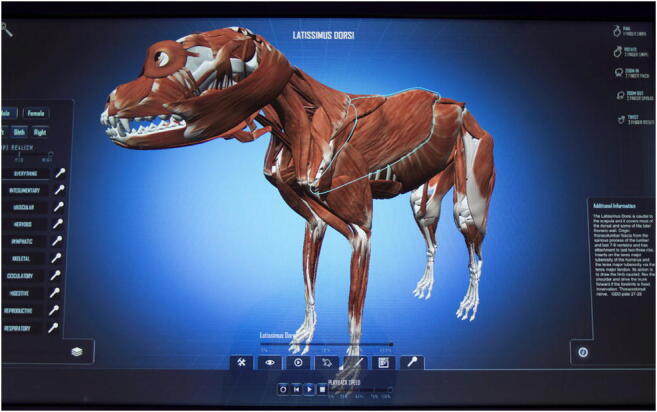
A 3D virtual canine model
The preliminary virtual canine cadaver images were produced by computer artists with no veterinary or anatomical expertise. All images were created by free-hand graphic design artists and without diagnostic imaging techniques to use as a template. Consequently, many structural representations were anatomically inaccurate and/or inappropriately labeled, while other structures were completely missing. The development of the APEX prototype-engaged computer animators in creating rudimentary anatomy structures with anticipation of future refinement by anatomists. The images for the 3D assets of the APEX table (virtual dog) were created with Autodesk 3DS Max© and/or Autodesk Maya© to develop the shape, and Adobe Photoshop© to create the texture and color. All of the 2D assets (buttons and other flat user interface items) were created in Adobe Photoshop© and/or Adobe Illustrator©.
Participants
All participants were asked to read and sign an informed consent form prior to the start of the study. Standards, guidelines, and approval for the use of student cohorts relative to this research were administered by the RUSVM institutional review board (IRB).
Student Participants
During the summer semester at RUSVM (May 9–August 18, 2016), eight third-semester students were hired as research assistants from a volunteer applicant pool who had successfully completed both of the RUSVM gross anatomy courses with a grade of 80% or higher. Success in RUSVM anatomy curriculum was used as selection criteria.
Faculty Surveyed
All RUSVM Faculty with a DVM or VMD degree, as well as those with a PhD that routinely apply veterinary anatomy were invited via email to provide survey perceptions of the APEX table. Thirty-two faculty members initially volunteered but only 29 completed both surveys.
Assessment of Student Critique
At the start of this study, students were informed that the anatomic accuracy of the virtual canine was not dependable. The students were asked to critique the APEX program by providing the following: (1) identification of an assigned anatomic structure, (2) description of the structure as found in their provided texts, and (3) a critique of the virtual structure’s anatomic accuracy in relation to size, shape, location, and association to other surrounding anatomic features. Structures which students were tasked to label were from a list of required learning objectives for the musculoskeletal system of the fore and hind limbs for the first semester RUSVM veterinary anatomy course. Each student was randomly allocated 50 structures to label as time permitted in the study. Each student devoted a minimum of 15 total hours to the labeling process. The number of labels produced per student, time spent on each label, and accuracy of the 3D representation and description were evaluated. Student-submitted corrections of anatomic structures were incorporated into an improved version of APEX by computer programmers, digital artists, and one the authors (WBL).
Survey Tools
Faculty were asked to complete a survey following each of their interactions with APEX, where their opinions of the prototype and the student-improved version were assessed. Faculty surveys collected perceptions via Likert style statements regarding the following: (1) general attitude and experience surrounding CAL tools, (2) perceived educational value of CAL tools compared to cadaver dissection, and (3) accuracy, fidelity and content of APEX table.
Students were asked to complete pre and post APEX interaction survey. The surveys consisted of Likert style statements about their opinion and interaction with the APEX table in four categories of interest: (1) general attitude and experience surrounding CAL tools, (2) perceived educational value of CAL compared to other learning resources, (3) consequences of the APEX prototype’s lack of accuracy, and (4) educational benefits associated with the APEX table.
All surveys were adapted from those developed to evaluate another computerized anatomy education tool by Linton et al. [31]. Surveys were administered online over a secure RUSVM website using E*Value™ software [32]. Although our student cohort critiqued APEX as editors rather than utilization for anatomic education, the authors shared the objective with Linton et al. to quantify student perceptions.
Statistical Analysis
Quantitative data entry of Likert responses and label scores were composed using Microsoft Excel™ in which descriptive results such as means, median, and standard deviations were calculated for each question amongst the cohort. Due to the small sample size (eight students), the median Likert response was utilized for aggregate answers. Likert scale responses were considered to be in disagreement with the statement if the median score was 2 or less, neutral if the median score was 3 and in agreement with the statement if the median score was 4 or more. To compare faculty perceptions of the initial prototype versus the student-improved APEX table, a Paired Wilcoxon test was performed on Likert survey responses. A value of p < 0.05 was considered to be statistically significant.
Each student-derived label was graded by one researcher (WBL) using a four-point scale where 4 was excellent. The scale reflected the accuracy of the student’s textual description of each specific anatomic structure, as well as the student’s critique of its 3D anatomic location, size, and shape. The grading rubric is shown in Table 1.
Table 1.
Student label evaluation rubric
| Description of student label | ||
|---|---|---|
| Score | Structure identification is anatomically accurate | Text description of structure is thorough and correct |
| 4.0 | Yes | Yes |
| 3.0 | Student label needs minor modification to identification or text description to be complete and accurate. | |
| 2.0 | Either Identification or text description is wrong (but not both) | |
| 1.0 | No | No |
Results
Student Labels
Students evaluated 306 structures on the prototype APEX table. The ability of students to correctly critique the accuracy of the prototype virtual cadaver anatomy was excellent with an average score of 3.7 out of 4.0 (Table 2).
Table 2.
Student average time required for each label produced and mean score
| Number of structures | Time (min) | Mean score | Standard deviation | |
|---|---|---|---|---|
| Thoracic limb bones | 100 | 8.3 | 3.9 | 0.46 |
| Thoracic limb muscles | 56 | 10.0 | 3.7 | 0.59 |
| Pelvic limb bones | 111 | 9.2 | 3.8 | 0.57 |
| Pelvic limb muscles | 39 | 16.0 | 3.6 | 0.63 |
| All structures | 306 | 9.9 | 3.8 | 0.55 |
Student Pre Interaction Survey
All students (n = 8) responded to the pre- and post-interaction surveys. A summary of pre-interaction Likert scale survey responses is provided in Table 3. Prior to their interaction with the APEX table, students generally enjoyed CAL (median = 4), and were excited about using the APEX table (median = 5). They reported strong disagreement with using CAL in place of hands on dissection (median= 1), and overwhelmingly agreed that CAL cannot completely replace cadaver-assisted anatomy education (median = 4.5).
Table 3.
Student Likert response for Pre APEX table interaction survey (N = 8)
| Likert scale question | |||
|---|---|---|---|
| Category | What is the general attitude and experience with CAL educational tools and the virtual anatomy table? | Median | Range |
| 1 | Computer assisted learning was commonplace in my undergraduate studies. | 2 | 1–5 |
| 1 | I relied heavily on computer assisted resources during my previous education. | 2 | 1–5 |
| 1 | I enjoy computer assisted learning. | 4 | 3–5 |
| 1 | I could happily do without computer assisted learning. | 2 | 1–4 |
| 1 | I think students would benefit by having all learning resources computerized. | 2.5 | 1–5 |
| 1 | I am excited about using the APEX table. | 5 | 5 |
| Category | What is the perceived educational value of the virtual anatomy table in relation to other learning resources? | Median | Range |
| 2 | I would rather use computer assisted learning than paper text. | 3 | 2–5 |
| 2 | I would rather use computer assisted learning than attend live lectures. | 2 | 1–5 |
| 2 | I would rather use computer assisted learning than hands on experience of cadaver dissection. | 1 | 1–2 |
| 2 | I wish I could have more of my educational resources provided in computerized from, rather than more traditional forms like text, live lectures and laboratory exercises. | 2 | 1–5 |
| 2 | I wish that all of my learning resources were in hard copy text supplemented with hands on experience. | 3 | 1–5 |
| 2 | I think that there is no substitute for hands on experience no matter how realistic a computerized simulation may be for learning. | 4.5 | 4–5 |
| 2 | I think that computerized resources should be used as a supplement to traditional learning resources such as text, live lectures and laboratory exercises, but should never replace them. | 4 | 2–5 |
| 2 | I think that computerized resources should replace some, but not all, traditional learning resources like text, live lectures and laboratory exercises. | 4 | 3–5 |
| 2 | I would like the APEX table (or a similar program) to completely replace traditional cadaver assisted anatomy learning in the future. | 1 | 1–2 |
| 2 | I do not believe the APEX table (or a similar program) could ever completely replace cadaver assisted anatomy education. | 4.5 | 3–5 |
| 2 | I believe a mixture of traditional cadaver assisted anatomy lessons parallel and complementary to computer assisted learning would be the best possible experience for my education. | 5 | 4–5 |
Likert scale; 1 = strongly disagree, 2 = disagree, 3=neutral, 4=agree, 5=strongly agree
Student Post-Interaction Survey
Survey results are summarized in Table 4. Utilization of the APEX table was found to be enjoyable (median = 4), beneficial to their knowledge of anatomy (median = 4), and agreed that it would have been helpful to have an APEX table application for their mobile device to study from while enrolled in the anatomy courses (median = 4). Students indicated that they would recommend use of the APEX table to future anatomy classes (median = 4.5). Once more, students strongly disagreed with the ability of CAL to completely replace the use of canine cadavers for anatomic education in the future (median = 1).
Table 4.
Student Likert response for post APEX table interaction survey (n = 8)
| Likert scale question | |||
|---|---|---|---|
| Category | 1) What is the general attitude and experience with CAL educational tools and the virtual anatomy table? | Median | Range |
| 1 | I enjoyed using the “APEX” table. | 4 | 3–5 |
| 1 | I appreciate that the “APEX” table could help to reduce the number of canine cadavers needed for anatomy instruction. | 3 | 1–5 |
| 1 | I appreciate the ability of the “APEX” table in reducing the environmental consequences of chemically preserved cadavers for anatomy education. | 3 | 2–5 |
| 1 | I would recommend using the “APEX” table in future RUSVM anatomy classes. | 4.5 | 1–5 |
| 1 | If I had access to the “APEX” table when I was taking the anatomy courses, I would not have had enough time to spend with the “APEX” table because there are so many other anatomy learning resources provided in the veterinary anatomy course curriculum. | 2 | 1–3 |
| Category | 2) What is the perceived educational value of the virtual anatomy table in relation to other learning resources? | Median | Range |
| 2 | I believe that computerized learning models and simulation should completely replace the use of canine cadavers for anatomic education in the future. | 1 | 1–2 |
| 2 | There are already enough computerized anatomy learning resources available for study and I feel that the addition of the “APEX” table is an unnecessary burden to the anatomy study load. | 1.5 | 1–3 |
| 2 | I found the “APEX” table more useful than some of the other electronic anatomy learning resources provided in the course. | 4 | 4 |
| 2 | I would have preferred to spend more time with the “APEX” table rather than some of the other computer assisted resources provided when I was taking the veterinary anatomy course. | 3 | 1–4 |
| Category | 3) What is the consequence of the prototype virtual anatomy table’s lack of accuracy? | Median | Range |
| 3 | I predict the “APEX” table will be more useful when the detail, accuracy and fidelity are increased with advancement in software. | 4.5 | 4–5 |
| 3 | I would like to see the “APEX” table brought to a higher level of fidelity and accuracy. | 5 | 4–5 |
| 3 | Even though the fidelity and accuracy of the “APEX” table was not perfect, I found its use to be beneficial to my overall knowledge. | 4 | 4–5 |
| 3 | The “APEX” table obviously lacks the detail of a cadaver canine specimen and I found these imperfections to be a significant hindrance to my learning. | 2 | 2–5 |
| 3 | The anatomic inaccuracies of the table made it difficult to increase my anatomic knowledge from the “APEX” table. | 2.5 | 1–4 |
| 3 | I believe I would have learned more anatomy if I could have interacted with a “APEX” table that was anatomically perfect. | 4 | 2–5 |
| Category | 4) Will the virtual anatomy table be an educational benefit? | Median | Range |
| 4 | The “APEX” table was beneficial to my knowledge of veterinary anatomy. | 4 | 3–5 |
| 4 | I believe the “APEX” table and other virtual learning tools will be an essential part of anatomy education in the future. | 4 | 3–4 |
| 4 | It would have been helpful to have a “APEX” table application for my mobile device when I took the RUSVM anatomy courses. | 4 | 3–5 |
| 4 | If there were an application for my mobile device that was similar to the “APEX” table, I would use it as a study aid for future veterinary courses. | 4 | 4–5 |
| 4 | After spending over 15 h with the “APEX” table, I feel my canine anatomic knowledge is significantly improved. | 4 | 3–5 |
| 4 | Discovering and rectifying the anatomic inaccuracies within the “APEX” table improved my anatomic knowledge. | 5 | 4–5 |
| 4 | I believe I learned more anatomy from discovering and fixing inaccuracies within the table, than I would have if the table were anatomically perfect. | 4 | 2–5 |
| 4 | I believe my experience with this version of the “APEX” table will be helpful for my future education and career. | 4 | 4–5 |
Likert scale; 1 = strongly disagree, 2 = disagree, 3 = neutral, 4 = agree, 5 = strongly agree
Faculty Survey Post-Interaction with the Prototype APEX Table
Survey responses have been cumulated in Table5. Likert scale responses reveal that faculty were in disagreement with the statement that the prototype version of the APEX table was ready to be integrated into the RUSVM curriculum (mean = 2.0). Despite faculty’s general discontent with the initial prototype, they believed that this type of 3D, virtual anatomy model could be an excellent benefit to anatomy students in the future (mean = 4.3). Faculty agreed that the “APEX” table (or a similar program) could never completely replace cadaver based anatomy education (mean = 4.00).
Table 5.
Faculty survey Likert response and p value comparisons of means between prototype and improved APEX perceptions
| Perceptions of prototype (n = 32) | Perceptions after students’ improvements (n = 29) | |||||
|---|---|---|---|---|---|---|
| Category | Likert scale question | Mean | Standard deviation | Mean | Standard deviation | p value (paired Wilcoxon of means) |
| Category | Category 1) What is the general attitude and experience of the APEX table? | |||||
| 1 | I enjoyed using the virtual anatomy table. | 3.6 | 0.8 | 4.3 | 0.6 | < 0.001 |
| 2 | Category 2) What is the perceived educational value of CAL and the APEX table in relation to other learning resources? | |||||
| 2 | I do not believe that the “APEX” table (or a similar program) could ever completely replace cadaver based anatomy education. | 4.0 | 1.1 | 4.4 | 0.8 | 0.04 |
| 2 | I believe that a mixture of traditional cadaver based anatomy lessons parallel and complementary to computer based learning would be the best possible option for promoting anatomy education. | 4.6 | 0.6 | 4.7 | 0.6 | 0.6 |
| 3 | Category 3) Is the accuracy, fidelity and content of APEXL table suitable for this anatomy course? | |||||
| 3 | I believe the “APEX” table is ready to enter into the RUSVM curriculum, in its current state of fidelity, anatomic accuracy and content. | 2.0 | 1.2 | 3.9 | 0.7 | < 0.001 |
| 3 | I believe this type of three-dimensional, virtual anatomy model will be an excellent benefit to anatomy students in the future. | 4.3 | 0.7 | 4.5 | 0.6 | 0.4 |
| 3 | I am concerned that a student could learn incorrect anatomy because of the “APEX” table’s inaccuracies. | 3.2 | 0.9 | 2.2 | 0.7 | < 0.001 |
| 3 | The “APEX” table has great potential; however it is not ready for implementation into the RUSVM curriculum. | 3.8 | 1.0 | Not applicable | ||
| 3 | The virtual anatomy table looked realistic enough to learn the thoracic and pelvic limb anatomy effectively. | Not applicable | 4.0 | 4.4 | 0.6 | |
| 3 | This new version of the virtual anatomy table exhibits more accurate anatomy of the thoracic and pelvic musculoskeletal structures than the previous version. | Not applicable | 5.0 | 4.6 | 0.5 | |
Likert scale; 1 = strongly disagree, 2 = disagree, 3 = neutral, 4 = agree, 5 = strongly agree
Faculty Survey Post-Interaction with Student-Improved APEX Table
The second survey was completed by 29 (90.63%) of the original volunteers. Faculty noted that the improved APEX table exhibited more accurate anatomy of the canine thoracic and pelvic limb musculoskeletal structures (mean = 4.6), and that the represented anatomy looked realistic enough to effectively promote learning (mean = 4.4). Concerns that students could learn incorrect anatomy because of the “APEX” table’s inaccuracies significantly decreased (from mean 3.2 to 2.2, p < 0.001).
Faculty indicated that the updated version of the virtual anatomy table was ready to be integrated into the RUSVM curriculum to supplement teaching of musculoskeletal structures of the thoracic and pelvic limb (mean = 3.9), which was significantly improved over their perceptions of the prototype version (p < 0.001).
Discussion
The results of this study demonstrate the value of involving veterinary students in the development of the RUSVM APEX table. Students’ efforts in identifying and describing inaccurate anatomical structures resulted in the correction of 306 anatomic structures on the APEX prototype with excellent precision. This improvement in accuracy achieved by the students can potentially alleviate some of the aforementioned issues surrounding the production of learning tools recognized previously, including investment of time, money, and expertise [33].
Students and faculty reported a generally positive experience with the APEX table. Furthermore, participants reported a positive perception toward its accuracy, content, and overall benefit toward facilitating the learning of veterinary anatomy. Statistically significant improvements were noted in faculty perceptions of the improved version of APEX table with respect to readiness for incorporation into the curriculum (p = < 0.001) and decreased concern for inaccuracies (p = 0.001) in comparison to the prototype. These findings are similar to other studies which have shown CAL to be enjoyable by students [4, 34, 35], effective [18], and well accepted in delivery of anatomic information [36–38]. This type of learning technology may facilitate discovery of important material, mental organization of the subject, and assimilation of knowledge [39].
Although student and faculty perceptions regarding the effectiveness of the APEX table were positive, participants in this study were opposed to the concept of replacing cadaveric dissection with a virtual learning resource. The forward-looking survey questions in this study were created to discover the extent of student and faculty acceptance in supplementing traditional cadaveric dissection with virtual anatomy tools. Computer models have been shown to rely heavily on the user’s own innate visuospatial capabilities [40] making the design of a universally effective computer-based resource problematic [41]. In agreement with previous literature [38], it is the authors’ opinion that CAL programs should not replace traditional methods of teaching anatomy. We believe that hands on dissection is an essential part of veterinary anatomy education.
Virtual anatomic simulation offers an attractive option to circumvent some of the vast challenges affecting traditional veterinary anatomy education [10]. As there is no single anatomy teaching strategy proven to be superior to all others, instructors must enhance students’ academic achievements by maintaining flexibility in their teaching techniques [17]. The goal of educational research should not be to determine higher acceptance or effectiveness of one teaching methodology over another but rather to most efficiently utilize the learning benefits available from each teaching method [42]. This premise may explain the great variation in responses received when students were asked if they would have learned more had they interacted with an APEX table that was anatomically perfect. These results also suggest that involving students in the development of teaching resources may be a useful learning technique.
A few limitations associated with this project exist. This study used a relatively small cohort of paid student assistants, and by definition may not have been completely representative of the class as a whole. We acknowledge that these results do not address the effectiveness of the APEX tool in enhancing student understanding or exam outcomes and emphasize that the focus of the current study is to determine the feasibility of student efforts in the development of virtual anatomy educational tools.
Although the results of this study may not be unexpected regarding students’ ability to correctly identify anatomic inaccuracies, it is noteworthy to describe a proven mechanism by which students may be used to assist in the development of CAL and how to objectively measure their accuracy. We understand that CAL technology may be financially limiting as a developmental venture as well as acquisition of commercially available products. By utilizing student efforts during development of CAL, perhaps some financial burden may be limited.
Judgements about computer-based learning tools vary widely, and it is clear that much work is left to be done, both in developing and in evaluating new instructional systems [20, 43, 44]. Future work is necessary to assess the effect of the APEX table interaction on student anatomy examination outcomes. Continued evaluation is also warranted to assess if virtual anatomy tools can decrease student dependence upon instructors during laboratory dissection, if knowledge attained with this resource is retained over longer periods of time, and if that knowledge is transferable to other courses of study.
Conclusions
Involving veterinary student assistants offers a novel approach to the development of veterinary anatomy teaching resources. Although students have been co-producers of their educational tools in the past, this study specifically addresses student evaluation as the driving force of CAL development. Students accurately identified and documented where the virtual cadaver required improvement. In regard to their experience with the APEX table, students overwhelmingly reported that the experience was enjoyable as well as helpful to their education.
RUSVM faculty perceptions overwhelmingly supported the use of the APEX table indicating that the student-improved version of the software exhibited accurate anatomy and fidelity and was ready to be incorporated into the RUSVM anatomy curriculum. Virtual simulation offers an ethical, safe and novel method of enhancing the teaching of veterinary gross anatomy. Further research is indicated to assess students’ examination outcomes following the use of APEX table.
Acknowledgements
APEX table development was facilitated by Gear Learning development studio.
Funding Information
Funding for this project was provided by an intramural grant from Ross University School of Veterinary Medicine Center-4, Research and Innovation in Veterinary and Medical Education.
Compliance with Ethical Standards
All participants were asked to read and sign an informed consent form prior to the start of the study. Standards, guidelines, and approval for the use of student cohorts relative to this research were administered by the RUSVM institutional review board (IRB).
Conflict of Interest
The authors declare that they have no conflict of interest.
This manuscript does not contain images, tables or figures which are under copyright. All authors have read and accept responsibility for this manuscript.
Footnotes
Publisher’s Note
Springer Nature remains neutral with regard to jurisdictional claims in published maps and institutional affiliations.
References
- 1.Cottam WW. Adequacy of medical school gross anatomy education as perceived by certain postgraduate residency programs and anatomy course directors. Clin Anat. 1999;12(1):55–65. doi: 10.1002/(SICI)1098-2353(1999)12:1<55::AID-CA8>3.0.CO;2-O. [DOI] [PubMed] [Google Scholar]
- 2.Aziz MA, Mckenzie JC, Wilson JS, Cowie RJ, Ayeni SA, Dunn BK. The human cadaver in the age of biomedical informatics. Anat Rec. 2002;269(1):20–32. doi: 10.1002/ar.10046. [DOI] [PubMed] [Google Scholar]
- 3.Cake MA. Deep dissection: motivating students beyond rote learning in veterinary anatomy. J Vet Med Educ. 2006;33(2):266–271. doi: 10.3138/jvme.33.2.266. [DOI] [PubMed] [Google Scholar]
- 4.Nicholson DT, Chalk C, Funnell WRJ, Daniel SJ. Can virtual reality improve anatomy education? A randomised controlled study of a computer-generated three-dimensional anatomical ear model. Med Educ. 2006;40(11):1081–1087. doi: 10.1111/j.1365-2929.2006.02611.x. [DOI] [PubMed] [Google Scholar]
- 5.Adams CM, Wilson TD. Virtual cerebral ventricular system: an MR-based three-dimensional computer model. Anat Sci Educ. 2011;4(6):340–347. doi: 10.1002/ase.256. [DOI] [PubMed] [Google Scholar]
- 6.Abdelhamid TM. The multidimensional learning model: A novel cognitive psychologybased model for computer assisted instruction in order to improve learning in medical students. Medical Education Online. 1999;4(1):4302.
- 7.Trelease RB. Anatomical informatics: millennial perspectives on a newer frontier. Anat Rec. 2002;269(5):224–235. doi: 10.1002/ar.10177. [DOI] [PubMed] [Google Scholar]
- 8.Sugand K, Abrahams P, Khurana A. The anatomy of anatomy: a review for its modernization. Anat Sci Educ. 2010;3(2):83–93. doi: 10.1002/ase.139. [DOI] [PubMed] [Google Scholar]
- 9.McLachlan JC, Bligh J, Bradley P, Searle J. Teaching anatomy without cadavers. Med Educ. 2004;38(4):418–424. doi: 10.1046/j.1365-2923.2004.01795.x. [DOI] [PubMed] [Google Scholar]
- 10.Hart LA, Wood MW, Weng H-Y. Mainstreaming alternatives in veterinary medical education: resource development and curricular reform. J Vet Med Educ. 2005;32(4):473–480. doi: 10.3138/jvme.32.4.473. [DOI] [PubMed] [Google Scholar]
- 11.Arráez-Aybar LA, Casado-Morales MI, Castaño-Collado G. Anxiety and dissection of the human cadaver: an unsolvable relationship? Anat Rec B New Anat. 2004;279(1):16–23. doi: 10.1002/ar.b.20022. [DOI] [PubMed] [Google Scholar]
- 12.Jones DG. Reassessing the importance of dissection: a critique and elaboration. Clin Anat. 1997;10(2):123–127. doi: 10.1002/(SICI)1098-2353(1997)10:2<123::AID-CA9>3.0.CO;2-W. [DOI] [PubMed] [Google Scholar]
- 13.Issenberg S, Petrusa ER, McGaghie WC, Felner JM, Waugh RA, Nash IS, et al. Effectiveness of a computer-based system to teach bedside cardiology. Acad Med. 1999;74(10):S93–S95. doi: 10.1097/00001888-199910000-00051. [DOI] [PubMed] [Google Scholar]
- 14.Patronek GJ, Rauch A. Systematic review of comparative studies examining alternatives to the harmful use of animals in biomedical education. J Am Vet Med Assoc. 2007;230(1):37–43. doi: 10.2460/javma.230.1.37. [DOI] [PubMed] [Google Scholar]
- 15.Salazar I. Coming changes in veterinary anatomy: what is or should be expected? J Vet Med Educ. 2002;29(3):126–130. doi: 10.3138/jvme.29.3.126. [DOI] [PubMed] [Google Scholar]
- 16.Drake RL, McBride JM, Lachman N, Pawlina W. Medical education in the anatomical sciences: the winds of change continue to blow. Anat Sci Educ. 2009;2(6):253–259. doi: 10.1002/ase.117. [DOI] [PubMed] [Google Scholar]
- 17.Al-Mohrej OA, Al-Ayedh NK, Masuadi EM, Al-Kenani NS. Learning methods and strategies of anatomy among medical students in two different Institutions in Riyadh, Saudi Arabia. Medical Teacher. 2017;39(sup1):S15–S21. [DOI] [PubMed]
- 18.Nieder GL, Scott JN, Anderson MD. Using QuickTime virtual reality objects in computer-assisted instruction of gross anatomy: Yorick—the VR skull. Clin Anat. 2000;13(4):287–293. doi: 10.1002/1098-2353(2000)13:4<287::AID-CA9>3.0.CO;2-L. [DOI] [PubMed] [Google Scholar]
- 19.Tam M, Hart A, Williams S, Holland R, Heylings D, Leinster S. Evaluation of a computer program (‘disect’) to consolidate anatomy knowledge: a randomised-controlled trial. Med Teach. 2010;32(3):e138–ee42. doi: 10.3109/01421590903144110. [DOI] [PubMed] [Google Scholar]
- 20.Tam M, Hart A, Williams S, Heylings D. Leinster S. is learning anatomy facilitated by computer-aided learning? A review of the literature. Med Teach. 2009;31(9):e393–e3e6. doi: 10.1080/01421590802650092. [DOI] [PubMed] [Google Scholar]
- 21.Yammine K, Violato C. A meta-analysis of the educational effectiveness of three-dimensional visualization technologies in teaching anatomy. Anat Sci Educ. 2015;8(6):525–538. doi: 10.1002/ase.1510. [DOI] [PubMed] [Google Scholar]
- 22.Motola I, Devine LA, Chung HS, Sullivan JE, Issenberg SB. Simulation in healthcare education: a best evidence practical guide. AMEE Guide No 82. Med Teach. 2013;35(10):e1511–e1e30. doi: 10.3109/0142159X.2013.818632. [DOI] [PubMed] [Google Scholar]
- 23.Brophy J. Motivating students to learn. Ch 1. Introduction to Student Motivation. Abingdon: Routledge; 2013. p. 1–26.
- 24.Reidenberg JS, Laitman JT. The new face of gross anatomy. Anat Rec. 2002;269(2):81–88. doi: 10.1002/ar.10076. [DOI] [PubMed] [Google Scholar]
- 25.Abutarbush SM, Naylor JM, Parchoma G, D'Eon M, Petrie L, Carruthers T. Evaluation of traditional instruction versus a self-learning computer module in teaching veterinary students how to pass a nasogastric tube in the horse. J Vet Med Educ. 2006;33(3):447–454. doi: 10.3138/jvme.33.3.447. [DOI] [PubMed] [Google Scholar]
- 26.Berg D, Raugi G, Gladstone H, Berkley J, Weghorst S, Ganter M, Turkiyyah G. Virtual reality simulators for dermatologic surgery: measuring their validity as a teaching tool. Dermatol Surg. 2001;27(4):370–374. doi: 10.1046/j.1524-4725.2001.00323.x. [DOI] [PubMed] [Google Scholar]
- 27.Spitzer VM, Scherzinger AL. Virtual anatomy: an anatomist’s playground. Clin Anat. 2006;19(3):192–203. doi: 10.1002/ca.20330. [DOI] [PubMed] [Google Scholar]
- 28.Balcombe J. Student/teacher conflict regarding animal dissection. Am Biol Teach. 1997;59(1):22–25.
- 29.Dyce KM, Sack WO, Wensing CJG. Textbook of veterinary anatomy. Amsterdam: Elsevier Health Sciences; 2009.
- 30.Evans HE, De Lahunta A. Guide to the dissection of the dog. Amsterdam: Elsevier Health Sciences; 2016.
- 31.Linton A, Schoenfeld-Tacher R, Whalen LR. Developing and implementing an assessment method to evaluate a virtual canine anatomy program. J Vet Med Educ. 2005;32(2):249–254. doi: 10.3138/jvme.32.2.249. [DOI] [PubMed] [Google Scholar]
- 32.Medhub. E*Value http://www.medhub.com/evalue/evalue-product/. Accessed 19 July 2017.
- 33.Inwood MJ, Ahmad J. Development of instructional, interactive, multimedia anatomy dissection software: a student-led initiative. Clin Anat. 2005;18(8):613–617. doi: 10.1002/ca.20140. [DOI] [PubMed] [Google Scholar]
- 34.Francis NR, Lewis W. What price dissection? Dissection literally dissected. Med Humanit. 2001;27(1):2–9. doi: 10.1136/mh.27.1.2. [DOI] [PubMed] [Google Scholar]
- 35.Nieder GL, Nagy F. Analysis of medical students' use of web-based resources for a gross anatomy and embryology course. Clin Anat. 2002;15(6):409–418. doi: 10.1002/ca.10067. [DOI] [PubMed] [Google Scholar]
- 36.Jastrow H, Vollrath L. Teaching and learning gross anatomy using modern electronic media based on the visible human project. Clin Anat. 2003;16(1):44–54. doi: 10.1002/ca.10062. [DOI] [PubMed] [Google Scholar]
- 37.Khalil M, Lamar C, Johnson T. Using computer-based interactive imagery strategies for designing instructional anatomy programs. Clin Anat. 2005;18(1):68–76. doi: 10.1002/ca.20049. [DOI] [PubMed] [Google Scholar]
- 38.Codd AM, Choudhury B. Virtual reality anatomy: is it comparable with traditional methods in the teaching of human forearm musculoskeletal anatomy? Anat Sci Educ. 2011;4(3):119–125. doi: 10.1002/ase.214. [DOI] [PubMed] [Google Scholar]
- 39.Frick CMRTW, Reigeluth CM. Formative research: A methodology for creating and improving design theories. Instructional-design theories and models, 1999;2.
- 40.Estevez ME, Lindgren KA, Bergethon PR. A novel three-dimensional tool for teaching human neuroanatomy. Anat Sci Educ. 2010;3(6):309–317. doi: 10.1002/ase.186. [DOI] [PMC free article] [PubMed] [Google Scholar]
- 41.Khot Z, Quinlan K, Norman GR, Wainman B. The relative effectiveness of computer-based and traditional resources for education in anatomy. Anat Sci Educ. 2013;6(4):211–215. doi: 10.1002/ase.1355. [DOI] [PubMed] [Google Scholar]
- 42.Turney B. Anatomy in a modern medical curriculum. Ann R Coll Surg Engl. 2007;89(2):104–107. doi: 10.1308/003588407X168244. [DOI] [PMC free article] [PubMed] [Google Scholar]
- 43.McLachlan JC, Patten D. Anatomy teaching: ghosts of the past, present and future. Med Educ. 2006;40(3):243–253. doi: 10.1111/j.1365-2929.2006.02401.x. [DOI] [PubMed] [Google Scholar]
- 44.Winkelmann A. Anatomical dissection as a teaching method in medical school: a review of the evidence. Med Educ. 2007;41(1):15–22. doi: 10.1111/j.1365-2929.2006.02625.x. [DOI] [PubMed] [Google Scholar]