Case description
A 27-years-old male presented with progressively worsening dysphagia for four years. The dysphagia was worse for solids, and for the past three months, the patient had restricted his diet to liquids and pureed foods. He had also developed some hoarseness in his voice during this time. Past medical history was significant for gastroesophageal reflux disease, hypertension and hyperlipidemia. Physical examination was unremarkable.
Esophagogastroduodenoscopy (EGD) was performed that revealed a pulsating impression at 27 cm from the incisors, causing narrowing in the esophagus.
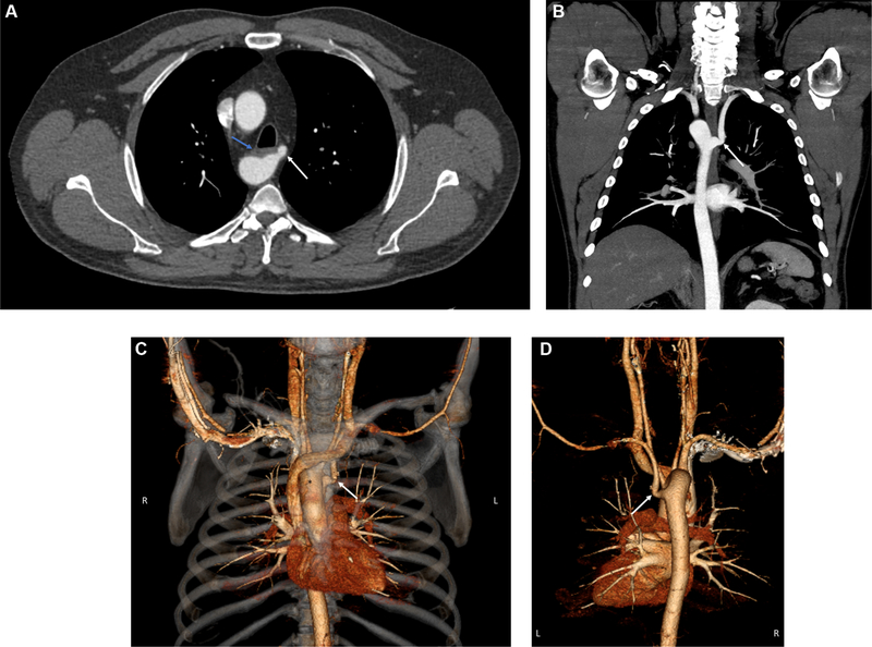
Following which, CT angiogram of the chest was performed, and is depicted in Fig. 1A and B. 3D reconstruction images are described in 1C and 1D. What is the cause of this patient’s symptoms?
Fig. 1.
CT angiogram of the chest (Fig. 1A and B) along with 3D reconstruction images (Fig. 1C and D) demonstrates right-sided aortic arch with aberrant left subclavian artery (white arrow) causing external compression of the esophagus (blue arrow), the condition is known as dysphagia lusoria.
Diagnosis:
Dysphagia lusoria for aberrant subclavian artery
Discussion
CT angiogram of the chest (Fig. 1A and B) and 3D reconstruction image (Fig. 1C and D), shows right-sided aortic arch with aberrant left subclavian artery (white arrow) causing external compression of the esophagus (blue arrow), a condition known as dysphagia lusoria.
Right-sided aortic arch with associated left subclavian artery occurs from left aortic arch dorsal segment interruption between left common carotid and left subclavian arteries. It is found in 0.05–0.2% of the population [1].
Patient was managed with surgical left carotid subclavian bypass and stent graft coverage of the aberrant subclavian was performed. He is doing well and reports improvement in dysphagia at a two year follow up.
Dysphagia lusoria is a rare condition caused by an aberrant right or left subclavian artery. The diagnosis is often missed on an EGD, although occasionally a pulsating impression may be seen on EGD. Patients are usually diagnosed with a barium upper GI contrast study or a contrast enhanced CT scan. In many cases, the finding is incidental, and patients are managed conservatively [2]. Symptoms arise if the aberrant subclavian artery extrinsically compresses the esophagus, leading to dysphagia. Surgical correction is considered for patients with intractable symptoms [3].
Footnotes
Disclosures
The authors have no conflicts of interest to disclose.
Submission declaration and verification
The authors have not published, posted, or submitted any related papers from this same study.
References
- [1].Türkvatan A, Büyükbayraktar FG, Olçer T, et al. Congenital anomalies of the aortic arch: evaluation with the use of multidetector computed tomography. Korean J Radiol 2009;10:176–84. [DOI] [PMC free article] [PubMed] [Google Scholar]
- [2].Janssen M, Baggen MG, Veen HF, et al. Dysphagia lusoria: clinical aspects, manometric findings, diagnosis, and therapy. Am J Gastroenterol 2000;95:1411–6. [DOI] [PubMed] [Google Scholar]
- [3].Carrizo GJ, Marjani MA. Dysphagia lusoria caused by an aberrant right subclavian artery. Tex Heart Inst J 2004;31:168–71. [PMC free article] [PubMed] [Google Scholar]


