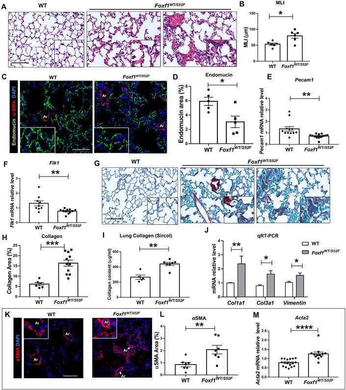
Figure 2. Adult Foxf1WT/S52F mice exhibit lung remodeling and vascular abnormalities.
(A-B) H&E staining of lung sections shows diffuse pulmonary inflammation and alveolar simplification in Foxf1WT/S52F adult mice. Mean linear intercept (MLI) was determined using 5 random images from each of 3 H&E-stained lung sections per mouse (n=6 mice). Scale bars are 50μm. (C-D) Immunostaining for Endomucin (green) and αSMA (red) shows reduced capillary density in lungs from Foxf1WT/S52F mice. Lung sections were counterstained with DAPI (blue). Five random lung images per mouse (n=5 mice per group) were used for quantification. Scale bars are 25μm. (E-F) qRT-PCR shows that Pecam1 and Flk1 mRNAs are decreased in Foxf1WT/S52F lungs. mRNAs were normalized using β-actin mRNA (n=9-13 mice per group). (G-H) Sirius red staining shows increased collagen deposition in Foxf1WT/S52F lungs. Scale bars are 50μm. Data were quantified using 5 random lung images per mouse (n=6-11 mice per group). (I) Sircol assay shows increased collagen content in the left lobe of Foxf1WT/S52F lungs (n=5-7 mice per group). (J) qRT-PCR shows that Col1a1, Col3a1 and Vimentin mRNAs are increased in Foxf1WT/S52F lungs (n=6-9 mice per group). (K-L) Images show increased αSMA staining (red) with small artery muscularization (inserts) in lungs from Foxf1WT/S52F mice. Lung sections were counterstained with DAPI (blue). Data were quantified using 5 random lung images per mouse (n=7 mice per group). Scale bars are 50μm. (M) qRT-PCR shows that Acta2 mRNA is increased in lungs from Foxf1WT/S52F mice (n=12-17 mice per group). * indicates p < 0.05, ** indicates p < 0.01, *** indicates p < 0.001, **** indicates p < 0.0001. Abbreviations: MLI, Mean linear intercept; Ar, artery; V, vein.

