Abstract
Covid-19 pandemic reveals that the virus causes Covid-19 associated coagulopathy and it is well known that thrombotic risk is associated with ethnicity. To describe the Covid-19 associated coagulopathy in Indian population and to correlate it with the disease severity and survivor status. A cross sectional descriptive study of 391 confirmed Covid-19 cases was carried out over a period of 1.5 months. Patients were categorised as mild to moderate, severe and very severe and also labelled as survivors and non survivors. Prothrombin time (PT), International normalised ratio (INR), activated partial thromboplastin time, D dimer, Fibrin degradation products (FDP), fibrinogen and thrombin time and platelet counts were investigated among the subgroups. Mean age was higher in patients with severe disease (57.62 ± 13.08) and among the non survivors (56.54 ± 12.78). Statistically significant differences in D dimer, FDP, PT, INR and age were seen among the 3 subgroups and survivors. Strong significant positive correlation was noted between D dimer and FDP (r = 0.838, p < .001), PT and INR (r = 0.986, p < 0.001). D dimer was the best single coagulation parameter as per the area under curve (AUC: 0.762, p < 0.001) and D dimer + FDP was the best combination parameter (AUC: 0.764, p = 0) to differentiate mild moderate from severe disease. Raised levels of D dimer, FDP, PT, PT INR and higher age correlated positively with disease severity and mortality in Indian Population.
Keywords: Covid-19, Coagulopathy, D dimer, FDP, Indian population
Introduction
A Pneumonia of obscure origin was first noted in Wuhan district of Hubei province of China in Dec 2019 with high rate of spread among humans and was traced to the wet market [1, 2]. The disease, since named COVID-19, has emerged as a global pandemic that has affected 213 countries and devasted almost the entire world in an unforeseen way. In the first 6 months it has infected more than 19.43 million people globally with more than 0.72 million deaths. In India itself more than 2.15 million people are affected with more than 43,000 deaths [3].
The causative agent was identified as a novel betacorona virus and was named as Severe acute respiratory syndrome coronavirus 2 (SARS COV 2) [2]. It was similar to Severe acute respiratory syndrome coronavirus (SARS COV) and Middle East Respiratory Syndrome (MERS) viruses [2, 4]. Spike proteins on the surface envelope of this RNA virus appeared like studs under electron microscope giving it the typical corona appearance and hence the name. It showed tropism with specific angiotensin converting enzyme 2 (ACE-2) receptors which are present on the respiratory epithelium mainly the vascular endothelial cells and Type II pneumocytes [2, 5].
Similar to SARS and MERS, SARS COV 2 causes mainly respiratory infection in humans but the disease severity is much less [6]. Symptoms are usually mild but rarely few patients develop serious respiratory infections including viral Pneumonia, acute respiratory distress syndrome (ARDS) or systemic infection causing multiorgan failure leading to death [7].
Host inflammatory reaction releases proinflammatory cytokines which activates the coagulation system and host defence mechanisms leading to disseminated intravascular coagulation (DIC) [8]. Cytokine storm resulting from excess inflammatory response also causes rise in coagulation parameters and D dimer [9].
Increased disease severity enhances host defence mechanism causing rise in proinflammatory cytokines like Il 6, IL-1beta, TNF alpha and activation of complement system to initiate coagulopathy. The proinflammatory cytokines activates the acute phase reactants (APR) like CRP, ferritin, fibrinogen and procalcitonin which take part in coagulation and defence [10]. The systemic inflammatory markers are also increased [7, 10, 11]. Recent studies on COVID 19 also demonstrates comparable results substantiating SARS COV 2 causes severe inflammation and deranged coagulation [12–15].
Fibrinogen, an acute phase reactant and a pro coagulant is seen to be raised initially in all patients of Covid-19 [13]. IL-6, a pro inflammatory marker is also increased at admission in these patients proving that a connection exists between inflammation and procoagulant change [14].
Studies have supported the fact that coagulation changes are present in these patients [13, 15, 16]. This is referred to as Covid-19 associated coagulopathy (CAC) which is distinct from classical disseminated intravascular coagulation (DIC) which follows sepsis. CAC shows minimum changes in PT, APTT and platelet count, although D dimer and fibrinogen levels are high in the initial stages [8].
Also, recent autopsy findings of Covid-19 patients reveals pulmonary intravascular coagulopathy (PIC) as a crucial pathogenic mechanism which is characterised by micro vascular thrombosis of the pulmonary vessels. It is an immune thrombosis and is confined to the lungs [17].
Current research on coagulopathy in Covid-19 is mainly dominated by Chinese cohort but data on people of other races, especially from Indians are scarce. Studies on Chinese population have shown D dimer levels are elevated while Platelet counts and PT are usually normal [18–22].
The coagulation changes are likely to show variability in people of different ethnicities as thrombotic risk is known to be dependent on race and ethnicity [21].
Development of coagulopathy and elevated levels of D dimer have been implicated as a lone predictor of poor prognosis in Covid-19 patients of both Chinese and Caucasians cohort [5, 13, 16, 21].
Hence, it becomes imperative to study the difference in the coagulation parameters in Indians which might explain the inter-ethnic variation in the disease presentation and mortality.
To the best of our knowledge this is the first study on coagulation parameters on Covid-19 positive Indian population to predict the disease severity and progression.
Materials and Methods
A cross sectional descriptive study on 391 confirmed positive Covid-19 patients by real time Reverse Transcriptase Polymerase Chain reaction (RT PCR) who were admitted to ESIC MC and Hospital, Faridabad was conducted in the Departments of Pathology from 1st June to 15th July 2020. Prior approval of Institutional Ethics Committee and written consent from patients were taken.
Data Collection
Detailed clinical history, symptoms, socio demographic profile, travel history and comorbidities from the study population were obtained.
Sample Collection
All the samples were collected within day 1–3 of the admission to intensive care unit (ICU) or isolation ward. Venous blood samples (3–4 ml each) were collected using vacutainer in citrated vial for coagulation profile and EDTA vial for platelets. Fully automated coagulation analyser (Stago STA compact max) was used for the coagulation tests while Sysmex XN 1000 haematology analyser was used for platelet counts.
The samples were collected, processed and discarded by trained technical staff wearing proper personal protective equipment (PPE) kits and taking all necessary precautions as per the standard guidelines and protocols for Covid-19 sample handling.
Inclusion Criteria
All those individuals who tested positive by real time reverse transcriptase polymerase chain reaction (RT-PCR) on nasopharyngeal or oropharyngeal swabs were considered as Covid-19 positive cases. They presented as asymptomatic or with mild or moderate or severe or very severe symptoms.
Based on disease severity and symptoms the cases were categorised into mild moderate, severe and very severe.
Mild to moderate Covid-19 were the ones presenting with fever, cough, diarrhoea, body ache, headache but having normal oxygen saturation and no features of severe or very severe disease.
Severe Covid-19 is defined as having either one of the following criteria: (1) Respiratory distress with respiratory rate more than 30 times/min; (2) Oxygen saturation ≤ 93% in resting state; (3) PaO2/FiO2 ≤ 300 mmHg (1 mmHg = 0.133 kPa). Very severe Covid-19 is defined as having either one of the following criteria: (1) Respiratory failure in need of mechanical ventilation; (2) Shock, (3) Other organ dysfunction [16].
Exclusion Criterion
All asymptomatic cases and patients whose complete set of all above coagulation parameters and platelet counts were not available were not considered. The patients with history of cardiovascular disease, metabolic syndromes, inflammatory bowel disease, haematological disorders, trauma or surgeries in last six months or non-ambulatory patients, those having thromboembolic disorders, pregnant females or on drugs known to alter the coagulation profile/platelet count, kidney or liver disease, neoplasms, were excluded from the study.
Statistical Analysis
All the lab data were tabulated along with age, sex, disease severity and survivor status. Computer based data entry was done. SPSS statistics software (version 21.0) (IBM, NY) was used for statistical data analysis. The continuous data were presented as mean and standard deviation. The test of significance applied to test difference between groups for continuous variables was Student’s t test and ANOVA. The categorical data were presented as proportions and analysed using chi square test. Pearson’s correlation coefficient was calculated for studying association between lab parameters. Binary logistic regression analysis was used to combine various parameters. The diagnostic values of valuable parameters for differentiating mild moderate and severe cases of Covid-19 patients were assessed by receiver operating characteristic (ROC) and area under the ROC curve (AUC). The significance level was set at 5%.
Results
Age: (Table 1 and 2)
Table 1.
Comparison of various coagulation parameters and age of COVID-19 patients in accordance with disease severity
| Clinical status | Age | PT | INR | APTT | Ddimer | FDP | FIB | TT | PLT |
|---|---|---|---|---|---|---|---|---|---|
| Mild moderate | |||||||||
| Mean | 40.45 | 13.89 | 1.08 | 30.28 | 1002.57 | 6.37 | 463.16 | 18.45 | 213.50 |
| N | 273 | 273 | 273 | 273 | 273 | 273 | 273 | 273 | 273 |
| Std. deviation | 15.36 | 2.41 | .23 | 4.58 | 1317.36 | 6.83 | 164.33 | 3.15 | 49.77 |
| Severe | |||||||||
| Mean | 48.60 | 15.06 | 1.20 | 30.10 | 3324.25 | 13.92 | 548.20 | 18.78 | 204.73 |
| N | 79 | 79 | 79 | 79 | 79 | 79 | 79 | 79 | 79 |
| Std. deviation | 16.34 | 3.12 | .32 | 3.31 | 4542.37 | 15.96 | 193.88 | 3.57 | 46.59 |
| Very severe | |||||||||
| Mean | 57.62 | 16.52 | 1.34 | 30.42 | 5585.05 | 23.88 | 599.25 | 19.94 | 201.66 |
| N | 39 | 39 | 39 | 39 | 39 | 39 | 39 | 39 | 39 |
| Std. deviation | 13.08 | 5.22 | .57 | 6.64 | 5507.67 | 20.91 | 257.04 | 3.81 | 60.54 |
| P value | < 0.001 | < 0.001 | < 0.001 | 0.928 | < 0.001 | < 0.001 | < 0.001 | 0.03 | 0.20 |
| Total | |||||||||
| Mean | 43.81 | 14.39 | 1.14 | 30.26 | 1928.73 | 9.64 | 493.92 | 18.67 | 210.55 |
| N | 391 | 391 | 391 | 391 | 391 | 391 | 391 | 391 | 391 |
| Std. deviation | 16.32 | 3.06 | .31 | 4.60 | 3259.51 | 12.55 | 187.51 | 3.33 | 50.40 |
*Normal reference range; Fibrinogen: 200–400 mg/ml, D dimer: < 500 ng/ml, FDP: < 5 µg/ml, Platelet count: 1.5–4.5 lacs/mm3, PT: 11–15 s, APTT: 24–36 s, INR: 1.01, TT: 14–20 s
Table 2.
Comparative analysis of coagulation parameters among survivors and non survivors
| Parameters | Survival status | Total number of cases | Mean values | Std. deviation from mean | P value |
|---|---|---|---|---|---|
| AGE | Dead | 37 | 56.541 | 12.7858 | < 0.001 |
| Survivor | 354 | 42.480 | 16.0910 | ||
| PT | Dead | 37 | 17.2946 | 5.44931 | < 0.001 |
| Survivor | 354 | 14.0928 | 2.52813 | ||
| INR | Dead | 37 | 1.42676 | .601780 | < 0.001 |
| Survivor | 354 | 1.10664 | .246598 | ||
| APTT | Dead | 37 | 31.373 | 7.1045 | 0.308 |
| Survivor | 354 | 30.143 | 4.2575 | ||
| D dimer | Dead | 37 | 5461.973 | 4992.1869 | < 0.001 |
| Survivor | 354 | 1559.444 | 2784.0987 | ||
| FDP | Dead | 37 | 23.3997 | 18.99868 | < 0.001 |
| Survivor | 354 | 8.2100 | 10.74543 | ||
| FIB | Dead | 37 | 615.324 | 246.9285 | 0.003 |
| Survivor | 354 | 481.229 | 175.8409 | ||
| TT | Dead | 37 | 19.5243 | 3.87810 | 0.102 |
| Survivor | 354 | 18.5823 | 3.26091 | ||
| PLT | Dead | 37 | 205.081 | 58.0858 | 0.488 |
| Survivor | 354 | 211.127 | 49.5885 |
The mean age of the all patients was 43.81 ± 16.32. The mean age showed a proportional increase as the severity escalated from Mild Moderate to severe to very severe. For very severe group the mean age was much higher than moderate ones (57.62 ± 13.08 vs 40.45 ± 15.36) and was statistically significant (p < 0.01). Non survivors had a greater mean age than survivors (56.54 ± 12.78 vs 42.48 ± 16.09) and also was statistically significant (p < 0.01).
Out of 391 hospital admitted patients enrolled for the present study, 60.1% (235/391) were males while 29.9% (156/235) were females. No significant difference in the coagulation parameters nor age were noted based on gender.
Coagulation Parameters: (Table 1 and 2)
Mean PT was shown to be increasing with disease severity (severe vs mild moderate, 16.52 ± 5.22 vs 13.89 ± 2.41) It was higher in the non survivors than survivors. (17.29 ± 5.44vs 14.09 s ± 2.52) and was statistically significant (p < 0.01) in both the situations.
Mean INR was also shown to higher in very severe disease compared to mild moderate ones (1.34 ± 0.57 vs 1.08 ± 0.23). Also, mean INR was higher in non-survivors than in survivors (1.42 ± 0.60 vs 1.10 ± 0.24, p < 0.01). The raised level of PT and INR was noted in 13.5% of total cases.
Mean D Dimer values were much higher in very severe cases (5585.05 ± 5507.67 vs 1002.5 ± 1317.36) than moderate ones and also was much higher in those who did not survive (5461.97 ± 4992.18 vs 1559.444 ± 2784.0987) and these values were statistically significant (p < 0.01). Raised d dimer values were seen in 68.8% of total cases.
Mean FDP values were also much higher in very severe compared to moderate cases (23.88 ± 20.9 vs 6.37 ± 6.83) and values were higher in non survivors than in survivors (23.39 ± 18.99 vs 8.21 ± 10.74) and showed statistical significance (p < 0.01). Out of the total 391 cases, 57.55% patients had elevated levels of FDP.
Fibrinogen (FIB) was raised in very severe cases compared to mild moderate ones and was statistically significant. (599.25 ± 257.04 vs 463.16 ± 164.33, p < 0.01) and values were higher in non survivors than survivors (615.32 ± 246.92 vs 481.22 ± 175.84). Raised levels of FIB was noted in 63.9% of total cases.
TT values were increased in very severe compared to mild moderate cases (19.94 ± 3.81 vs 18.45 ± 3.15) and was statistically significant (p = 0.03) and values were higher in non survivors (19.52 ± 3.87 vs 18.58 ± 3.26) but was not statistically significant.
No significant difference in platelet count and APTT were noted with progression of disease severity and in non survivors compared to survivors.
Platelet count and APTT were in the normal range in 93.6% and 97.9% of cases.
Survivor status: (Table 2)
Of 391 patients, 345 were admitted in the covid general wards and 46 required ICU admission comprising 11.76% cases. Mortality was noted 9.7% of total cases (37/391). Mortality in ICU patients were 80.4% (37/391). A significant difference was noted in PT, INR, D dimer, FDP and FIB between with the survivors and non-survivors. All these parameters were higher in the dead patients and statistically significant. p value was < 0.001 for all parameters and p = 0.003 for FIB.
Significant Strong positive correlation was noted between FDP and D dimer and PT and INR. Moderately significant correlation was seen between PT and APTT and APTT and INR (Table 3).
Table 3.
Correlation of various parameters
| PT | INR | APTT | Ddimer | FDP | FIB | TT | PLT | |
|---|---|---|---|---|---|---|---|---|
| PT | ||||||||
| Pearson correlation | 1 | 0.986 | 0.483 | 0.218 | 0.228 | 0.015 | 0.111 | − 0.047 |
| Sig. (2-tailed) | < 0.001 | < 0.001 | < 0.001 | < 0.001 | .764 | .029 | .352 | |
| INR | ||||||||
| Pearson correlation | .986 | 1 | .489 | .212 | .222 | .008 | .098 | − .054 |
| Sig. (2-tailed) | < 0.001 | < 0.001 | < 0.001 | < 0.001 | .872 | .054 | .284 | |
| APTT | ||||||||
| Pearson correlation | .483 | .489 | 1 | − .026 | .005 | − .041 | − .010 | .064 |
| Sig. (2-tailed) | < 0.001 | < 0.001 | .607 | .922 | .418 | .837 | .208 | |
| Ddimer | ||||||||
| Pearson correlation | .218 | .212 | − .026 | 1 | .838 | .191 | .069 | − .148 |
| Sig. (2-tailed) | < 0.001 | < 0.001 | .607 | < 0.001 | < 0.001 | .174 | .003 | |
| FDP | ||||||||
| Pearson correlation | .228 | .222 | .005 | .838 | 1 | .128 | .092 | − .118 |
| Sig. (2-tailed) | < 0.001 | < 0.001 | .922 | < 0.001 | .011 | .069 | .020 | |
| FIB | ||||||||
| Pearson correlation | .015 | .008 | − .041 | .191 | .128* | 1 | − .092 | .072 |
| Sig. (2-tailed) | .764 | .872 | .418 | < 0.001 | .011 | .071 | .153 | |
| TT | ||||||||
| Pearson correlation | .111 | .098 | − .010 | .069 | .092 | − .092 | 1 | .003 |
| Sig. (2-tailed) | .029 | .054 | .837 | .174 | .069 | .071 | .953 | |
| PLT | ||||||||
| Pearson correlation | − .047 | − .054 | .064 | − .148 | − .118 | .072 | .003 | 1 |
| Sig. (2-tailed) | .352 | .284 | .208 | .003 | .020 | .153 | .953 |
*Pearson correlation—r, Sig. (2-tailed)—p
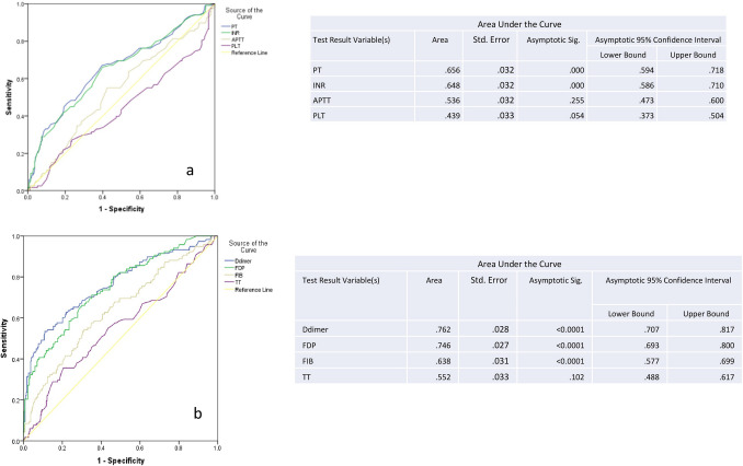
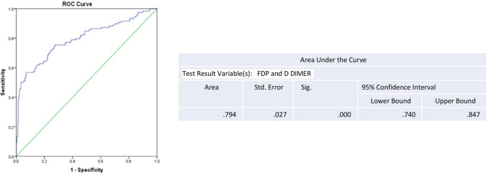
Receiver operating curve (ROC) were calculated for all the coagulation parameters (Fig. 1a, b). Positive sample is a result of severe and very severe cases. Negative sample is a result of mild to moderate cases. D dimer emerged as the best predictive marker to assess the disease severity based on the area under the curve (AUC) which was the highest. D dimer (AUC: 0.762, p < 0.001) and FDP (AUC: 0.746, p < 0.0001) demonstrated better predictive power to differentiate mild moderate from severe and very severe cases as per the AUC of ROC as compared to other parameters (Fig. 1b). The cut off values for D dimer was 697 ng/ml at sensitivity of 85% and specificity of 52.4%. FDP cut off value was 5.11 mg/value at sensitivity of 85% and specificity of 53.5%. The combined ROC curve for the best two parameters (FDP and D dimer) had an area under the curve of 0.794 which was higher than the individual values (Fig. 2).
Fig. 1.
ROC analysis of various coagulation parameters to diagnose severe COVID cases. a Plot of single parameters (PT, INR, APTT, Platelets). b Plot of single parameters (D dimer, Fibrinogen, TT)
Fig. 2.
Plot of combination parameters—D dimer and FDP
Platelet count did not emerge as a good predictor of disease severity in the present study (Fig. 1a).
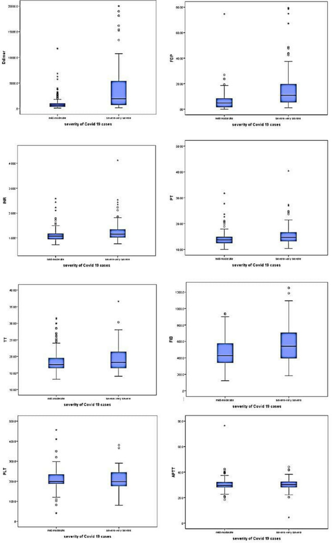
The box plots shows the difference in coagulation parameters between mild, moderate and severe disease (Fig. 3).
Fig. 3.
Box plot of various coagulation parameters
D dimer alone and the combined use of the two parameters D dimer and FDP are useful to monitor the disease severity.
Discussion
SARS-COV 2 is the third coronavirus causing pandemic in the twenty-first century [23, 24]. The pandemic has challenged the entire medical fraternity, globally, to discover a treatment for alleviating the suffering of millions of infected patients.
Recent studies indicate Covid-19 differs from other coronavirus diseases in clinical symptoms and presentation [25]. In comparison to others, SARS COV 2 showed larger number of patients with no history of fever, less severe respiratory and milder GIT symptoms with frequent multi organ injury. Also fewer patients had severe lung injury and the fatalities too were lower. Different types of blood cytokines were noted in Covid-19 indicating a different mechanism [26].
The current study covered 391 patients, having a mean age of 43.81 years. The mean age increased as the disease progressed from mild moderate to very severe. The mean age in the non survivors were more than in survivor group highlighting that the disease is more severe in the elderly, who usually have associated comorbidities and succumb to the illness. This was similar to findings in other studies [7, 19, 22, 24, 26, 27]. There was no significant difference in mean age among males and females in the present study. The male to female ratio was 1.5: 1. Male were seen to outnumber the females in most of the studies [7, 13, 22].
Prothrombin time and INR values were seen to show an increasing trend from mild moderate to severe to very severe cases and also was raised in non survivors compared to survivors. The PT was greater than 16.5 secs in 13.5% of total cases and in 35.1% of the non-survivors. This finding was consistent with other studies on Chinese cohort [13, 19, 22, 28]. Shiyu Yin [18] reported PT was increased in Covid-19 patients compared to controls.
However, study by Hans et al showed no difference in PT INR between the three subgroups based on disease severity. The PT was also not shown to increase in a study on Caucasian patients by Fogarty [21]. Rise in PT indicates activation of the extrinsic pathway of coagulation on exposure to the virus, pathogen associated molecular pathways (PAMP) [8, 12].
D dimer and FDP values showed an increasing trend with disease severity. This finding was similar to studies by Hans et al., Tang et al. and Long et al. [13, 22, 28]. The dimer levels were above 3 times the normal (> 1800 ng/ml) in 75.6% of deceased (28/37) while FDP values were 2 times above normal (> 10 mg/dl) in 81% (30/37). Higher values of D dimer and FDP were also noted in other studies [13, 19, 21, 22].
Tang and Long both acknowledged increase in PT and D dimer similar to the present study [13, 28].
Raised levels of D dimer and FDP suggests there is onset of fibrinolysis following activation of coagulation in Covid-19 patients. Its onset correlates with poor clinical course. High levels of D dimers can also be explained as a feature of hypercoagulable state due to excess thrombin generation and absence of fibrinolysis early in the disease due to direct endothelial injury by viral infection [11, 29, 30]. or due to hypoxia induced thrombosis due to increase in blood viscosity [31]. D dimer is also raised due to local pulmonary micro vascular and venous thrombosis as evident from autopsy studies [32].
Fibrinogen levels were seen to increase with disease severity and was more in the non survivors. Similar findings were noted by Tang et al., Fogarty et al. and long H et al. [13, 21, 28].
This could be attributed to the fact that fibrinogen is an acute phase reactant and a procoagulant and increases with progressive disease and onset of coagulopathy [10]. However, study by Hans et al. [20] showed no change in fibrinogen levels with increase in disease severity.
Thrombin time (TT) was seen to be increasing with disease progression and was increased in deceased. Thrombin causes conversion of fibrinogen to fibrin. Increased thrombin time (> 20 s) was seen in 40% of deaths in the present study. Long et al. also noted increase in TT with disease severity. However, Hans et al. could not establish any such increasing trend [22, 28]. There is not extensive published literature yet on this parameter in other Covid-19 studies.
The APTT and platelet values in the present study did not show marked difference with disease progression nor with the survivor status. The present study showed only 2.3% of the total patients had platelet (9/391) count below 100,000/ml. Most of the studies have shown usually normal platelet count in Covid-19 patients [7, 13, 22]. The thrombopoietin-released following pulmonary inflammation may stimulate platelet production in Covid-19 pts [33]. This directs towards local lung injury and elevated platelet count in these pts [17].
Zheng et al. [26] however noted thrombocytopenia and lymphocytopenia in both severe and non-severe cases. Tang [19] also noted thrombocytopenia in severe disease when D dimer values were 6 times higher than normal levels. Thrombocytopenia was seen in 20% of non survivors in study by Zhou et al. [16]. This could be attributed to the fact that DIC like coagulopathy is seen in few of the Covid-19 cases in later stages [17].
No significant difference in APTT were noted varying with disease severity in the present study. This was similar to finding by Hans et al. [22]. However, Tang et al. [13] noted APTT was raised in non survivors on admission. Studies have revealed raised APTT are noted in moderate and severe disease [32].
The present study showed coagulation profile of Covid-19 patients was distinct from classical DIC. The levels of D dimer, FDP, PT INR and Fibrinogen and TT were raised but the Platelet count was not lowered and APTT was not prolonged. It differs from few Chinese [18, 22] and Caucasian studies [21] by having elevated PT INR in non survivors and in the severe group. Few other studies also suggest that Covid-19 patients presents with coagulopathy but do not meet the criteria of DIC following sepsis [19–22, 34]. However, a strong association of DIC and deaths were noted in one single Chinese study by Tang [13]. These findings indicate the possibility of racial differences in the coagulation profile among Covid-19 patients.
Autopsy findings demonstrate Pulmonary intravascular coagulopathy, a type of immune thrombosis which is distinct from DIC, maybe a critical underlying pathogenic mechanism. It was first mentioned by McGonagle et al. [35]. Direct endothelial cell and type 2 pneumocyte injury causes extensive alveolar damage which activates the coagulation system and fibrin deposition. Fibrinolysis occurs as a compensatory mechanism early in disease but later plasminogen ceases to act which results in high levels of D dimer. This causes pulmonary micro vascular thrombosis extending into the pulmonary veins causing pulmonary thrombosis [17]. This may explain the rise in PT, PT INR, FDP, D dimer, fibrinogen and TT with the disease onset and progression but without any evidence of bleeding.
A close association between inflammation and thrombosis [36, 37] results in cytokine storm releasing proinflammatory circulatory markers causing acute respiratory distress syndrome (ARDS) and multiorgan dysfunction in the systemic phase of the disease [34, 38]. Covid-19 s disease starts with hyper coagulation and Pulmonary intravascular coagulation (PIC), which may progress to DIC with the onset of sepsis.
Studies have established a positive correlation between disease severity and elevated D dimer or progressive increase of D dimer. The value has been found to be high amongst ICU patients and non survivors [13, 19, 22, 28]. An Italian study also revealed raised levels of D dimer is a predictor of ARDS or ICU admission and is a marker of poor prognosis [39].
As the thrombotic risk varies with the race and Covid-19 causes derangement of coagulation, this initial PIC may be the basis of variation of the coagulation parameters seen in different population.
Our present study also shows D dimer level alone as best single coagulation parameter and useful predictor of disease severity. Also the combination of D dimer and FDP is the best combined indicator amongst coagulation parameters. The cut off values of these markers may vary among different populations.
Limitations
The study was limited by the fact that it was confined to one centre and controls could not be collected as it was declared an exclusive Covid hospital with the emergence of the pandemic. The validity of the study could be improved by including controls in the future studies from other larger centres. Serial sampling was not possible due to the lack of adequate manpower and practical discomfort of obtaining the samples wearing the PPE kit. Platelets count did not emerge as a good predictor of disease severity in the present study which may need more extensive research.
The mortality of the present study is 9.7% which is much higher than the death rate globally and in India which at the time of writing the study is 2.0% and 3.7% respectively [3]. This can also be attributed to the limitation of the study as it included only hospital admitted patients. The less severe, home quarantine and asymptomatic patients were out of the scope of the study, thereby enhancing the probability of mortality.
Conclusion
The rapidly progressing pandemic has accelerated the research regarding the pathogenicity and behaviour of the SARS COV2 virus. It is noted from existing studies that severe cases who succumb to the illness have deranged coagulation profile. The index study suggests D dimer alone or D dimer-FDP combination can serve as useful indicators of disease severity. Higher values of PT, INR, D dimer, FDP, Fibrinogen suggests Covid-19 disease is a hypercoagulable state. As the number of cases are still rising in India, larger studies may be conducted to substantiate this observation that coagulopathy in Indians is CAC and is distinct from the classical DIC.
Author Contributions
Idea and design: Dr. SR, Dr. MP, Dr. MJ, Dr. HK. Data acquisition: Dr. VC, Dr. AS, Dr. NV, Dr. SA, Dr. RM. Analysis: Dr. MS, Dr. SR. Interpretation of findings: Dr. SR, Dr. MP. Preparation of manuscript: Dr. SR, Dr. MP, Dr. VC. Critical revision: Dr. SR, Dr. MP, Dr. MJ, Dr. RKC.
Declarations
Conflict of interest
There is no conflict of interest.
Institutional Ethics Committee Clearance
Taken.
Footnotes
Publisher's Note
Springer Nature remains neutral with regard to jurisdictional claims in published maps and institutional affiliations.
Contributor Information
Sujata Raychaudhuri, Email: sujatabulu@yahoo.co.in.
Mukta Pujani, Email: drmuktapujani@gmail.com.
Reetika Menia, Email: reetika830@gmail.com.
Nikhil Verma, Email: drnick.ver@gmail.com.
Mitasha Singh, Email: mitasha.17@gmail.com.
Varsha Chauhan, Email: varsha.chauhan3@gmail.com.
Manjula Jain, Email: drmanjulajain@gmail.com.
R. K. Chandoke, Email: drchandoke@gmail.com
Harnam Kaur, Email: drharnam@gmail.com.
Snehil Agrawal, Email: drsnehil26@gmail.com.
Aparna Singh, Email: aparnasingh515@gmail.com.
References
- 1.Li Q, Guan X, Wu P, Wang X, Zhou L, Tong Y, Ren R, Leung KSM, Lau EHY, Wong JY, et al. Early transmission dynamics in wuhan, china, of novel coronavirus-infected pneumonia. N Engl J Med. 2020;2020:10–1056. doi: 10.1056/NEJMoa2001316. [DOI] [PMC free article] [PubMed] [Google Scholar]
- 2.Zhu N, Zhang D, Wang W, Li X, Yang B, Song J, Zhao X, Huang B, Shi W, Lu R, et al. A novel coronavirus from patients with pneumonia in China. N Engl J Med. 2019;2020:10–1056. doi: 10.1056/NEJMoa2001017. [DOI] [PMC free article] [PubMed] [Google Scholar]
- 3.World Health Organization. Novel Coronavirus (2019-nCoV) situation reports. https://www.who.int/emergencies/diseases/novel-coronavirus-2019/situation-reports. Accessed: 9 Aug 2020
- 4.Zhou P, Yang XL, Wang XG, et al. A pneumonia outbreak associated with a new coronavirus of probable bat origin. Nature. 2020;579(7798):270–273. doi: 10.1038/s41586-020-2012-7. [DOI] [PMC free article] [PubMed] [Google Scholar]
- 5.Lu R, Zhao X, Li J, et al. Genomic characterisation and epidemiology of 2019 novel coronavirus: implications for virus origins and receptor binding. Lancet. 2020;395:565–574. doi: 10.1016/S0140-6736(20)30251-8. [DOI] [PMC free article] [PubMed] [Google Scholar]
- 6.Huang C, Wang Y, Li X, Ren L, Zhao J, Hu Y, et al. Clinical features of patients infected with 2019 novel coronavirus in Wuhan. China Lancet. 2020;395:497–506. doi: 10.1016/S0140-6736(20)30183-5. [DOI] [PMC free article] [PubMed] [Google Scholar]
- 7.Yang AP, Liu JP, Tao WQ, Li HM. The diagnostic and predictive role of NLR, d-NLR and PLR in COVID-19 patients. Int Immunopharmacol. 2020;84:106504. doi: 10.1016/j.intimp.2020.106504. [DOI] [PMC free article] [PubMed] [Google Scholar]
- 8.Connors JM, Levy JH. COVID-19 and its implications for thrombosis and anticoagulation. Blood. 2020;135(23):2033–2040. doi: 10.1182/blood.2020006000. [DOI] [PMC free article] [PubMed] [Google Scholar]
- 9.Mehta P, McAuley DF, Brown M, et al. COVID-19: consider cytokine storm syndromes and immunosuppression. Lancet. 2020;395:1033–1034. doi: 10.1016/S0140-6736(20)30628-0. [DOI] [PMC free article] [PubMed] [Google Scholar]
- 10.Gulhar R, Ashraf MA, Jialal I (2020) Physiology, acute phase reactants. [Updated 2020 May 4]. In: StatPearls [Internet]. Treasure Island (FL): StatPearls Publishing. https://www.ncbi.nlm.nih.gov/books/NBK519570/ [PubMed]
- 11.Iba T, Levy JH, Connors JM, Warkentin TE, Thachil J, Levi M. The unique characteristics of COVID-19 coagulopathy. Crit Care. 2020;24(1):360. doi: 10.1186/s13054-020-03077-0. [DOI] [PMC free article] [PubMed] [Google Scholar]
- 12.Chen G, Wu D, Guo W, et al. Clinical and immunologic features in severe and moderate Coronavirus Disease 2019. J Clin Invest. 2020;20:2020. doi: 10.1172/JCI137244. [DOI] [PMC free article] [PubMed] [Google Scholar]
- 13.Tang N, Li D, Wang X, Sun Z. Abnormal coagulation parameters are associated with poor prognosis in patients with novel coronavirus pneumonia. J Thromb Haemost. 2020;18:844–847. doi: 10.1111/jth.14768. [DOI] [PMC free article] [PubMed] [Google Scholar]
- 14.Ranucci M, Ballotta A, Di Dedda U, et al. The procoagulant pattern of patients with COVID-19 acute respiratory distress syndrome. J Thromb Haemost. 2020;18:1747–1751. doi: 10.1111/jth.14854. [DOI] [PMC free article] [PubMed] [Google Scholar]
- 15.Wang D, Hu B, Hu C, et al. Clinical characteristics of 138 hospitalized patients with 2019 novel coronavirus-infected pneumonia in Wuhan, China. JAMA. 2020;323:1061. doi: 10.1001/jama.2020.1585. [DOI] [PMC free article] [PubMed] [Google Scholar]
- 16.Zhou F, Yu T, Du R, et al. Clinical course and risk factors for mortality of adult inpatients with COVID-19 in Wuhan, China: a retrospective cohort study. Lancet. 2020;395(10229):1054–1062. doi: 10.1016/S0140-6736(20)30566-3. [DOI] [PMC free article] [PubMed] [Google Scholar]
- 17.Belen-Apak FB, Sarıalioğlu F. Pulmonary intravascular coagulation in COVID-19: possible pathogenesis and recommendations on anticoagulant/thrombolytic therapy. J Thromb Thrombolysis. 2020;50(2):278–280. doi: 10.1007/s11239-020-02129-0. [DOI] [PMC free article] [PubMed] [Google Scholar]
- 18.Yin S, Huang M, Li D, Tang N. Difference of coagulation features between severe pneumonia induced by SARS-CoV2 and non-SARS-CoV2. J Thromb Thrombolysis. 2020;3:1–4. doi: 10.1007/s11239-020-02105-8. [DOI] [PMC free article] [PubMed] [Google Scholar]
- 19.Tang N, Bai H, Chen X, Gong J, Li D, Sun Z. Anticoagulant treatment is associated with decreased mortality in severe coronavirus disease 2019 patients with coagulopathy. J Thromb Haemost. 2020;18(5):1094–1099. doi: 10.1111/jth.14817. [DOI] [PMC free article] [PubMed] [Google Scholar]
- 20.Guan WJ, Ni ZY, Hu Y, Liang WH, Ou CQ, He JX, Liu L, Shan H, Lei CL, Hui DS, et al. China medical treatment expert group for COVID-19. Clinical characteristics of Coronavirus Disease 2019 in China. N Engl J Med. 2020;382:1708–1720. doi: 10.1056/NEJMoa2002032. [DOI] [PMC free article] [PubMed] [Google Scholar]
- 21.Fogarty H, Townsend L, Ni Cheallaigh C, et al. COVID19 coagulopathy in Caucasian patients. Br J Haematol. 2020;189(6):1044–1049. doi: 10.1111/bjh.16749. [DOI] [PMC free article] [PubMed] [Google Scholar]
- 22.Han H, Yang L, Liu R, Liu F, Wu KL, Li J, et al. Prominent changes in blood coagulation of patients with SARS-CoV-2 infection. Clin Chem Lab Med. 2020 doi: 10.1515/cclm-2020-0188. [DOI] [PubMed] [Google Scholar]
- 23.Munster VJ, Koopmans M, van Doremalen N, van Riel D, de Wit E. A novel coronavirus emerging in China—key questions for impact assessment. N Engl J Med. 2020 doi: 10.1056/NEJMp2000929. [DOI] [PubMed] [Google Scholar]
- 24.Mattiuzzi CG. Which lessons shall we learn from the 2019 novel coronavirus outbreak? Ann Transl Med. 2020;8:48. doi: 10.21037/atm.2020.02.06. [DOI] [PMC free article] [PubMed] [Google Scholar]
- 25.Guan W-j, Ni Z-y, Hu Y et al Clinical characteristics of 2019 novel coronavirus infection in China. medRxiv. 10.1101/2020.02.10.20021675
- 26.Zheng Y, Huang Z, Ying G et al (2020) Study of the lymphocyte change between COVID-19 and non-COVID-19 pneumonia cases suggesting other factors besides uncontrolled inflammation contributed to multi-organ injury. medRxiv; 10.1101/2020.02.19.20024885
- 27.Wang C, Deng R, Gou L, Fu Z, Zhang X, Shao F, et al. Preliminary study to identify severe from moderate cases of COVID-19 using combined hematology parameters. Ann Transl Med. 2020;8(9):593. doi: 10.21037/atm-20-3391. [DOI] [PMC free article] [PubMed] [Google Scholar]
- 28.Long H, Nie L, Xiang X, et al. D-dimer and prothrombin time are the significant indicators of severe COVID-19 and poor prognosis. Biomed Res Int. 2020;2020:6159720. doi: 10.1155/2020/6159720. [DOI] [PMC free article] [PubMed] [Google Scholar]
- 29.Levi M, Van Der Poll T. Coagulation and sepsis. Thromb Res. 2017;149:38–44. doi: 10.1016/j.thromres.2016.11.007. [DOI] [PubMed] [Google Scholar]
- 30.Iba T, Levy JH, Raj A, Warkentin TE. Advance in the management of sepsis-induced coagulopathy and disseminated intravascular coagulation. J Clin Med. 2019;8:728. doi: 10.3390/jcm8050728. [DOI] [PMC free article] [PubMed] [Google Scholar]
- 31.Gupta N, Zhao YY, Evans CE (2019) The stimulation of thrombosis by hypoxia. Thromb Res 181:77–8312. Menter DG, Kopetz S, Hawk E et al (2017) Platelet “frst respond [DOI] [PubMed]
- 32.Marchandot B, Sattler L, Jesel L, Matsushita K, Schini-Kerth V, Grunebaum L, Morel O. COVID-19 related coagulopathy: a distinct entity? J Clin Med. 2020;9:1651. doi: 10.3390/jcm9061651. [DOI] [PMC free article] [PubMed] [Google Scholar]
- 33.Menter DG, Kopetz S, Hawk E, et al. Platelet “frst responders” in wound response, cancer, and metastasis. Cancer Metastasis Rev. 2017;36(2):199–213, 13. doi: 10.1007/s10555-017-9682-0. [DOI] [PMC free article] [PubMed] [Google Scholar]
- 34.Thachil J. The versatile heparin in COVID-19. J Thromb Haemost. 2020 doi: 10.1111/jth.14821. [DOI] [PMC free article] [PubMed] [Google Scholar]
- 35.McGonagle D, O’Donnell JS, Sharif K, Emery P, Bridgewood C. Why the immune mechanisms of pulmonary intravascular coagulopathy in COVID-19 pneumonia are distinct from macrophage activation syndrome with disseminated Intravascular coagulation. Autoimmun Rev. 2020 doi: 10.1016/j.autrevol2020.1025375.FoxSE,Akmatbek. [DOI] [Google Scholar]
- 36.Jackson SP, Darbousset R, Schoenwaelder SM, et al. Thromboinflammation: challenges of therapeutically targeting coagulation and other host defense mechanisms. Blood. 2019;133:906–918. doi: 10.1182/blood-2018-11-882993. [DOI] [PubMed] [Google Scholar]
- 37.Iba T, Levy JH. Inflammation and thrombosis: roles of neutrophils, platelets and endothelial cells and their interactions in thrombus formation during sepsis. J Thromb Haemost. 2018;16:231–241. doi: 10.1111/jth.13911. [DOI] [PubMed] [Google Scholar]
- 38.Yang X, Yu Y, Xu J, et al. Clinical course and outcomes of critically ill patients with SARS-CoV-2 pneumonia in Wuhan, China: a single-centered, retrospective, observational study. Lancet Respir Med. 2020;8:475–481. doi: 10.1016/S2213-2600(20)30079-5. [DOI] [PMC free article] [PubMed] [Google Scholar]
- 39.Marietta M, Ageno W, Artoni A, De Candia E, Gresele P, Marchetti M, Marcucci R, Tripodi A. COVID-19 and haemostasis: a position paper from Italian Society on Thrombosis and Haemostasis (SISET) Blood Transfus. 2020;18(3):167–169. doi: 10.2450/2020.0083-40. [DOI] [PMC free article] [PubMed] [Google Scholar]


