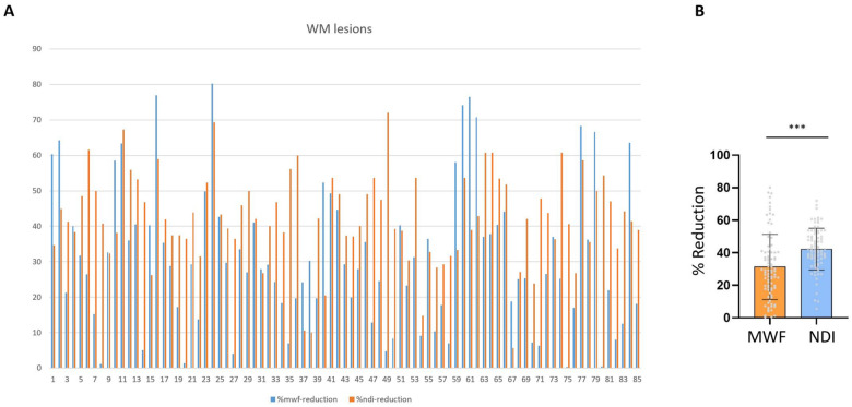
Figure 5.
Alteration in MWF versus NDI in multiple sclerosis white matter lesions. (A) Percentage of MWF and NODDI-NDI decline for individual white matter (WM) lesions relative to mirror region of interest in contralateral hemisphere (individual multiple sclerosis white matter lesions are shown with numbers). (B) Bar graph shows that NODDI-NDI decreases more than MWF in multiple sclerosis lesions (***P < 0.0001).

