Abstract
Background
Left main coronary artery (LMCA)–acute coronary syndrome (ACS) is a rare complication of a floating thrombus in the ascending aorta. However, diagnosing the aetiology of LMCA–ACS during an emergency situation is challenging. We present a rare case of LMCA–ACS caused by a large thrombus in the ascending aorta, confirmed by intravascular ultrasound (IVUS).
Case summary
A 90-year-old woman presented to the emergency department complaining of chest pain and syncope. On admission, her electrocardiogram showed normal sinus rhythm and a complete right bundle branch block with significant ST depression in the V3–V6 leads; hence, ACS was suspected. The first emergency angiogram of the left coronary artery showed filling defect in the proximal ascending aorta. IVUS revealed a large thrombus in the ascending aorta. The thrombus extended from the ascending aorta to the proximal left anterior descending coronary artery. IVUS confirmed that there was no dissection of the coronary artery or the proximal ascending aorta. Based on the IVUS findings, this case was diagnosed as ACS of the LMCA caused by a floating thrombus in the ascending aorta.
Discussion
This rare case of LMCA–ACS caused by a thrombus in the ascending aorta was confirmed by IVUS, which can be a useful imaging tool for diagnosing morphological abnormalities during emergencies.
Keywords: Intravascular ultrasound, Acute coronary syndrome, Floating thrombus, Ascending aorta, Left main coronary artery, Case report
Learning points
A floating thrombus in the ascending aorta is a rare cause of left main coronary artery (LMCA)–acute coronary syndrome.
Emergency use of intravascular ultrasound can assist in the immediate diagnosis of the aetiology of LMCA syndrome in an unstable haemodynamic condition.
Introduction
It is well known that acute coronary syndrome (ACS) occurs as a ruptured plaque resulting in a thrombus; however, in rare cases, it might be caused without ruptured plaque such as dissection or other embolic sources.1–9 On the other hand, the haemodynamic status of ACS is often dramatically compromised prior to recanalization. Intravascular ultrasound (IVUS) is an imaging tool that can be utilized together with emergency percutaneous intervention for recanalization of ACS with or without unstable vital signs. Here, we present a rare case of left main coronary artery (LMCA)–ACS caused by a large thrombus in the ascending aorta, confirmed by IVUS, while the patient was in cardiogenic shock.
Timeline
Case presentation
A 90-year-old woman presented to the emergency department complaining of chest pain and syncope. Her medical history included chronic kidney disease (stage G4) and heart failure with preserved ejection fraction. Arrhythmia, atrial fibrillation, or atrial flutter had not been previously detected. On admission, her blood pressure (149/77 mmHg) and heart rate (68 b.p.m.) were stable, physical examination revealed no heart murmur. Electrocardiogram showed normal sinus rhythm and a complete right bundle branch block, with significant ST depression in the V3–V6 leads. Bedside transthoracic echocardiogram showed diffuse hypokinesis of the left ventricular wall with an ejection fraction of 20%; no evidence of apical thrombi, severe mitral regurgitation, and ventricular septal rupture were observed. Troponin I level was slightly elevated, at 0.039 ng/mL (normal, <0.034 ng/mL). Subsequently, the patient’s blood pressure gradually decreased, and, as cardiogenic shock was expected, catecholamines (noradrenaline 0.2 γ) were initiated, and emergency coronary angiography was scheduled for suspected ACS. Before the procedure, 200 mg aspirin and 300 mg clopidogrel were administered, in addition, 8000 units of unfractionated heparin (∼200 units per kg) were administered by intravenous bolus.
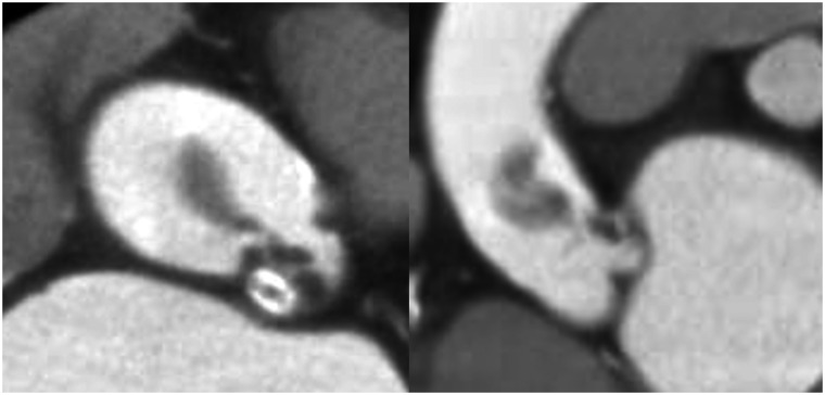
An intra-aortic balloon pump was inserted via the left common femoral artery in the catheter laboratory. The first angiogram of the left coronary artery showed filling defect in the proximal ascending aorta (Figure 1 and Video 1). A guiding catheter was inserted into the left coronary artery, and the angiogram revealed no enhanced area at the LMCA or the left anterior descending coronary artery (LAD) (Figure 2). IVUS revealed a large thrombus (Figure 3 and Video 2), extending from the ascending aorta to the proximal LAD, without dissection of the coronary artery or the proximal ascending aorta (Figure 4 and Video 3). Thus, the patient was diagnosed with ACS of the LMCA caused by a floating thrombus in the ascending aorta. Although we performed thrombectomy several times, the thrombus could not be completely removed. Consequently, we deployed a drug-eluting stent, and a final Thrombolysis in Myocardial Infarction flow grade of 3 was obtained. The patient’s vital signs were stable after the intervention. Contrast-enhanced computed tomography (CT) confirmed a large abnormal low-density area; thus, a large thrombus in the left coronary cusp without aneurysm was suspected. Additionally, contrast-enhanced CT confirmed that the ascending aorta was not dissected (Figure 5). We suggested surgical repair to remove the thrombus; however, the patient and her family opted for palliative care without invasive surgical intervention, as she was a nonagenarian. Therefore, antithrombotic (warfarin) and dual antiplatelet therapy (aspirin and clopidogrel) were initiated, together with unfractionated heparin by continuous intravenous infusion (therapeutic dosing targeting 2.0–3.0 times the activated partial thromboplastin time). Unfortunately, her general condition gradually worsened, and she died of heart failure and multiple organ failure on Day 7 of hospitalization after the percutaneous intervention. The family denied an autopsy request.
Figure 1.

Initial coronary angiogram showing the filling defect at the proximal ascending aorta.
Figure 2.

Angiogram using a guide catheter showing the filling defect at the left main coronary artery and the left anterior descending coronary artery.
Figure 3.

Intravascular ultrasound showing a large thrombus in the ascending aorta.
Figure 4.
Intravascular ultrasound showing no dissection of the coronary artery or proximal ascending aorta, and showing that the thrombus extended from the ascending aorta to the proximal left anterior descending artery. LAD, left anterior descending coronary artery; LMCA, left main coronary artery.
Figure 5.
Contrast-enhanced computed tomography showing a large, abnormal, low-density area suspected to be a large thrombus in the left coronary cusp without an aneurysm in the ascending aorta without a dissection.
Discussion
Here, we present a rare case of LMCA–ACS caused by a large thrombus in the ascending aorta, as confirmed by IVUS.
A floating thrombus in the aorta can be caused by many factors, such as age-related atherosclerosis, collagen disease, trauma, malignancy, and hypercoagulable conditions.3,7,10,11 These conditions constitute a very rare cause of embolic events in the coronary, cerebral, and peripheral arteries.11 In the present case, concerning an elder woman, we considered that the large thrombus was caused by age-related atherosclerosis, although we could not further investigate as her family refused additional examination. The patient survived until the age of 90 years without any history of significant systemic illnesses that might have led to the development of a hypercoagulable condition. In addition, contrast-enhanced CT did not reveal a malignant tumour.
Most cases of LMCA–ACS involve complicated, life-threatening haemodynamic status and recanalization should be performed immediately to prevent cardiac death. IVUS is a useful imaging tool for diagnosing morphological abnormalities, such as intramural haematoma and dissection. Moreover, IVUS can immediately assist in the diagnosis of unstable haemodynamic conditions.
LMCA–ACS is sometimes caused by diseases that require surgical treatment. It is well known that acute aortic dissection (AAD) extending to the LMCA constitutes a rare case of LMCA–ACS.5,6,9 Previous reports demonstrated that IVUS was useful to confirm haematoma and false lumen between the LMCA and the aorta.5,6 In the present case, we first suspected dissection of the aorta in the first angiogram; however, an additional IVUS procedure did not detect haematoma or dissection of the LMCA into the aorta. Instead, a massive thrombus in the left coronary cusp and ascending aorta was found. Consequently, we were able to diagnose the cause of the LMCA–ACS.
In conclusion, we encountered a case of a large floating thrombus in the ascending aorta complicated by LMCA–ACS. IVUS assisted in the diagnosis of LMCA–ACS, showing that it can be a valuable tool, particularly in unstable haemodynamic conditions during an emergency.
Lead author biography

Hiroya Takafuji, MD, is a chief physician of cardiology at Tokyo Bay Urayasu Ichikawa Medical Center in Japan. His main interests are coronary intervention, peripheral intervention, and structural heart disease intervention (especially, congenital heart disease such as atrial septal defect and patent foramen ovale).
Supplementary material
Supplementary material is available at European Heart Journal - Case Reports online.
Slide sets: A fully edited slide set detailing this case and suitable for local presentation is available online as Supplementary data.
Consent: The authors confirm that written consent for submission and publication of this case report including images and associated text has been obtained from the patient in line with COPE guidance.
Conflict of interest: T.N. is a consultant for Boston Scientific, Medtronic, and Century Medical. All other authors declared no conflict of interest.
Funding: None declared.
Supplementary Material
References
- 1.Bruno P, Massetti M, Babatasi G, Khayat A.. Catastrophic consequences of a free floating thrombus in ascending aorta. Eur J Cardiothorac Surg 2001;19:99–101. [DOI] [PubMed] [Google Scholar]
- 2.Yamawaki M, Shimoyama M, Furuse Y, Kinugasa Y, Ogino K, Enokida M. et al. Floating thrombus arising from left sinus of Valsalva induced intermittent occlusion of left coronary artery and caused cardiac shock. In J Cardiol 2007;114:272–273. [DOI] [PubMed] [Google Scholar]
- 3.Nishizaki F, Tomita H, Abe N, Kimura M, Higuma T, Osanai T. et al. Acute myocardial infarction caused by a floating thrombus in the ascending aorta: a role of CD34-positive endothelial cells. J Cardiol Cases 2013;8:e88–e90. [DOI] [PMC free article] [PubMed] [Google Scholar]
- 4.Ozaki N, Yuji D, Sato M, Inoue K, Wakita N.. A floating thrombus in the ascending aorta complicated by acute myocardial infarction. Gen Thorac Cardiovasc Surg 2017;65:213–215. [DOI] [PubMed] [Google Scholar]
- 5.Kim SH, Kim KH, Lim SW, Kim IJ, Moon JY, Cha DH.. IVUS images of the left main intramural hematoma from aortic dissection. Int J Cardiol 2014;173:e27–e30. [DOI] [PubMed] [Google Scholar]
- 6.Arao K, Naito R, Funayama H, Ako J, Momomura S.. The usefulness of an intravascular ultrasound in the diagnosis of left main coronary artery dissection. J Cardiol Cases 2012;5:e133–e136. [DOI] [PMC free article] [PubMed] [Google Scholar]
- 7.Kobayashi Y, Kobayashi Y, Hirohata A.. Left main coronary thrombotic occlusion due to secondary polycythemia in a normal sinus of Valsalva. J Invasive Cardiol 2013;25:421–422. [PubMed] [Google Scholar]
- 8.Eguchi K, Ohtaki E, Misu K, Aikawa M, Sumiyoshi T, Hosoda S. et al. Acute myocardial infarction caused by embolism of thrombus in the right coronary sinus of Valsalva: a case report and review of the literature. J Am Soc Echocardiogr 2004;17:173–177. [DOI] [PubMed] [Google Scholar]
- 9.Ohara Y, Hiasa Y, Hosokawa S.. Successful treatment in a case of acute aortic dissection complicated with acute myocardial infarction due to occlusion of the left main coronary artery. J Invasive Cardiol 2003;15:660–662. [PubMed] [Google Scholar]
- 10.Nader RG, Barr F, Rubin R, Hirshfeld JW Jr, Eisen HJ, Laposata E.. Aortic degenerative changes and thrombus formation: an unusual cause of massive myocardial infarction with normal coronary arteries. Am J Med 1989;86:718–722. [DOI] [PubMed] [Google Scholar]
- 11.Weiss S, Bühlmann R, von Allmen RS, Makaloski V, Carrel TP, Schmidli J. et al. Management of floating thrombus in the aortic arch. J Thorac Cardiovasc Surg 2016;152:810–817. [DOI] [PubMed] [Google Scholar]
Associated Data
This section collects any data citations, data availability statements, or supplementary materials included in this article.


