Abstract
Introduction:
The objective of the study was to measure the horizontal distance between the FA-WALA (Facial Axis Point-William Andrews and Larry Andrews) of posterior teeth in Angle's Class I, Class II, and Class III malocclusions and to assess the depth of the Curve of Spee, to find the correlation between intercanine FA and intercanine WALA and its significance.
Material and Methods:
Sixty pretreatment mandibular casts of patients with an age range of 18–35 years were included. A sample size of 20 was evaluated in Angle's Class I, Class II, and Class III, respectively. The WALA ridge and FA points were marked in the model and calibrated using the digital Vernier caliper.
Results:
There was an incremental increase in the horizontal distance from the FA-WALA in the posterior teeth. The mandibular intercanine FA-FA and intercanine WALA-WALA distance were greater in Angle's Class III group when compared to Angle's Class II. The Curve of Spee measurement was increased in Angle's Class II group, while Angle's Class III had a flat curve.
Conclusion:
The horizontal distance between FA-WALA increased incrementally in the posterior teeth in Angle's Class I, Class II, and Class III malocclusions. In Angle's Class II malocclusion, the Curve of Spee measurement was increased and had a narrower mandibular arch.
KEYWORDS: Curve of Spee, FA point, intercanine FA, intercanine WALA, WALA ridge
INTRODUCTION
The stability of orthodontic treatment depends on the mandibular arch form. WALA ridge serves as an anatomic guide for positioning the teeth as proposed by Andrews. WALA ridge is the strip of soft tissue immediately above the mucogingival junction of the mandible, at the level of the line that passes through the center of rotation of the teeth or close to it.[1] There are many variations in the dental arch forms among individuals and WALA ridge helps in customizing their arch forms.[2,3,4] Knowing the importance of WALA ridge, this study aimed at analyzing the horizontal distance between FA-WALA ridge in various malocclusions and to find its correlation with the Curve of Spee. Intercanine distance between FA-FA and WALA-WALA was evaluated to study the transverse discrepancies.
MATERIAL AND METHODS
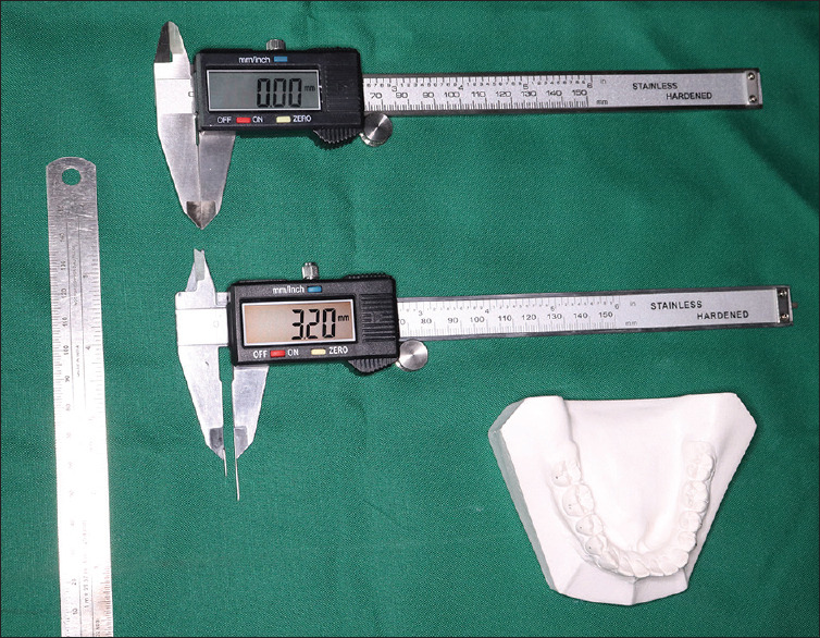
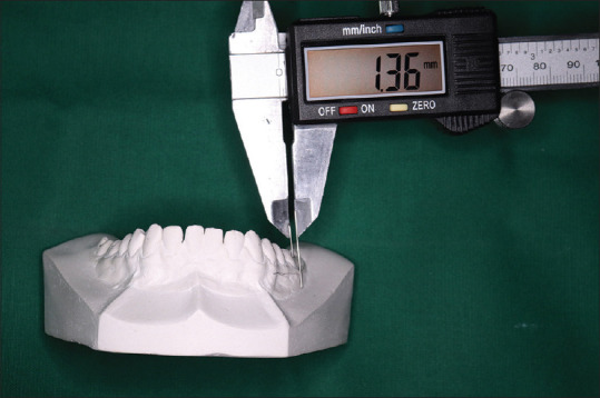
Pretreatment mandibular dental casts of 60 patients who reported to the department of orthodontics, were selected for the study. Samples consisted of 20 each in Angle's Class I, Class II, and Class III, respectively, with the age range of 18–30 years. Inclusion criteria were patients with permanent dentition and no previous history of orthodontic treatment. Casts with dental agenesis, supernumerary teeth, mutilated dentition, and patients with craniofacial syndromes, bruxism, and attrition were excluded from the study. A single examiner marked the FA point on the tooth and WALA ridge on the mandibular model from the occlusal aspect [Figure 1]. The horizontal distance between the FA point on the posterior teeth of the mandible and WALA ridge was assessed on both sides of the mandibular arch using a modified digital Vernier caliper parallel to the occlusal plane [Figure 2]. The Curve of Spee measurement was made on the right and left sides from the cusp tip of the first premolar. Intercanine width at the region of WALA and FA was also analyzed.
Figure 1.

Armamentarium (A. Measuring Scale, B. Digital vernier caliper, C. Modified Digital vernier caliper, D. Mandibular Dental cast)
Figure 2.

FA-WALA Ridge measurement using modified Digital Vernier Caliper (A. Modified Digital Vernier Caliper, B. Manibular Cast)
Statistics
The data were subjected to statistical analysis. Paired t-test was done to find the significance of the mean value between the intercanine width and WALA in Angle's Class I, Class II, and Class III malocclusions. Comparison of horizontal distance of FA-WALA in posterior teeth was done with one-way analysis of variance. Post Hoc test was done to find the significance of the Curve of Spee between Angle's Class I, Class II, and Class III malocclusions.
DISCUSSION
The primary goal of orthodontic treatment relies on establishing a stable relationship with the underlying basal bone.[5] Shape, size, and position of teeth, jaw size, facial pattern, musculature, and occlusion are various factors that can cause variation. The six elements of orofacial harmony developed by Andrews act as a guideline for achieving ideal occlusion, where the teeth are positioned in harmony with the basal bone and the surrounding soft tissue. Archwires shaped differently from the WALA ridge may cause adverse effects on the gingiva, root, and alveolus. In this study, the plaster models of the mandibular arch with Angle's Class I, Class II, and Class III were analyzed. The results showed that the horizontal distance from FA-WALA in mandibular posterior teeth increased gradually from 1st premolar to 2nd molar on both sides.[6] These were similar to the previously reported studies of Andrews and Andrews, Kanashiro and Vigorito, and Ronay et al. The values in the 1st molars were 2.38 mm and 2.41 mm, 2.30 mm in Angle's Class II and Class III, respectively [Table 1]. The values are higher than the studies reported by Andrews (2.0 mm) Kong-Zarate et al. (2.12 mm), and Trivino et al. (2.21 mm). This can be explained as there was a more lingual position of the mandibular teeth than with normal occlusion. Based on this study, there was more inclination and lingual position of teeth from premolars to 1st molars, while the 2nd molar was more stable irrespective of the class of malocclusion.
Table 1.
Comparison of FA–WALA distance in posterior teeth in Angle’s Class I, Class II, and Class III malocclusions
| Tooth | Mean±SD | P | ||
|---|---|---|---|---|
|
| ||||
| Angle’s Class I malocclusion | Angle’s Class II malocclusion | Angle’s Class III malocclusion | ||
| 1st premolar | 0.61±0.41 | 0.45±0.38 | 0.37±0.25 | 0.0115 (S) |
| 2nd premolar | 1.01±0.48 | 0.98±0.41 | 0.68±0.33 | 0.0006 (S) |
| 1st molar | 1.87±0.52 | 1.67±0.45 | 1.51±0.32 | 0.0015 (S) |
| 2nd molar | 2.38±0.57 | 2.41±0.58 | 2.30±0.4 | 0.06571 |
| Total | 1.47±0.85 | 1.38±0.86 | 1.35±0.82 | 0.0268 (S) |
SD: Standard deviation, S: Significant
Andrews described the six keys of occlusion and found that the Curve of Spee ranged from mild to flat in subjects with proper occlusion. The curve of Spee provides posterior disocclusion and anterior tooth guidance with mandibular forward movement.[7] The depth of the Curve of Spee8 increased with the eruption of second molars and slightly decreased during adolescence and remained stable in adulthood. The Curve of Spee and WALA-FA point is subjected to occlusal variations. The Curve of Spee was measured from the cusp tip of 1st premolars on both sides. Interpretation of the results of our study revealed that the depth of the Curve of Spee was greatest in the Angle's Class II malocclusion group, followed by Angle's Class I, and Angle's Class III having the least amount of depth. The increase in the depth of the Curve of Spee in Angle's Class II was attributed to the unopposed eruption of teeth in lower anteriors due to increased overjet.[9] The results were similar to the study by Veli, Ozturk, and Uysal et al.
Intercanine width FA-FA and intercanine WALA-WALA
Transverse dimensions of the mandibular arch form in subjects across different malocclusions were assessed in canine-canine FA and canine-canine WALA points.10 The arch form was established based on mandibular canines and molars.[11] The mandibular intercanine width was significantly larger in Angle's Class III group12 than in Angle's Class II which indicates a restricted growth in this region of Angle's Class II malocclusion.[13,14] Angle's Class I malocclusion had a wider width of mandibular arch than in Angle's Class II malocclusion.15 Increased intercanine width in Angle's Class III can be imputed to the increased growth potential of mandible even in transverse dimension.16 The disparity in age or severity of malocclusion may cause differences in studies.
CONCLUSION
The horizontal distance from the FA-WALA in posterior teeth increased incrementally in Angle's Class I, Class II, and Class III malocclusions. The WALA-FA distance showed the difference between classes for all teeth except 2nd molars. The Curve of Spee was deepest in Angle's Class II malocclusion[17] [Table 2] and intercanine FA and WALA width was widest for Angle's Class III malocclusion [Table 3].[18] The WALA ridge helps in determining the faciolingual position of the posterior teeth and identifies the transverse dimension.[19]
Table 2.
Comparison of the Curve of Spee between Angle’s Class I, Class II, and Class III malocclusions
| Parameter | Mean±SD | P | ||
|---|---|---|---|---|
|
| ||||
| Class I | Class II | Class III | ||
| Curve of Spee | 1.425±0.65 | 1.81±0.45 | 1.17±0.34 | 0.0466 (S) |
SD: Standard deviation, S: Significant
Table 3.
Comparison of intercanine width (FA-FA) and WALA-WALA (intercanine width) between Angle’s Class I, Class II, and Class III malocclusions
| Malocclusion | Mean±SD | |
|---|---|---|
|
| ||
| FA-FA (intercanine width) | WALA-WALA (intercanine width) | |
| Angle’s Class I | 30.97±2.78 | 31.76±2.82 (S) |
| Angle’s Class II | 28.59±3.58 | 29.61±3.43 (S) |
| Angle’s Class III | 35.98±1.69 | 36.78±1.62 (S) |
SD: Standard deviation, S: Significant
Financial support and sponsorship
Nil.
Conflicts of interest
There are no conflicts of interest.
REFERENCES
- 1.Andrews LF. The 6-elements orthodontic philosophy: Treatment goals, classification and rules for treating. Am J Orthod Dentofacial Orthop. 2015;148:883–7. doi: 10.1016/j.ajodo.2015.09.011. [DOI] [PubMed] [Google Scholar]
- 2.Triviño T, Siqueira DF, Scanavini MA. A new concept of mandibular dental arch forms with normal occlusion. Am J Orthod Dentofacial Orthop. 2008;133:10.e15–22. doi: 10.1016/j.ajodo.2007.07.014. [DOI] [PubMed] [Google Scholar]
- 3.Zou W, Jiang J, Xu T, Wu J. Relationship between mandibular dental and basal bone arch forms for severe skeletal class III patients. Am J Orthod Dentofacial Orthop. 2015;147:37–44. doi: 10.1016/j.ajodo.2014.08.019. [DOI] [PubMed] [Google Scholar]
- 4.Ronay V, Miner RM, Will LA, Arai K. Mandibular arch form: The relationship between dental and basal anatomy. Am J Orthod Dentofacial Orthop. 2008;134:430–8. doi: 10.1016/j.ajodo.2006.10.040. [DOI] [PubMed] [Google Scholar]
- 5.Kong-Zárate CY, Carruitero MJ, Andrews WA. Distances between mandibular posterior teeth and the WALA ridge in Peruvians with normal occlusion. Dental Press J Orthod. 2017;22:56–60. doi: 10.1590/2177-6709.22.6.056-060.oar. [DOI] [PMC free article] [PubMed] [Google Scholar]
- 6.Conti MF, Filho MV, Vedovello SA, Valdrighi HC, Kuramae M. Longitudinal evaluation of dental arches individualized by the WALA ridge method. Dental Press J Orthod. 2011;16:65–74. [Google Scholar]
- 7.Marshall SD, Caspersen M, Hardinger RR, Franciscus RG, Aquilino SA, Southard TE, et al. Development of the curve of spee. Am J Orthod Dentofacial Orthop. 2008;134:344–52. doi: 10.1016/j.ajodo.2006.10.037. [DOI] [PubMed] [Google Scholar]
- 8.Dindaroğlu F, Duran GS, Tekeli A, Görgülü S, Doğan S. Evaluation of the relationship between curve of spee, WALA-FA distance and curve of wilson in normal occlusion. Turk J Orthod. 2016;29:91–7. doi: 10.5152/TurkJOrthod.2016.1614. [DOI] [PMC free article] [PubMed] [Google Scholar]
- 9.Triviño T, Siqueira DF, Andrews WA. Evaluation of distances between the mandibular teeth and the alveolar process in Brazilians with normal occlusion. Am J Orthod Dentofacial Orthop. 2010;137:308.e1–4. doi: 10.1016/j.ajodo.2009.09.017. [DOI] [PubMed] [Google Scholar]
- 10.Tamburrino RK, Boucher NS, Vanarsdall R. The Transverse Dimension: Diagnosis and Relevance to Functional Occlusion RWISO. 2010 Sep [Google Scholar]
- 11.Kanashiro LK, Vigorito JW. Distance between the buccal aspects of the dental arches and alveolar ridge in different types of occlusion. SPO. 2007;40:115–23. [Google Scholar]
- 12.Suk KE, Park JH, Bayome M, Nam YO, Sameshima GT, Kook YA, et al. Comparison between dental and basal arch forms in normal occlusion and class III malocclusions utilizing cone-beam computed tomography. Korean J Orthod. 2013;43:15–22. doi: 10.4041/kjod.2013.43.1.15. [DOI] [PMC free article] [PubMed] [Google Scholar]
- 13.Gupta D, Miner RM, Arai K, Will LA. Comparison of the mandibular dental and basal arch forms in adults and children with class I and class II malocclusions. Am J Orthod Dentofacial Orthop. 2010;138:10.e1–8. doi: 10.1016/j.ajodo.2010.01.024. [DOI] [PubMed] [Google Scholar]
- 14.Sayin MO, Turkkahraman H. Comparison of dental arch and alveolar widths of patients with Class II division 1 malocclusion and subjects with Class I ideal occlusion. Angle Orthod. 2004;74:356–60. doi: 10.1043/0003-3219(2004)074<0356:CODAAA>2.0.CO;2. [DOI] [PubMed] [Google Scholar]
- 15.Lux CJ, Conradt C, Burden D, Komposch G. Dental arch widths and mandibular-maxillary base widths in class II malocclusions between early mixed and permanent dentitions. Angle Orthod. 2003;73:674–85. doi: 10.1043/0003-3219(2003)073<0674:DAWAMB>2.0.CO;2. [DOI] [PubMed] [Google Scholar]
- 16.Mitani H, Sato K, Sugawara J. Growth of mandibular prognathism after pubertal growth peak. Am J Orthod Dentofacial Orthop. 1993;104:330–6. doi: 10.1016/S0889-5406(05)81329-0. [DOI] [PubMed] [Google Scholar]
- 17.Veli I, Ozturk MA, Uysal T. Curve of spee and its relationship to vertical eruption of teeth among different malocclusion groups. Am J Orthod Dentofacial Orthop. 2015;147:305–12. doi: 10.1016/j.ajodo.2014.10.031. [DOI] [PubMed] [Google Scholar]
- 18.Al-Khateeb SN, Abu Alhaija ES. Tooth size discrepancies and arch parameters among different malocclusions in a Jordanian sample. Angle Orthod. 2006;76:459–65. doi: 10.1043/0003-3219(2006)076[0459:TSDAAP]2.0.CO;2. [DOI] [PubMed] [Google Scholar]
- 19.Ball RL, Miner RM, Will LA, Arai K. Comparison of dental and apical base arch forms in class II division 1 and class I malocclusions. Am J Orthod Dentofacial Orthop. 2010;138:41–50. doi: 10.1016/j.ajodo.2008.11.026. [DOI] [PubMed] [Google Scholar]


