Abstract
This protocol describes experimental procedures for in vitro dephosphorylation assay of human protein c-Myc. This protocol can be adapted to detect phosphatase activity of other Ser/Thr phosphatases.
Keywords: Dephosphorylation, c-Myc, SCP1
Background
Carboxy-terminal domain RNA polymerase II polypeptide A small phosphatase 1 (SCP1, also known as CTDSP1 or NLI-IF) belongs to the FCP/SCP phosphatase family and was originally reported to dephosphorylate the C-terminal domain (CTD) of RNA polymerase II ( Yeo et al., 2003 ). Smad2, 3 ( Wrighton et al., 2006 ), Snail ( Wu et al., 2009 ), PML ( Lin et al., 2014 ), and c-Myc ( Wang et al., 2016 ) have also been identified as substrates of SCP1.
Materials and Reagents
1.5 ml Eppendorf tubes
50 ml tube
100 mm dish
E. coli cells
HKE293T cell
HA-c-Myc plasmid
LB medium
IPTG
Glutathione sepharose 4B (GE Healthcare, catalog number: 17075601)
Glycerol (Sigma-Aldrich, catalog number: G5516)
PierceTM BCA Protein Assay Kit (Thermo Fisher Scientific, Thermo ScientificTM, catalog number: 23225)
Lipofectamine 2000
c-Myc antibody (Abcam, catalog number: ab32072)
Protein A/G beads (Santa Cruz Biotechnology, catalog number: sc-2003)
BSA
Coomassie blue R250 (Sigma-Aldrich, catalog number: 27816)
c-Myc Ser62 phosphorylation antibody ([Abnova, catalog number: MAB6763] or [Abcam, catalog number: ab78318])
Sodium hydroxide (NaOH) (Sigma-Aldrich, catalog number: 221465)
Sodium chloride (NaCl) (Sigma-Aldrich, catalog number: S7653)
Potassium chloride (KCl)
Disodium hydrogen phosphate (Na2HPO4)
Potassium phosphate monobasic (KH2PO4)
Tris base (C4H11NO3) (Sigma-Aldrich, catalog number: T1503)
EDTA (Sigma-Aldrich, catalog number: E5134)
Triton X-100 (Sigma-Aldrich, catalog number: X100)
Phenylmethanesμlfonyl fluoride (PMSF) (C7H7FO2S) (Sigma-Aldrich, catalog number: P7626)
cOmplete protease inhibitor cocktail (Roche Diagnostics, catalog number: 04693116001)
Dithiothreitol (DTT) (Sigma-Aldrich, catalog number: D0632)
PhosSTOP tablet (Roche Diagnostics, catalog number: 04906845001)
β-mercaptoethanol
Bromophenol blue
Sodium dodecyl sulfate (SDS)
Magnesium chloride (MgCl2) (Sigma-Aldrich, catalog number: 449172)
L-Glutathione reduced (Sigma-Aldrich, catalog number: G4251)
Phosphate-buffered saline (PBS) (see Recipes)
IP lysis buffer (see Recipes)
Phosphatase reaction buffer (see Recipes)
5x SDS loading dye (see Recipes)
GST elution buffer (see Recipes)
Equipment
250 ml flask
Shaker
Cell scraper
Incubator
Centrifuges
Procedure
-
Heterologous expression and purification of phosphatase
Inoculate a single colony of E. coli cells containing the GST-vector, GST-SCP1-WT (wild type) or GST-SCP1-DN (dominant-negative) plasmid into 10 ml LB medium and grow overnight (ON) at 37 °C.
Transfer 2 ml of ON culture to a 250 ml flask containing 48 ml of LB medium.
Incubate at 37 °C in a shaker at 200 rpm until OD600 reaches to 0.5. Induce with IPTG at a final concentration of 1 mM for 4 h.
Transfer the cell culture into a 50 ml tube.
Spin down at room temperature at 5 min, 3,000 × g to collect cells.
Discard supernatant, wash the cell pellet once with 1x PBS.
Resuspend the cell pellet in 500 μl IP lysis buffer without phosSTOP and then put the 50 ml tube on ice for 30 min. Vortex for 15 sec every 10 min.
Spin at 20,000 × g for 10 min at 4 °C. Collect the supernatant into a new Eppendorf tube.
Prepare a 50% (v/v) slurry of glutathione sepharose 4B in lysis buffer, and add 50 μl of slurry to the supernatant. Rotate for 1 h at 4 °C.
Collect the beads by spinning at 1,000 × g for 1 min at 4 °C. Discard the supernatant and wash the beads 3 times with 1x PBS. Store in 1 beads volume of PBS supplemented with 10% glycerol at 4 °C if not going on to the next step immediately.
To elute GST protein from beads, add 1 bead volume of GST elution buffer, vortex for 10 min at 4 °C. Collect supernatant, repeat one more time, and combine the supernatants. Use BCA Protein Assay Kit to quantify the concentration of protein.
-
Expression and purification of phosphorylated c-Myc
Transfect HKE293T cells (at a density of 1.5 x 10⁶ in a 100 mm dish) with 5 µg of HA-c-Myc plasmid using Lipofectamine 2000.
Harvest the cells in 1 ml of 1x PBS by using a cell scraper after a 36-48 h incubation.
Collect the cell pellet by spinning at 1,000 × g for 1 min at 4 °C.
Discard the supernatant, and lyse the cells in 500 µl lysis buffer on ice for 30 min.
Collect the lysate by spinning at 12,000 × g for 15 min at 4 °C, and then transfer the cell lysate into a new Eppendorf tube.
Use 1 μg of c-Myc antibody to immunoprecipitate HA-c-Myc protein from 1 mg/500 μl HEK293T cell lysate. Gently rotate for 1 h at 4 °C.
Wash protein A/G beads 3 times with IP lysis buffer. Add 30 μl (60 μl 50% slurry) to the cell lysate (step 2d) and rotate for 1 h at 4 °C.
Collect the beads by spinning at 1,000 × g for 1 min at 4 °C. Wash 3 times with 0.5 ml ice-cold IP lysis buffer.
Store the beads in 1 bead volume of PBS supplemented with 10% glycerol on ice before use.
Pick 5 µl solution and 1 µg BSA to run SDS-PAGE gel, and stain the gel with Coomassie blue, using 1 µg BSA as control, to determine HA-c-Myc immunocomplex concentration.
-
Dephosphorylation assay
Add the 0.1 µg HA-c-Myc, with 0.5 μg of bacterially-expressed GST-vector, GST-SCP1-WT (wild-type) or GST-SCP1-DN (dominant-negative) in 50 μl phosphatase buffer.
Incubate the mixture at 37 °C for 30 min.
Add 1 bead volume of 2x SDS loading buffer into the mixture. Stop the phosphatase reaction by heating at 100 °C for 5 min.
Half of each sample is fractioned by SDS-PAGE and detect phosphatase on the gel by Coomassie blue staining (Simpson, 2010).
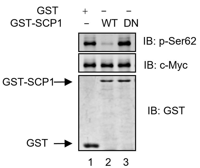
The other half of each sample is used to detect the phosphorylation status of c-MYC at Ser62 by Western blotting using anti-pSer62 antibody.
After phosphorylation detection, strip the membrane using 0.8 N NaOH for 30 sec. Detect total c-Myc protein substrate with anti-c-Myc antibody.
Data analysis
HA-c-Myc purified from HEK293T cells was incubated with purified GST, GST-SCP1-WT, and GST-SCP1-DN proteins at 37 °C for 30 min. The immunoprecipitates are analyzed using anti-pSer62, anti-c-Myc, or anti-GST antibodies. SCP1 dephosphorylates c-Myc at Ser62 in vitro, analyzed using Western blotting.
Figure 1. SCP1 dephosphorylates c-Myc at Ser62 in vitro .

Recipes
-
Phosphate-buffered saline (PBS)
137 mM NaCl
2.7 mM KCl
10 mM Na2HPO4
1.8 mM KH2PO4
Adjust the pH to 7.4
-
IP lysis buffer
50 mM Tris-HCl, pH 7.5
150 mM NaCl
0.5 mM EDTA
0.5% Triton X-100
Add 1 mM PMSF, Roche cOmplete protease inhibitor cocktail and phosSTOP tablet before use
Adjust NaCl concentration or/and Triton X-100 level to obtain optimum condition for different phosphatases and different antibodies
-
Phosphatase reaction buffer
50 mM Tris-HCl, pH 6.8
150 mM NaCl
1 mM EDTA, pH 8.0
10 mM DTT without phosphatase inhibitors
-
5x SDS loading buffer
5% β-mercaptoethanol
0.02% bromophenol blue
30% glycerol
10% sodium dodecyl sulfate (SDS)
250 mM Tris-Cl, pH 6.8
-
GST elution buffer
3.4 mM reduced dry glutathione
50 mM Tris-HCl pH 8.0
Acknowledgments
This work was supported by grants from National Natural Science Foundation of China (31501141).
Citation
Readers should cite both the Bio-protocol article and the original research article where this protocol was used.
References
- 1.Lin Y. C., Lu L. T., Chen H. Y., Duan X., Lin X., Feng X. H., Tang M. J. and Chen R. H.(2014). SCP phosphatases suppress renal cell carcinoma by stabilizing PML and inhibiting mTOR/HIF signaling. Cancer Res 74(23): 6935-6946. [DOI] [PubMed] [Google Scholar]
- 2.Simpson R. J.(2010). Rapid Coomassie blue staining of protein gels. Cold Spring Harb Protoc 2010(4): pdb prot5413. [DOI] [PubMed] [Google Scholar]
- 3.Wang W., Liao P., Shen M., Chen T., Chen Y., Li Y., Lin X., Ge X. and Wang P.(2016). SCP1 regulates c-Myc stability and functions through dephosphorylating c-Myc Ser62. Oncogene 35(4): 491-500. [DOI] [PubMed] [Google Scholar]
- 4.Wrighton K. H., Willis D., Long J., Liu F., Lin X. and Feng X. H.(2006). Small C-terminal domain phosphatases dephosphorylate the regulatory linker regions of Smad2 and Smad3 to enhance transforming growth factor-beta signaling. J Biol Chem 281(50): 38365-38375. [DOI] [PubMed] [Google Scholar]
- 5.Wu Y., Evers B. M. and Zhou B. P.(2009). Small C-terminal domain phosphatase enhances snail activity through dephosphorylation. J Biol Chem 284(1): 640-648. [DOI] [PMC free article] [PubMed] [Google Scholar]
- 6.Yeo M., Lin P. S., Dahmus M. E. and Gill G. N.(2003). A novel RNA polymerase II C-terminal domain phosphatase that preferentially dephosphorylates serine 5. J Biol Chem 278(28): 26078-26085. [DOI] [PubMed] [Google Scholar]


