Abstract
Objectives
To evaluate the reduction of artifacts from cardiac implantable electronic devices (CIEDs) by virtual monoenergetic images (VMI), metal artifact reduction (MAR) algorithms, and their combination (VMIMAR) derived from spectral detector CT (SDCT) of the chest compared to conventional CT images (CI).
Methods
In this retrospective study, we included 34 patients (mean age 74.6 ± 8.6 years), who underwent a SDCT of the chest and had a CIED in place. CI, MAR, VMI, and VMIMAR (10 keV increment, range: 100–200 keV) were reconstructed. Mean and standard deviation of attenuation (HU) among hypo- and hyperdense artifacts adjacent to CIED generator and leads were determined using ROIs. Two radiologists qualitatively evaluated artifact reduction and diagnostic assessment of adjacent tissue.
Results
Compared to CI, MAR and VMIMAR ≥ 100 keV significantly increased attenuation in hypodense and significantly decreased attenuation in hyperdense artifacts at CIED generator and leads (p < 0.05). VMI ≥ 100 keV alone only significantly decreased hyperdense artifacts at the generator (p < 0.05). Qualitatively, VMI ≥ 100 keV, MAR, and VMIMAR ≥ 100 keV provided significant reduction of hyper- and hypodense artifacts resulting from the generator and improved diagnostic assessment of surrounding structures (p < 0.05). Diagnostic assessment of structures adjoining to the leads was only improved by MAR and VMIMAR 100 keV (p < 0.05), whereas keV values ≥ 140 with and without MAR significantly worsened diagnostic assessment (p < 0.05).
Conclusions
The combination of VMI and MAR as well as MAR as a standalone approach provides effective reduction of artifacts from CIEDs. Still, higher keV values should be applied with caution due to a loss of soft tissue and vessel contrast along the leads.
Key Points
• The combination of VMI and MAR as well as MAR as a standalone approach enables effective reduction of artifacts from CIEDs.
• Higher keV values of both VMI and VMIMAR at CIED leads should be applied with caution since diagnostic assessment can be hampered by a loss of soft tissue and vessel contrast.
• Recommended keV values for CIED generators are between 140 and 200 keV and for leads around 100 keV.
Supplementary Information
The online version contains supplementary material available at 10.1007/s00330-021-07746-8.
Keywords: Tomography, X-ray computed; Artifacts; Pacemaker
Introduction
Cardiovascular implantable electronic devices (CIEDs) such as permanent pacemakers, implantable cardioverter defibrillators, and cardiac resynchronization therapy devices improve outcome of various cardiac diseases and are increasingly used in aging populations [1, 2]. Imaging of CIEDs is usually conducted after implantation and when complications—e.g., macrodislocation lead-dysfunctioning syndrome—are suspected. Conventional radiography represents the standard of care to evaluate physical integrity and positioning [2–4] with multidetector computed tomography (MDCT) of the chest being performed less frequently, e.g., for procedural planning of device lead extraction and assessment of lead perforation [5].
Metal artifacts arise as a combination of beam-hardening which results from absorption of low energetic photons [6, 7], photon starvation which is caused by an insufficient amount of photons reaching the detector [7, 8], and scatter artifacts [9]. In CT scans, the metallic generator and leads of implanted CIEDs may cause hypo- and hyperdense artifacts, which impede assessment of adjacent structures. For instance, artifacts surrounding the pectoral CIED generator can hamper assessment of surrounding vessels, soft tissue, and lymph nodes [2] with the latter being especially of importance in oncologic patients, in which detection of lymph node or muscle metastases is relevant [10]. Likewise, pacemaker leads can cause strong artifacts peaking at the lead tip, which impair the assessment of vessel lumen and image interpretation of cardiac structures, such as chambers, valves, myocardium, and major thoracic vessels [2]. Consequently, the detection of lead-associated thrombosis, coronary/valve calcification, myocardial hypertrophy, and pericardial effusion can thereby be hampered [11, 12].
Previous studies have demonstrated the use of virtual monoenergetic images (VMI) from spectral detector CT (SDCT) and metal artifact reduction algorithms (MAR) for reduction of artifacts from implanted metal material as standalone techniques and as combined approaches (VMIMAR) [12–17]. To date, in SDCT imaging the combination of both methods has not been evaluated to reduce artifacts arising from CIEDs.
The objective of the study was to investigate the potential of VMI, MAR, and VMIMAR derived from venous phase SDCT of the chest to reduce artifacts surrounding CIED generator and leads. To this end, artifact reduction was objectively evaluated by examining the attenuation, or Hounsfield units (HU), respectively, of specific regions of interests (ROIs) around the CIED generator and leads. Additionally, artifact reduction was subjectively rated by two independent readers.
Methods
This retrospective study was approved by the local institutional review board (reference number 20-1068) and conducted in accordance with the ethical regulations of the 1964 Helsinki declaration including later amendments. The local institutional review board waived the necessity for informed consent.
Patient population
Patient scans were retrospectively selected from our internal database including data between March and December 2019 applying the following inclusion criteria:
-
(i)
Contrast-enhanced, venous phase SDCT examinations of the thorax
-
(ii)
Presence of a CIED with pectoral placement of the CIED generator and at least one lead with the respective tip positioned in a heart chamber
-
(iii)
Availability of MAR reconstructions in addition to conventional and spectral image reconstructions
-
(iv)
Age: ≥ 18 years
There were no explicit exclusion criteria.
Imaging protocol
All patients were scanned head-first supine on a clinical SDCT (IQon, Philips Healthcare). No scans were explicitly performed for the purpose of this study. Iodinated contrast media (Accupaque 350 mg/ml, GE Healthcare) was administered into an antecubital vein with scans being initialized with a 20- (thorax) or 50-s delay (thorax and abdomen) after exceeding a threshold value of 150 HU in the descending aorta. The following scan parameters were used: matrix 512 × 512, collimation 64 × 0.625 mm, rotation time (thorax) 0.4 s / (thorax and abdomen) 0.33 s, pitch (thorax) 1.015 / (thorax and abdomen) 0.671, tube voltage 120 kVp. All examinations were conducted using automatic tube current modulation (DoseRight 3D-DOM, Philips Healthcare). Dose right index for the thorax examinations was 13 and for the thoracoabdominal examinations 17.
Image post-processing
Image reconstructions were performed using a hybrid-iterative reconstruction algorithm with a standard soft tissue kernel using the following specifications:
Conventional CT images (iDose4, level 3, filter B, Philips Healthcare; referred to as CI)
MAR (O-MAR, filter B, Philips Healthcare)
VMI (spectral B, level 3, Philips Healthcare; range: 100–200 kiloelectron volt (keV), increment of 10 keV)
Combination of MAR and VMI (spectral B, level 3, range: 100–200 keV, increment of 10 keV; referred to as VMIMAR).
For all reconstructions, slice thickness was set to 2 mm with an overlap of 50%. MAR reconstructions were only performed on demand when artifacts from CIED were impairing diagnostic assessment. On the contrary, VMI reconstructions can be derived from SDCT after every examination.
Objective analysis
Image assessment was performed by a radiologist with 3 years of experience in chest imaging using a ROI-based method with a consistent size of approximately 50 mm2. ROIs were placed in CI and copied to MAR, VMI, and VMIMAR using the vendor’s proprietary image viewer (IntelliSpace Portal, v9; Philips Healthcare).
ROIs were located in pronounced hypo- and hyperdense artifacts surrounding the CIED generator and the tip of CIED leads. Furthermore, for each ROI in artifact-impaired tissue, a ROI in the corresponding artifact-free reference tissue was also placed. For instance, when a hyperdense artifact impaired the ventricle lumen, an artifact-free region within the ventricle lumen was selected as the reference tissue. For each patient, seven ROIs were placed each in CI and MAR (reference tissue for artifact-free ventricle lumen could be used for the hypo- and hyperdense artifact around the tip of CIED leads) and then copied to the VMI at the reconstructed keV levels. The mean and standard deviation (SD) of attenuation within each ROI was recorded. We considered the SD in artifact-impaired tissue indicative for artifact burden [18], although it needs to be considered that SD depends on several factors that might bias artifact reduction and although changes in SD can be rather subtle even though changes in mean attenuation can be large.
The corrected attenuation for hypo- and hyperdense artifacts was calculated as the difference of attenuation in artifact-impaired and artifact-free reference tissue [13]. This method considers general changes in attenuation along altering keV values of VMI to minimize potential bias to only detect real artifacts and artifact reduction [19]. Furthermore, corrected image noise [20] was calculated as the difference between image noise in artifact-impaired and artifact-free reference tissue. This method accounts for the general lower image noise in VMI with higher keV [19].
Subjective analysis
Two radiologists with 2 and 3 years of experience in chest imaging independently evaluated images on the same Picture Archiving and Communication System workstation (IMPAX EE release 20; Agfa HealthCare N.V.). Readers were blinded to clinical and patient data as well as to the results of the objective analysis. Full blinding towards reconstructions appeared not feasible for a consistent evaluation because of their distinct visual characteristics. Therefore, in each patient, readers were given a complete set of images, including CI, VMI, and VMIMAR. KeV values for VMI and VMIMAR were 100, 140, and 200 keV. The larger increments compared to the objective analysis were chosen to allow for detection of relevant changes in image assessment. These might otherwise be obscured when images are rated that are too similar due to smaller keV value increment. Image parameters were as follows: axial plane, initial window level: 60, and window width: 360. Readers were allowed to adjust the window settings manually.
Readers were instructed to assess the extent of hypo- and hyperdense artifacts surrounding CIED generator and leads using a 5-point Likert scale (Table 1). Alike, they evaluated the diagnostic assessment of tissue adjacent CIED generator as well as cardiac structures and major vessels surrounding the CIED leads on a 5-point Likert scale (Table 1); readers considered artifact reduction capabilities but also a loss of soft tissue/vessel contrast that can appear at higher keV values in VMI and new artifacts that can be introduced by application of VMI as well as MAR [13, 21, 22].
Table 1.
Likert scale for subjective analysis of artifact reduction. CIED, cardiac implantable electric device; CI, conventional images; MAR, metal artifact reduction algorithm
| Extent of hypo- and hyperdense artifacts surrounding CIED generator and leads | (5) Artifacts are absent/almost absent |
| (4) Minor artifacts | |
| (3) Moderate artifacts | |
| (2) Pronounced artifacts | |
| (1) Massive artifacts | |
| Diagnostic assessment of pectoral soft tissue surrounding CIED generator, e.g., lymph nodes, muscles, fat, and vessels | (5) Fully diagnostic quality by no artifacts/almost no artifacts |
| (4) Marginally affected diagnostic interpretability by minor streaks | |
| (3) Hampered diagnostic interpretability by moderate artifacts | |
| (2) Restricted diagnostic interpretability by strong artifacts | |
| (1) Insufficient diagnostic interpretability | |
| Diagnostic assessment of the heart and major associated vessels adjacent to CIED leads regarding heart chambers, myocardium, and pericardium, also considering potential cardiac pathologies, e.g., thrombosis, calcification, myocardial hypertrophy, and pericardial effusion | (5) Full diagnostic quality/certainty without artifacts/almost no artifacts |
| (4) Marginally affected diagnostic quality/certainty by minor streaks | |
| (3) Hampered diagnostic quality/certainty by moderate artifacts | |
| (2) Restricted diagnostic quality/certainty by strong artifacts | |
| (1) Insufficient diagnostic quality/certainty |
Statistical analysis
Statistical analysis was performed with JMP software (release 14.1.0, SAS Institute). Quantitative results are shown as mean ± SD with qualitative results being displayed as median with 10/90 percentile. The Shapiro-Wilk test was used to test for normal distribution. The Wilcoxon test with Steel adjustment for multiple comparisons was performed to test for significant differences. The statistical significance value was defined as p < 0.05. Interreader agreement was assessed by calculating the intraclass correlation coefficient (ICC). Agreement was considered excellent when ICC > 0.74, good when ICC = 0.60–0.74, fair when ICC = 0.40–0.59, and poor when ICC < 0.4 [23].
Results
Study population and baseline characteristics
Thirty-four patients were included in this study (mean age 74.6 ± 8.6 years, 11 females). Seven patients had a single-chamber implanted cardioverter defibrillator (ICD), 26 patients a dual-chamber ICD, and one patient had a cardiac resynchronization therapy device placed. The manufacturers and models of the CIED generator and leads are provided in Table 1 of the supplementary material. CT scans of the thorax were performed in one patient, and of the abdomen and thorax in 33 patients.
Objective analysis
CIED generator
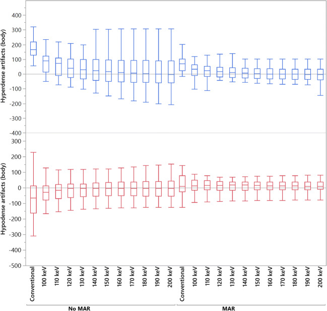
Results of objective analysis of artifact reduction at CIED generator are provided in Table 2 and Fig. 1. Compared to CI, in higher keV of VMI, the corrected attenuation within hypodense artifacts was increased (e.g., CI/VMI200keV: − 78.9 ± 129.5/4.3 ± 119.6 HU, p > 0.05) and decreased within hyperdense artifacts (e.g., CI/VMI200keV: 171.4 ± 165.4/32.3 ± 189.8 HU, p < 0.05), for the latter with statistical significant differences at ≥ 100 keV. MAR and VMIMAR ≥ 100 keV significantly increased/decreased corrected attenuation in hypo-/hyperdense artifacts (p < 0.05).
Table 2.
Objective analysis of artifact reduction and surrounding tissues at CIED generator. Data is reported as mean ± SD. CI, conventional images; VMI, virtual monoenergetic images; MAR, metal artifact reduction algorithm; VMIMAR, combination of MAR and VMI. Bold indicates significant changes in HU values compared to CI
| Corrected attenuation | Corrected image noise | |||
|---|---|---|---|---|
| Hypodense artifacts | Hyperdense artifacts | Hypodense artifacts | Hyperdense artifacts | |
| CI | − 78.9 ± 129.5 | 171.4 ± 165.4 | 69.9 ± 54.9 | 55.2 ± 92.0 |
| VMI | ||||
| 100 keV | − 37.9 ± 97.8 | 96.6 ± 145.6 | 56.9 ± 62.2 | 50.7 ± 57.8 |
| 140 keV | − 8.8 ± 110.2 | 52.3 ± 173.5 | 50.6 ± 64.9 | 46.4 ± 53.8 |
| 200 keV | 4.3 ± 119.6 | 32.3 ± 189.8 | 49.4 ± 66.3 | 46.3 ± 52.5 |
| MAR | 18.4 ± 69.5 | 70.1 ± 50.0 | 13.3 ± 13.2 | 17.0 ± 13.2 |
| VMIMAR | ||||
| 100 keV | 10.2 ± 46.2 | 28.3 ± 50.0 | 8.5 ± 9.0 | 13.0 ± 11.2 |
| 140 keV | 10.1 ± 40.7 | 10.5 ± 56.2 | 6.5 ± 8.4 | 10.8 ± 10.8 |
| 200 keV | 10.0 ± 39.8 | 2.5 ± 59.7 | 5.8 ± 8.3 | 10.1 ± 10.7 |
| p value | ||||
| CI vs. VMI 100–200 keV | > 0.05 | < 0.05 | > 0.05 | > 0.05 |
| CI vs. MAR | < 0.05 | < 0.05 | < 0.05 | < 0.05 |
| CI vs. VMIMAR 100–200 keV | < 0.05 | < 0.05 | < 0.05 | < 0.05 |
Fig. 1.
Box-plot diagram displaying corrected attenuation values within hypo- and hyperdense artifacts adjacent to CIED generator in conventional CT images (conventional), virtual monoenergetic images (VMI, 100–200 keV), metal artifact reduction (MAR) algorithms, and their combination. HU, Hounsfield units
VMI provided decreased corrected image noise in hypo- and hyperdense artifacts at all keV values, albeit this effect was not statistically significant (p > 0.05). However, MAR (p < 0.05) and VMIMAR ≥ 100 keV (p < 0.05) enabled a significant reduction of corrected image noise in hypo- and hyperdense artifacts.
CIED leads
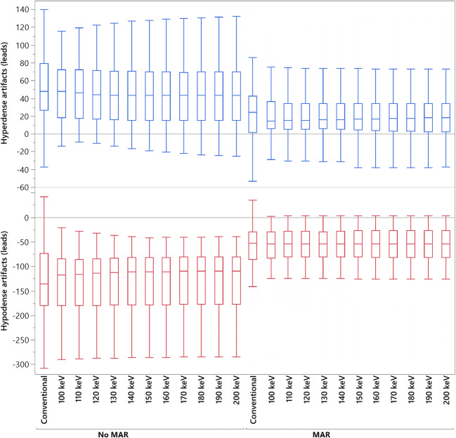
Results of objective analysis of artifact reduction around CIED leads are given in Table 3 and Fig. 2. Compared to CI in VMI ≥ 100 keV, corrected attenuation in hypo- and hyperdense artifacts was comparable without yielding statistical significance (p > 0.05). MAR alone provided a significant increase of attenuation in hypodense (CI/MAR: − 127.5 ± 77.3/− 59.7 ± 50.4 HU; p < 0.05) and reduction in hyperdense (CI/MAR: 51.8 ± 37.7/22.3 ± 30.5; p < 0.05) artifacts. Likewise, VMIMAR ≥ 100 keV yielded a significant increase and decrease in corrected attenuation for hypo- and hyperdense artifacts, respectively (p < 0.05).
Table 3.
Objective analysis of artifact reduction and surrounding tissues at CIED leads. Data is reported as mean ± SD. CI, conventional images; VMI, virtual monoenergetic images; MAR, metal artifact reduction algorithm; VMIMAR, combination of MAR and VMI. Bold indicates significant changes in HU values compared to CI
| Corrected attenuation | Corrected image noise | |||
|---|---|---|---|---|
| Hypodense artifacts | Hyperdense artifacts | Hypodense artifacts | Hyperdense artifacts | |
| CI | − 127.5 ± 77.3 | 51.8 ± 37.7 | 66.5 ± 46.1 | 29.3 ± 24.3 |
| VMI | ||||
| 100 keV | − 128.2 ± 64.9 | 47.6 ± 32.9 | 65.4 ± 41.7 | 30.1 ± 24.4 |
| 140 keV | − 127.2 ± 62.6 | 47.0 ± 35.5 | 64.9 ± 41.8 | 29.3 ± 24.8 |
| 200 keV | − 126.8 ± 61.9 | 46.7 ± 37.0 | 64.7 ± 42.1 | 29.1 ± 25.1 |
| MAR | − 59.7 ± 50.4 | 22.3 ± 30.5 | 23.6 ± 23.2 | 9.3 ± 12.9 |
| VMIMAR | ||||
| 100 keV | − 58.2 ± 39.4 | 19.7 ± 24.6 | 21.2 ± 21.4 | 10.6 ± 10.7 |
| 140 keV | − 57.1 ± 38.0 | 18.8 ± 24.3 | 20.4 ± 22.1 | 10.0 ± 10.1 |
| 200 keV | − 56.5 ± 37.8 | 18.4 ± 24.7 | 20.1 ± 22.4 | 9.7 ± 9.9 |
| p value | ||||
| CI vs. VMI 100–200 keV | > 0.05 | > 0.05 | > 0.05 | > 0.05 |
| CI vs. MAR | < 0.05 | < 0.05 | < 0.05 | < 0.05 |
| CI vs. VMIMAR 100–200 keV | < 0.05 | < 0.05 | < 0.05 | < 0.05 |
Fig. 2.
Box-plot diagram displaying corrected attenuation values within hypo- and hyperdense artifacts adjacent to CIED leads in conventional CT images (conventional), virtual monoenergetic images (VMI, 100–200 keV), metal artifact reduction (MAR) algorithms, and their combination. HU, Hounsfield units
Corrected image noise in CI and VMI ≥ 100 keV was comparable in hypo- and hyperdense artifacts (p < 0.05). However, MAR and VMIMAR ≥ 100 keV provided a significant decrease of image noise in both hypo- and hyperdense artifacts.
Subjective analysis
CIED generator
Results of subjective analysis of artifact reduction and surrounding tissue at CIED generator are given in Table 4. VMI ≥ 100 keV, MAR, and VMIMAR provided significant reduction of hypo- and hyperdense artifacts compared to CI (p < 0.05). Diagnostic assessment of tissue adjacent to CIED generator significantly improved in VMI ≥ 100 keV, MAR, and VMIMAR at 140 keV and 200 keV.
Table 4.
Subjective analysis of artifact reduction and presence of new artifacts at CIED generator. Data is reported as median with 10/90 percentile. CI, conventional images; VMI, virtual monoenergetic images; MAR, metal artifact reduction algorithm; VMIMAR, combination of MAR and VMI; ICC, intraclass correlation coefficient. Bold indicates significant changes in scores compared to CI
| Artifact extent | Diagnostic assessment of surrounding tissue | ||
|---|---|---|---|
| Hypodense | Hyperdense | ||
| CI | 3 (1–3) | 3 (1–4) | 3 (1–3) |
| VMI | |||
| 100 keV | 3 (2–4) | 3 (2–5) | 3.5 (2–4) |
| 140 keV | 4 (2–5) | 4 (2–5) | 4 (2–5) |
| 200 keV | 4 (2–5) | 4 (2–5) | 4 (2–5) |
| MAR | 3 (2–4) | 4 (3–4) | 3 (3–4) |
| VMIMAR | |||
| 100 keV | 4 (3–5) | 4 (3–5) | 4 (3–5) |
| 140 keV | 5 (3–5) | 5 (4–5) | 5 (4–5) |
| 200 keV | 5 (3–5) | 5 (4–5) | 5 (4–5) |
| p value | |||
| CI vs. VMI 100 keV | < 0.05 | < 0.05 | < 0.05 |
| CI vs. VMI 140 keV | < 0.05 | < 0.05 | < 0.05 |
| CI vs. VMI 200 keV | < 0.05 | < 0.05 | < 0.05 |
| CI vs. MAR | < 0.05 | < 0.05 | < 0.05 |
| CI vs. VMIMAR 100–200 keV | < 0.05 | < 0.05 | < 0.05 |
CIED leads
Table 5 displays the results of subjective analysis of artifact reduction and surrounding tissue at CIED leads. Only MAR and VMIMAR ≥ 100 keV provided significant reduction of hypodense artifacts (p < 0.05), whereas for hyperdense artifacts, all three techniques (VMI ≥ 100 keV, MAR, and VMIMAR) enabled significant decrease of artifacts (p < 0.05). Only MAR, VMI at 100 keV, and VMIMAR at 100 keV yielded a significant improvement for diagnostic assessment of tissue and cardiac structures adjacent to the leads. KeV values of 140 or higher in VMI and VMIMAR led to worsened diagnostic assessment.
Table 5.
Subjective analysis of artifact reduction and presence of new artifacts at CIED leads. Data is reported as median with 10/90 percentile. CI, conventional images; VMI, virtual monoenergetic images; MAR, metal artifact reduction algorithm; VMIMAR, combination of MAR and VMI; ICC, intraclass correlation coefficient. Bold indicates significant changes in scores compared to CI
| Artifact extent | Diagnostic assessment of surrounding tissue | ||
|---|---|---|---|
| Hypodense | Hyperdense | ||
| CI | 3 (3–4) | 2 (2–3) | 2 (2–3) |
| VMI | |||
| 100 keV | 3 (3–4) | 3 (2–4) | 3 (2–3) |
| 140 keV | 3 (3–4) | 3 (2–4) | 2 (1–4) |
| 200 keV | 3 (3–4) | 3 (3–4) | 1 (1–4) |
| MAR | 4 (3–5) | 3 (2–4) | 3 (2–4) |
| VMIMAR | |||
| 100 keV | 4 (3–5) | 4 (3–4) | 3 (2–4) |
| 140 keV | 4 (3–5) | 4 (3–5) | 2 (1–4) |
| 200 keV | 4 (3–5) | 4 (3–5) | 1 (1–4) |
| p value | |||
| CI vs. VMI 100 keV | > 0.05 | < 0.05 | < 0.05 |
| CI vs. VMI 140 keV | > 0.05 | < 0.05 | < 0.05 |
| CI vs. VMI 200 keV | > 0.05 | < 0.05 | < 0.05 |
| CI vs. MAR | < 0.05 | < 0.05 | < 0.05 |
| CI vs. VMIMAR 100–200 keV | < 0.05 | < 0.05 | < 0.05 |
Interreader agreement was good (ICC = 0.66).
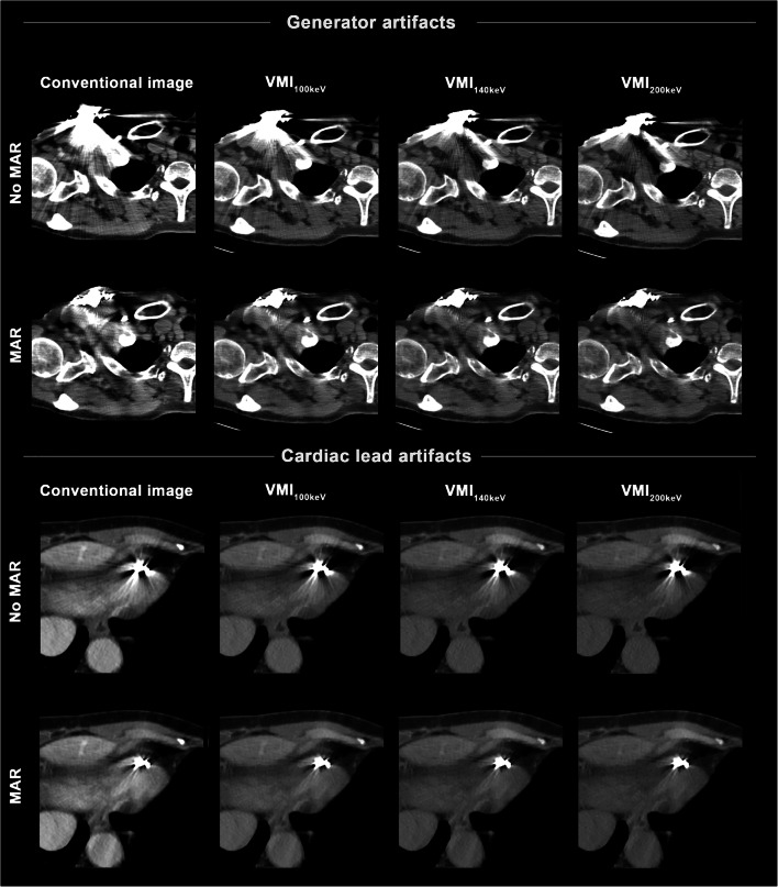
An illustrative case of artifact reduction around CIED generator and leads is presented in Fig. 3.
Fig. 3.
Conventional images, virtual monoenergetic images (VMI, 100–200 keV), MAR, and their combination (VMIMAR) in a 65-year-old female patient with a dual-chamber implanted cardioverter defibrillator in place. For the CIED generator, VMI as a standalone (top row) approach allow for reduction of hyperdense and hypodense artifacts with best performance at higher keV values, although relevant residual artifacts remain. These can be further reduced by MAR and VMIMAR (second row). For the CIED leads, VMI alone (third row) provide only minimal benefit in artifact reduction, whereas performance by VMIMAR (bottom row) is a lot stronger
Discussion
In this study, we assessed the performance of VMI, MAR, and their combination (VMIMAR) for reduction of artifacts from CIED generator and leads in SDCT imaging.
As major findings of this study, MAR and VMIMAR provided the most efficient artifact reduction in subjective and objective analysis while yielding improved diagnostic assessment of soft tissue and cardiac structures adjacent to CIED generator and leads. VMI at higher keV levels provided significant reduction of artifacts at CIED generator and leads (with exception of VMI for hypodense artifacts at the leads) in the subjective assessment, whereas a significant decrease of artifacts in the objective analysis was only observed for hyperdense artifacts at CIED generator. Of note, VMI and VMIMAR led to relevant and significant decrease of diagnostic assessment at ≥ 140 keV at CIED leads.
Prior studies have investigated the use of VMI, MAR, and their combination (VMIMAR) from dual-energy CT for reduction of artifacts arising from high density materials, such as orthopedic hardware [24], dental implants [13], deep brain stimulation leads [25], iodinated contrast agent [26], and coils and clips for intracranial aneurysm treatment [21, 27, 28]. More recent studies focusing on the reduction of artifacts impairing the assessment of the heart and surrounding structures (e.g., from pacemaker devices or other cardiac hardware) showed promising results for the application of artifact reduction algorithms and reconstruction techniques to improve image quality [12, 14–17]. For instance, Tatsugami et al. demonstrated that artifact reduction algorithms allow for a superior assessment of coronary arteries in patients with CIEDs [12]. Also, the application of a convolutional network has been successfully tested for the reduction of artifacts from pacemaker leads [16]. Van Hedent et al. investigated the application of VMI for the reduction of artifacts in chest and abdominal imaging of patients including artifacts from pacemakers [17].
Considering these previous studies, there has been uncertainty whether either method on its own (VMI or MAR) can provide sufficient artifact reduction. Particularly for stronger artifacts, e.g., those arising from CIED generator and leads, MAR and especially VMI as standalone approaches might yield suboptimal artifact reduction [13, 25, 29–31]. In line with these findings, our study demonstrated that, although VMI reduced artifacts in the visual assessment, it failed to significantly reduce artifacts in quantitative measurements, except for hyperdense artifacts at CIED generator. In contrast, MAR and VMIMAR enabled good artifact reduction for both kinds of artifacts and compartments of CIED as outlined in subjective and objective results. Still, this positive effect did not reflect on the diagnostic assessment of cardiac structures surrounding the CIED leads at higher keV values.
In line with previous studies, subjective artifact reduction at CIED generator and leads improved at higher keV levels in VMI and VMIMAR [13, 32]. However, this effect does not necessarily result in improved diagnostic assessment. This is due to the physical properties of VMI that accompanies higher keV values by the greater distance to the k-edge of iodine (~33 keV) [19], which at higher keV levels reduce contrast of soft tissue, vessel lumen, and cardiac chambers [19, 33]. This loss of contrast was found less severe around the CIED generator; therefore, diagnostic assessment was best at high keV values of ≥140 keV. Hence, we recommend keV values between 140 and 200 keV to assess structures around the CIED generator. On the contrary, the loss of contrast of vessels and heart chambers significantly hampers the diagnostic assessment at higher keV values at the leads. Therefore, we recommend 100 keV for optimal diagnostic assessment of structures adjacent to CIED leads. Nevertheless, as the optimal keV value differs between patients, these settings may need to be adjusted individually. A known limitation of VMI and MAR algorithms is that they can cause/produce new artifacts, which can impact the diagnostic quality of CT images [13, 21, 22]. Hence, we recommend the use of VMI and MAR only when artifacts are present, and CI should remain the standard of care. Of note, MAR was initially intended to be applied for orthopedic hardware. Still, as eluded above, several studies have shown its effectiveness also for non-orthopedic hardware [13, 21, 25, 34], e.g., deep brain stimulating electrodes [25]. However, in its white paper [22], the vendor does not recommend to apply MAR when metal is near air or low density tissue, e.g., in pacemakers, as the proximity to the lung can induce new artifacts [22]. Also, in smaller metal objects such as cardiac stents, the MAR algorithm might not alter image information as no or minimal metal is present [22, 25]. To this point, the vendor even disables MAR in dedicated cardiac examinations. Still, Große Hokamp et al. showed the dedicated value of MAR for reduction of artifacts from deep brain stimulating electrodes [25].
With the varying degrees of effectiveness for artifact reduction depending on implant type and its location within the human body, VMI and MAR algorithms for CT imaging are available from all major vendors [19, 35]. However, their combined use is only possible in dual-energy CT. Nevertheless, single-energy CT scanners are by far much more common than DECT in clinical routine; thereby, MAR as a standalone post-processing approach has the advantage of a higher availability in conventional CT systems [24, 35]. Given the results of this study, MAR, not only in combination with VMI but also standalone, enables good reduction of artifacts arising from CIED generator and leads, which allows usage on conventional non-dual-energy CT systems.
Limitations
Besides its retrospective, single-center setting, our study has the following additional limitations that should be considered. First, the method to measure artifacts needs to be discussed. Due to its high feasibility, a relatively simple and standard approach in artifact reduction studies is ROI-based measurement of mean and standard deviation of attenuation [13, 36, 37]. Nevertheless, more elaborate methods using dedicated artifact quantification algorithms might produce more precise results [25, 38]. Second, given their differing physical properties, general changes in terms of attenuation and image noise appear in different keV levels of VMI [19]. To this end, we applied an intra-individual comparison between artifact-impaired tissue and correspondent unimpaired reference tissue resulting in corrected attenuation and corrected image noise, which enables the detection of real artifact reduction [13]. Third, full blinding of readers did not seem feasible since images (CI, VMI, and VMIMAR) are distinguishable by their appearance. Furthermore, we aimed to encourage readers to also detect more subtle differences between reconstructions next to a qualitative rating of images; therefore, a full image set of one patient at a time was presented to the readers.
Conclusions
In presence of artifacts from CIED, the combination of VMI and MAR provides the most effective artifact reduction and improves diagnostic assessment of surrounding structures; however, MAR as a standalone approach also provides good artifact reduction. Still, higher keV values at CIED leads should be applied with caution due to a loss of soft tissue and vessel contrast, which impedes diagnostic accuracy. Based on our data, we recommend keV values between 140 and 200 keV for artifacts surrounding the CIED generator and of 100 keV for artifacts adjacent to the leads.
Supplementary information
(DOCX 19 kb)
Acknowledgements
Clinician Scientist position supported by the Deans Office, Faculty of Medicine, University of Cologne.
Abbreviations
- CI
Conventional CT images
- CIED
Cardiovascular implantable electronic devices
- ICC
Intraclass correlation coefficient
- ICD
Implanted cardioverter defibrillator
- keV
Kiloelectron volt
- MAR
Metal artifact reduction
- MDCT
Multidetector computed tomography
- ROI
Region of interest
- SD
Standard deviation
- SDCT
Spectral detector CT
- VMI
Virtual monoenergetic images
- VMIMAR
Combination of MAR and VMI
Funding
Open Access funding enabled and organized by Projekt DEAL.
Declarations
Guarantor
The scientific guarantors of this publication are Lenhard Pennig and Kai Roman Laukamp.
Conflict of interest
The authors of this manuscript declare relationships with the following companies: Philips Healthcare, Guerbet GmbH
Lenhard Pennig: Study leave unrelated to this project as part of a research contract between Philips Healthcare and University Hospital Cologne.
David Zopfs: Received exemption from clinical duties for research outside this project as a part of a research agreement between Philips Healthcare and University Hospital Cologne.
Roman Gertz: Study leave unrelated to this project as part of a research contract between Philips Healthcare and University Hospital Cologne.
Nils Große Hokamp: Speaker’s bureau, Philips Healthcare. Research support, Philips Healthcare.
Marcel Langenbach: Research grant, Guerbet GmbH.
Simon Lennartz: Research support, Philips Healthcare.
Jan Borggrefe: Speaker’s bureau, Philips Healthcare.
Statistics and biometry
No complex statistical methods were necessary for this paper.
Informed consent
Written informed consent was waived by the Institutional Review Board.
Ethical approval
Institutional Review Board approval was obtained.
Methodology
• retrospective
• observational
• performed at one institution
Footnotes
Publisher’s note
Springer Nature remains neutral with regard to jurisdictional claims in published maps and institutional affiliations.
Change history
4/19/2021
The funding note was missing: Open Access funding enabled and organized by Projekt DEAL.
References
- 1.Wong JA, Devereaux PJ. Cardiac device implantation complications: a gap in the quality of care? Ann Intern Med. 2019;171:368–369. doi: 10.7326/M19-1895. [DOI] [PubMed] [Google Scholar]
- 2.Aguilera AL, Volokhina YV, Fisher KL. Radiography of cardiac conduction devices: a comprehensive review. Radiographics. 2011;31:1669–1682. doi: 10.1148/rg.316115529. [DOI] [PubMed] [Google Scholar]
- 3.Aissa J, Boos J, Sawicki LM et al (2017) Iterative metal artefact reduction (MAR) in postsurgical chest CT: comparison of three iMAR-algorithms. Br J Radiol 90. 10.1259/bjr.20160778 [DOI] [PMC free article] [PubMed]
- 4.Lewis RK, Ehieli WL, Hegland DD, et al. Preprocedural computed tomography before cardiac implanted electronic device lead extraction: indication, technique, and approach to interpretation. J Cardiovasc Electrophysiol. 2020;31:723–732. doi: 10.1111/jce.14353. [DOI] [PubMed] [Google Scholar]
- 5.Mak GS, Truong QA. Cardiac CT: imaging of and through cardiac devices. Curr Cardiovasc Imaging Rep. 2012;5:328–336. doi: 10.1007/s12410-012-9150-8. [DOI] [PMC free article] [PubMed] [Google Scholar]
- 6.Fayad LM, Patra A, Fishman EK (2009) Value of 3D CT in defining skeletal complications of orthopedic hardware in the postoperative patient. AJR Am J Roentgenol 193:1155–1163 [DOI] [PubMed]
- 7.Lee M-J, Kim S, Lee S-A, et al. Overcoming artifacts from metallic orthopedic implants at high-field-strength MR imaging and multi-detector CT. Radiographics. 2007;27:791–803. doi: 10.1148/rg.273065087. [DOI] [PubMed] [Google Scholar]
- 8.Mori I, Machida Y, Osanai M, Iinuma K. Photon starvation artifacts of X-ray CT: their true cause and a solution. Radiol Phys Technol. 2013;6:130–141. doi: 10.1007/s12194-012-0179-9. [DOI] [PubMed] [Google Scholar]
- 9.Boas FE, Fleischmann D. CT artifacts: causes and reduction techniques. Imaging Med. 2012;4:229–240. doi: 10.2217/iim.12.13. [DOI] [Google Scholar]
- 10.Lennartz S, Große Hokamp N, Abdullayev N, et al. Diagnostic value of spectral reconstructions in detecting incidental skeletal muscle metastases in CT staging examinations. Cancer Imaging. 2019;19:50. doi: 10.1186/s40644-019-0235-3. [DOI] [PMC free article] [PubMed] [Google Scholar]
- 11.Kalisz K, Buethe J, Saboo SS, Abbara S, Halliburton S, Rajiah P (2016) Artifacts at cardiac CT: physics and solutions. Radiographics 36:2064–2083 [DOI] [PubMed]
- 12.Tatsugami F, Higaki T, Sakane H, et al. Coronary CT angiography in patients with implanted cardiac devices: initial experience with the metal artefact reduction technique. Br J Radiol. 2016;89:20160493. doi: 10.1259/bjr.20160493. [DOI] [PMC free article] [PubMed] [Google Scholar]
- 13.Laukamp KR, Zopfs D, Lennartz S, et al. Metal artifacts in patients with large dental implants and bridges: combination of metal artifact reduction algorithms and virtual monoenergetic images provides an approach to handle even strongest artifacts. Eur Radiol. 2019;29:4228–4238. doi: 10.1007/s00330-018-5928-7. [DOI] [PubMed] [Google Scholar]
- 14.Aissa J, Thomas C, Sawicki LM, et al. Iterative metal artefact reduction in CT: can dedicated algorithms improve image quality after spinal instrumentation? Clin Radiol. 2017;72:428.e7–428.e12. doi: 10.1016/j.crad.2016.12.006. [DOI] [PubMed] [Google Scholar]
- 15.Kikuchi N, Yanagawa M, Enchi Y, et al. The effect of the reconstruction algorithm for the pulmonary nodule detection under the metal artifact caused by a pacemaker. Medicine (Baltimore) 2020;99:e20579. doi: 10.1097/MD.0000000000020579. [DOI] [PMC free article] [PubMed] [Google Scholar]
- 16.Lossau (née Elss) T, Nickisch H, Wissel T, Morlock M, Grass M (2020) Learning metal artifact reduction in cardiac CT images with moving pacemakers. Med Image Anal 61. 10.1016/j.media.2020.101655 [DOI] [PubMed]
- 17.Van Hedent S, Kessner R, Große Hokamp N, Baran TZ, Kosmas C, Gupta A (2019) Metal artifact reduction in routine chest and abdominal examinations using virtual monoenergetic images from spectral detector computed tomography. J Comput Assist Tomogr 43:713–717 [DOI] [PubMed]
- 18.Große Hokamp N, Laukamp KR, Lennartz S, et al. Artifact reduction from dental implants using virtual monoenergetic reconstructions from novel spectral detector CT. Eur J Radiol. 2018;104:136–142. doi: 10.1016/j.ejrad.2018.04.018. [DOI] [PubMed] [Google Scholar]
- 19.McCollough CH, Leng S, Yu L, Fletcher JG. Dual- and multi-energy CT: principles, technical approaches, and clinical applications. Radiology. 2015;276:637–653. doi: 10.1148/radiol.2015142631. [DOI] [PMC free article] [PubMed] [Google Scholar]
- 20.Große Hokamp N, Neuhaus V, Abdullayev N, et al. Reduction of artifacts caused by orthopedic hardware in the spine in spectral detector CT examinations using virtual monoenergetic image reconstructions and metal-artifact-reduction algorithms. Skeletal Radiol. 2017;47:195–201. doi: 10.1007/s00256-017-2776-5. [DOI] [PubMed] [Google Scholar]
- 21.Zopfs D, Lennartz S, Pennig L et al (2020) Virtual monoenergetic images and post-processing algorithms effectively reduce CT artifacts from intracranial aneurysm treatment. Sci Rep 10. 10.1038/s41598-020-63574-8 [DOI] [PMC free article] [PubMed]
- 22.(2012) Philips CT Clinical Science PHU. Metal Artifact Reduction for Orthopedic Implants (O-MAR). Accessed December 2020. Availabel at: http://clinical.netforum.healthcare.philips.com/us_en/Explore/White-Papers/CT/Metal-Artifact-Reduction-for-O
- 23.Cicchetti DV. Guidelines, criteria, and rules of thumb for evaluating normed and standardized assessment instruments in psychology. Psychol Assess. 1994;6:284–290. doi: 10.1037/1040-3590.6.4.284. [DOI] [Google Scholar]
- 24.Wellenberg RHH, Hakvoort ET, Slump CH, Boomsma MF, Maas M, Streekstra GJ (2018) Metal artifact reduction techniques in musculoskeletal CT-imaging. Eur J Radiol 107:60–69 [DOI] [PubMed]
- 25.Große Hokamp N, Hellerbach A, Gierich A, et al. Reduction of artifacts caused by deep brain stimulating electrodes in cranial computed tomography imaging by means of virtual monoenergetic images, metal artifact reduction algorithms, and their combination. Invest Radiol. 2018;53:424–431. doi: 10.1097/RLI.0000000000000460. [DOI] [PubMed] [Google Scholar]
- 26.Laukamp KR, Gupta A, Große Hokamp N et al (2019) Role of spectral-detector CT in reduction of artifacts from contrast media in axillary and subclavian veins: single institution study in 50 patients. Acta Radiol 028418511986890 [DOI] [PubMed]
- 27.Pan YN, Chen G, Li AJ et al (2019) Reduction of metallic artifacts of the post-treatment intracranial aneurysms: effects of single energy metal artifact reduction algorithm. Clin Neuroradiol. 10.1007/s00062-017-0644-2 [DOI] [PubMed]
- 28.Fitsiori A, Martin SP, Juillet De Saint Lager A, et al. Iterative algorithms applied to treated intracranial aneurysms. Clin Neuroradiol. 2019;29:741–749. doi: 10.1007/s00062-018-0701-5. [DOI] [PubMed] [Google Scholar]
- 29.Dunet V, Bernasconi M, Hajdu SD, Meuli RA, Daniel RT, Zerlauth JB (2017) Impact of metal artifact reduction software on image quality of gemstone spectral imaging dual-energy cerebral CT angiography after intracranial aneurysm clipping. Neuroradiology 59:845–852 [DOI] [PubMed]
- 30.Pan Y-N, Chen G, Li A-J et al (2017) Reduction of metallic artifacts of the post-treatment intracranial aneurysms: effects of single energy metal artifact reduction algorithm. Clin Neuroradiol 1–8 [DOI] [PubMed]
- 31.Sunwoo L, Park S-W, Rhim JH, et al. Metal artifact reduction for orthopedic implants: brain CT angiography in patients with intracranial metallic implants. J Korean Med Sci. 2018;33:e158. doi: 10.3346/jkms.2018.33.e158. [DOI] [PMC free article] [PubMed] [Google Scholar]
- 32.Laukamp KR, Zopfs D, Wagner A, et al. CT artifacts from port systems: virtual monoenergetic reconstructions from spectral-detector CT reduce artifacts and improve depiction of surrounding tissue. Eur J Radiol. 2019;121:108733. doi: 10.1016/j.ejrad.2019.108733. [DOI] [PubMed] [Google Scholar]
- 33.Lennartz S, Laukamp KR, Neuhaus V, et al. Dual-layer detector CT of the head: initial experience in visualization of intracranial hemorrhage and hypodense brain lesions using virtual monoenergetic images. Eur J Radiol. 2018;108:177–183. doi: 10.1016/j.ejrad.2018.09.010. [DOI] [PubMed] [Google Scholar]
- 34.Gupta A, Obmann VC, Jordan M, et al. CT artifacts after contrast media injection in chest imaging: evaluation of post-processing algorithms, virtual monoenergetic images and their combination for artifact reduction. Quant Imaging Med Surg. 2021;11:226–239. doi: 10.21037/qims-20-435. [DOI] [PMC free article] [PubMed] [Google Scholar]
- 35.Huang JY, Kerns JR, Nute JL, et al. An evaluation of three commercially available metal artifact reduction methods for CT imaging. Phys Med Biol. 2015;60:1047–1067. doi: 10.1088/0031-9155/60/3/1047. [DOI] [PMC free article] [PubMed] [Google Scholar]
- 36.Kidoh M, Nakaura T, Nakamura S, et al. Reduction of dental metallic artefacts in CT: value of a newly developed algorithm for metal artefact reduction (O-MAR) Clin Radiol. 2014;69:11–16. doi: 10.1016/j.crad.2013.08.008. [DOI] [PubMed] [Google Scholar]
- 37.Bisschop R, Boomsma M, Van Raay JJA, Tiebosch ATMG, Maas M, Gerritsma CLE (2013) High prevalence of pseudotumors in patients with a Birmingham hip resurfacing prosthesis. J Bone Joint Surg Am 95:1554–1560 [DOI] [PubMed]
- 38.Mangold S, Gatidis S, Luz O, et al. Single-source dual-energy computed tomography: use of monoenergetic extrapolation for a reduction of metal artifacts. Invest Radiol. 2014;49:788–793. doi: 10.1097/RLI.0000000000000083. [DOI] [PubMed] [Google Scholar]
Associated Data
This section collects any data citations, data availability statements, or supplementary materials included in this article.
Supplementary Materials
(DOCX 19 kb)


