Abstract
Each year the American Thoracic Society (ATS) Conference brings together scientists who conduct basic, translational and clinical research to present on the recent advances in the field of respirology. Due to the Coronavirus Disease of 2019 (COVID-19) pandemic, the ATS2020 Conference was held online in a series of virtual meetings. In this review, we focus on the breakthroughs in pulmonary hypertension research. We have selected 11 of the best basic science abstracts which were presented at the ATS2020 Assembly on Pulmonary Circulation mini-symposium “What’s New in Pulmonary Arterial Hypertension (PAH) and Right Ventricular (RV) Signaling: Lessons from the Best Abstracts,” reflecting the current state of the art and associated challenges in PH. Particular emphasis is placed on understanding the mechanisms underlying RV failure, the regulation of inflammation, and the novel therapeutic targets that emerged from preclinical research. The pathologic interactions between pulmonary hypertension, right ventricular function and COVID-19 are also discussed.
Keywords: Translational research, pulmonary hypertension, COVID-19, ATS, basic science research
Introduction
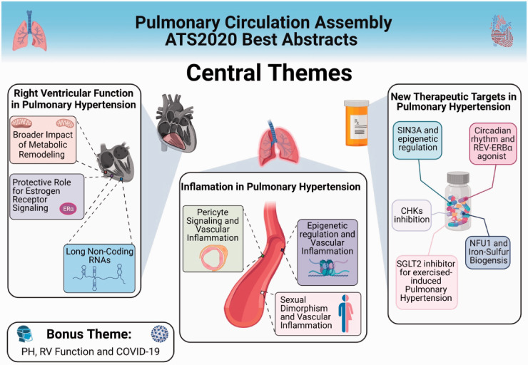
Due to the Coronavirus Disease of 2019 (COVID-19) pandemic, the American Thoracic Society (ATS) has cancelled the International Conference, originally scheduled for 15–20 May 2020 in Philadelphia. Founded in 1905 to combat tuberculosis, the ATS has brought together experts from around the world together to discuss challenges and opportunities to tackle various pulmonary diseases, including pulmonary hypertension (PH). PH is a progressive and often fatal illness presenting with nonspecific symptoms of dyspnea, lower extremity edema, and decreased exercise tolerance. Pathologically, endothelial dysfunction leads to abnormal intimal and smooth muscle proliferation along with reduced apoptosis, resulting in increased pulmonary vascular resistance (PVR), elevated pulmonary pressures, right ventricular (RV) dysfunction/failure and ultimately death. According to the 6th World Symposium on PH (Nice, 2018), PH is redefined as mean pulmonary arterial pressure (mPAP) > 20 mmHg and is subclassified into pre-capillary PH, isolated post-capillary (IpcPH) and combined pre- and post-capillary PH (CpcPH) based on pulmonary arterial wedge pressure (PAWP) and PVR.1 Regarding clinical classification, PH is subcategorized into five World Health Organization (WHO) groups based on pathophysiological mechanisms, clinical presentation, hemodynamic characteristics, and therapeutic management.2 Pulmonary arterial hypertension (PAH, Group 1 PH) specifically refers to disease processes, which result in vasoconstriction and stiffening of the small arteries in the lungs secondary to cell proliferation, fibrosis, as well as the development of in situ thrombi or plexiform lesions. This pathology both defines PAH and unifies the multiple etiologies, which may lead to the development of the disease. PAH can be idiopathic, heritable, and can be associated with connective tissue disease, HIV, drug use, etc. There are other pathologies in which PH presents as a secondary disease, including left heart disease (Group 2), chronic lung diseases and/or hypoxia (Group 3), chronic thromboembolic pulmonary hypertension (CTEPH, Group 4), and miscellaneous or multi-factorial etiologies (Group 5). While there are U.S. Food and Drug Administration (FDA)-approved medications for the treatment of PAH and CTEPH, the morbidity and mortality remain high. Moreover, there are no approved therapies for Groups 2, 3, and 5 PH at present. In this review, we present an overview of the recent advancements in PH, specifically focusing on RV function and inflammatory pathways, and highlighting new therapeutic targets for the treatment of PH from selected abstracts of the ATS2020 Conference (Fig. 1). An overview of the recent clinical findings in PH are discussed in the article “What’s New in Pulmonary Hypertension Clinical Research: Lessons from the Best Abstracts at the 2020 ATS International Conference” in this issue.
Fig. 1.
Overview of the central research themes presented at the ATS2020 Assembly on Pulmonary Circulation ATS2020 Assembly on Pulmonary Circulation mini-symposium “What’s New in PAH and RV Signaling: Lessons from the Best Abstracts”. Particular emphasis is placed on understanding the mechanisms underlying right ventricular failure, the regulation of inflammation, and the novel therapeutic targets in pulmonary hypertension.
SIN3A: switch-independent 3a; REV-ERBα: transcription repressor in cell-autonomous circadian transcriptional/translational feedback loop; CHK: checkpoint kinases; NFU1: mitochondrial iron-sulfur scaffold protein; SGLT2: sodium glucose co-transporter 2 inhibitors. (Note: Figure created with BIoRender.com)
Central Theme 1: RV Function in PH – Tsukasa Shimauchi, MD, PhD, Andrea Frump, PhD, and Junichi Omura, MD, PhD
RV function has long been recognized as a reliable prognostic indicator of survival, morbidity, and mortality in all groups of PH.3,4 Despite the recognized clinical importance of RV function, no RV-targeted therapies exist.5 Furthermore, the molecular processes underpinning the transition from a compensated (adaptive) to a decompensated (maladaptive) RV remain poorly understood. Consequently, the pathobiology of RV failure and whether its progression can be halted or reversed has emerged as a major focus of preclinical and clinical investigations.5 A growing body of work has begun to elucidate and characterize the pathophysiological changes that occur during RV failure. These dynamic studies represent a broad range of scientific interests: from understanding the shift in metabolism and the repercussions from those changes6; to the role of biological sex and sex hormone signaling in the RV7; to the influence of long non-coding RNAs (lncRNAs) on epigenetic regulation.8 Together, these RV-focused studies explore vital questions about the pathophysiological changes occurring in RV failure, whether these changes can be blocked or reversed, and if these processes can be harnessed therapeutically or prognostically for PH patients.
The broader impact of metabolic remodeling on RV failure
Under homeostatic conditions, the RV is estimated to derive 60–90% of its energy through fatty acid oxidation and the remaining 10–40% through glucose oxidation and glycolysis.9 One defining characteristic of RV failure is the increased reliance on glycolysis to meet energy demands, known as the Warburg effect.10 A recent study by Shimauchi et al. examined the broader repercussions of this metabolic shift on DNA damage and inflammation in the human RV.6 Pyruvate kinase muscle isozyme M2 (PKM2) is a key regulator of metabolism and has been linked to PH-induced changes in metabolism, proliferation, and fibrosis in the pulmonary vasculature11,12; however, until recently, PKM2 remained unexplored in RV failure.
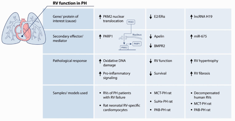
Using autopsy and surgical samples from PH patients with RV failure, investigators identified that compensated and decompensated RVs had an increased PKM2/PKM1 (pyruvate kinase muscle isozyme M1) ratio, indicative of uncoupled glycolysis. These RVs also exhibited increased poly ADP-ribose polymerase 1 (PARP1) expression, suggesting sustained activation of DNA damage signaling pathways. One pathway that might be increasing PARP1 expression is through the nuclear translocation of PKM2. Once localized to the nucleus, PKM2 can bind to and upregulate PARP1, eliciting downstream effects such as pro-inflammatory signaling and oxidative DNA damage.13 Indeed, DNA damage promoted the nuclear translocation and co-localization of PKM2 with PARP1 in rat neonatal RV-specific cardiomyocytes, which in turn stimulated oxidative DNA damage response and pro-inflammatory pathways. Nuclear co-localization between PKM2 and PARP1 was blocked when cells were treated with a PARP1 inhibitor and pro-inflammatory and oxidative DNA damage responses were abrogated. In sum, these studies suggest that the induction and subsequent nuclear localization of PKM2 and PARP1 may be associated with a trigger from RV compensation to RV decompensation, in particular through stimulation of pro-inflammatory and DNA damage signaling responses (Fig. 2).
Fig. 2.
Right ventricular (RV) function in pulmonary hypertension (PH).
PKM2: pyruvate kinase muscle isozyme M2; PARP 1: poly ADP-ribose polymerase 1; E2: 17β-Estradiol; ERα: estrogen receptor α; BMPR2: bone morphogenetic protein receptor type 2; MCT: monocrotaline; SuHx: sugen/hypoxia; PAB: pulmonary artery banding; lncRNA: long non-coding RNA; miR: microRNA.
A protective role for estrogen receptor signaling in RV failure
17β-Estradiol (E2) is the most predominant female sex hormone in women of reproductive age. It exerts complex, context and cell-dependent effects on the pulmonary vasculature in preclinical models of PH, but has been shown to be predominantly protective in the RV, which replicates clinical data where female patients with PH have better RV function and survival compared to males.14,15 While E2 signals through one of three receptors, estrogen receptor α (ERα) has been linked to beneficial effects in the systemic vasculature.16,17 However, the role of ERα in the cardiopulmonary system is largely unknown.
A recent study by Frump et al. explored the effects ERα in the development of RV failure by using two approaches: first they examined the loss of ERα in the development of monocrotaline (MCT) or sugen/hypoxia (SuHx)-PH using male and female wild type (WT) and ERα mutant rats; second, they examined the effects of stimulating ERα signaling in established MCT-PH or pulmonary artery banding (PAB)-RV failure.7 In the first approach, loss of ERα was associated with more severe MCT or SuHx-PH compared to WT animals. Interestingly, this difference in phenotype appeared to be driven by more severe changes in female animals, while changes in ERα mutant males were less pronounced. This more severe phenotype was associated with decreased RV expression of homeostatic regulators and ERα targets apelin and bone morphogenetic protein receptor type 2 (BMPR2)18 (Fig. 2). Meanwhile, administration of an ER agonist or E2 reversed MCT- and PAB-induced changes in RV function, restored RV apelin and BMPR2 expression and increased survival. Taken together, these data suggest that ERα exerts protective effects against RV failure and alterations in ERα signaling may contribute to the female bias in PH.
LncRNAs and RV failure
LncRNAs are encoded by the genome, are over 200 nucleotides long, and typically bind proteins or RNA where they enact epigenetic, transcriptional, and post-transcriptional gene regulation.19 Studying lncRNA biology is typically challenging due to poor conservation across mammalian species; however, the lncRNA H19 is among the most conserved lncRNAs in mammals and is abundantly expressed during development as well as in etiologies including left heart failure and cancer, where it and its embedded microRNA, miR-675, regulate proliferation.20–22 In left heart failure, H19 and miR-675 promote epigenetic changes that stimulate cardiomyocyte hypertrophy and fibrosis.21 Whether H19 and miR-675 play similar roles in PH and RV failure is unknown.
A recent study by Omura et al. identified H19 as a novel target and prognostic factor in PH-induced RV failure.8 H19 and miR-675 were significantly increased in decompensated human RVs compared to control biopsies. This finding was corroborated in both MCT and PAB rat models. Furthermore, H19 expression correlated with RV hypertrophy and fibrosis in human and rat RV, suggesting H19 may be an epigenetic driver of both processes in RV failure. Indeed, silencing H19 in vivo improved RV function, decreased fibrosis and hypertrophy, and increased the expression of epigenetic regulator enhancer of zeste homolog 2 (EZH2). Using rat neonatal cardiomyocytes, overexpression of H19 or a miR-675 mimic was able to reduce the expression of EZH2 while silencing H19 had the opposite effect on EZH2 expression. Together, these studies suggest an epigenetic mechanism driving RV failure. Subsequent studies found that circulating H19 levels in plasma not only correlated with RV function but also differentiate PH patients from controls and could be used to predict long-term patient survival offering a new potential prognostic marker for to identify PH-induced RV failure23 (Fig. 2).
Central Theme 2: Inflammation in PH – David Condon, MD, Francois Potus, PhD, and Christine Cunningham, BS
In the past decade, pro-inflammatory phenotype has emerged as a hallmark of PAH. It is now widely recognized that increased inflammation contributes to disease etiology, promotes adverse pulmonary vascular remodeling and RV dysfunction. Moreover, the increased level of circulating inflammatory cytokines correlates with PAH severity and predicts mortality in PAH patients.24–26 Pre-clinical studies have established inflammation as a promising therapeutic target, which led to the development of clinical trials assessing anti-inflammatory therapies in PAH (e.g. Anakinera NCT03057028, Tocilizumab NCT02676947).27 Surprisingly, the origin or mechanisms leading to this pro-inflammatory phenotype are not yet fully understood in PAH. The following work presented at the ATS 2020 provided some novel insight on the molecular, genetic, and epigenetic origin of inflammation in PAH.
Pericyte signaling and vascular inflammation
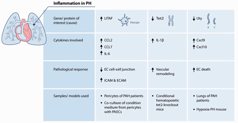
Condon and colleagues investigated the role of pericytes as a source of inflammation in PAH. Pericytes are cells that provide mural support and maintain endothelial homeostasis, which also contributes to the production of cytokines and regulation of endothelial response to injury. This group has previously demonstrated that pericytes contribute to vascular remodeling and vessel loss in PAH.28 At the ATS2020 Conference, Condon and colleagues identified increased expression of pro-inflammatory genes and proteins (including CCL2, CCL7, IL-6) in vitro in serum-starved pericytes of PAH patients related to that of healthy subjects. Using network analysis, they identified lipopolysaccharide-induced tumor necrosis factor-α factor (LITAF), a transcription factor known to be involved in the regulation of inflammatory response, as a potential regulator of this PAH pericyte expression pattern. LITAF expression was also found to be elevated in lungs and cultured pericytes from PAH patients compared to healthy controls. To evaluate whether PAH pericytes can trigger activation of inflammatory markers in healthy pulmonary vascular endothelial cells, they incubated endothelial cells with serum-starved media from either healthy or PAH pericytes. Compared to healthy pericytes, PAH pericytes media resulted in a reduction of endothelial cell–cell junctions and upregulation of inflammatory-related adhesion molecules ICAM and ECAM. However, this effect was reversed by silencing LITAF in PAH pericytes, which was also associated with decreased cytokine mRNA production in PAH pericyte. These studies suggest that LITAF-dependent pericyte signaling plays a role in the local pro-inflammatory milieu known to perpetuate vascular damage and remodeling in PAH (Fig. 3). Moreover, this ongoing work may lead to identification of LITAF as a potential new therapeutic target in PAH.
Fig. 3.
Inflammation in PH.
LITAF: lipopolysaccharide-induced tumor necrosis factor-α factor; CCL2: C–C motif chemokine ligand 2; CCL7: C–C motif chemokine ligand 7; IL-6: interleukin 6; EC: endothelial cell; ICAM: intercellular adhesion molecule; ECAM: epithelial cell adhesion molecule; PAECs: pulmonary arterial endothelial cells; Tet2: Tet-methylcytosine-dioxygenase-2; IL-1β = interleukin 1 beta; Uty: ubiquitously transcribed TPR gene on Y chromosome; Cxcl9: C-X-C motif chemokine ligand 9; Cxcl10: C-X-C motif chemokine ligand 10.
Epigenetic regulation and vascular inflammation
Tet-methylcytosine-dioxygenase-2 (TET2) is an epigenic regulator that contributes to the regulation of inflammation.29 Somatic mutations of TET2 occur in cardiovascular disease and are associated with clonal hematopoiesis, inflammation, and adverse vascular remodeling.30 Potus and colleagues performed a gene-specific rare variant association tests in 1832 unrelated European PAH patients from the PAH biobank and 7509 non-Finnish European controls subjects from the GnomAD depository.31 The authors found an increased burden of rare, predicted deleterious, germline variants in TET2 in PAH patients associated with an increased relative risk of 6. Interestingly, patients carrying TET2 mutations were older, unresponsive to vasodilator challenge, had lower PVR, and had increased levels of circulating inflammatory markers (including elevation of IL-1β). In an independent cohort of 140 patients, circulating TET2 expression was decreased in >86% of PAH patients and predicted the functional and hemodynamic severity of PAH. Using conditional hematopoietic tet2-knockout mice, Potus and colleagues observed that tet2-depleted mice spontaneously developed PH associated with decreased lung perfusion and increased pulmonary vascular remodeling. Tet2-knockout mice also exhibited a pro-inflammatory phenotype with elevated levels of cytokines in the lungs, including IL-1β. Targeting inflammation using a chronic therapy with IL-1β-specific antibody regressed PAH phenotype in tet2 mutated mice. Overall, these data demonstrate that mutations in the epigenetic regulator TET2 contribute to PAH etiology and pro-inflammatory phenotype (Fig. 3). Additionally, this work suggests that a specific IL-1β-targeted therapy might be beneficial for patients with TET2 mutation.
Sexual dimorphism and vascular inflammation
In an interesting study which linked sexual dimorphism to inflammation in PH, Cunningham and colleagues have found a Y-chromosome gene, Uty, protects against PH by reducing pro-inflammatory cytokines and endothelial cell death. Building on their recent work on Y-chromosome-mediated protection against PH,32 Cunningham and colleagues investigated the contribution of four key candidate Y-chromosome genes (Uty, Kdm5d, Ddx3y, Eif2s3y) in PH.32 In a series of experiments, they independently knocked down each of the four candidate Y-chromosome genes via sequential intratracheal instillation of siRNA in male mice exposed to hypoxia. Interestingly, knockdown of Uty, but none of the other genes, resulted in the development of significant PH. Lung RNAseq data revealed increase in pro-inflammatory cytokines, including Cxcl9 and Cxcl10, in Uty-knockdown mice compared to controls. Additionally, Cxcl9 and Cxcl10 were also significantly upregulated in human PAH lungs vs. healthy controls and in female PAH lungs vs. male (GSE15197). Cunningham and colleagues also observed co-localization of Uty with CD68+ macrophages, Cxcl9, and Cxcl10 in the lungs. Upon stimulation, macrophages derived from bone marrow of male Uty-knockout mice displayed increased expression of Cxcl9 and Cxcl10 compared to wildtype mice. Furthermore, treatment of human pulmonary artery endothelial cells with Cxcl9 and Cxcl10 exhibited significantly increased cell death in vitro. Finally, pharmacologic inhibition of Cxcr3 (the receptor for Cxcl9 and Cxcl10) reduced PH in MCT-treated female rats. Together, these findings demonstrate that Uty protects against PH development and may explain Y-chromosome-mediated protection in PH (Fig. 3). This work also suggests that inhibition of Cxcl9 and Cxcl10 activity may be a novel therapy for the treatment of PH.
Central Theme 3: New Therapeutic targets in PH – Malik Bisserier, PhD, Joel James, PhD, Roxane Paulin, PhD, Wen Wu, PhD, and Taijyu Satoh, MD, PhD
Over the past 30 years, scientific discovery has led to FDA-approved drugs for the treatment of PAH and CTEPH. However, mortality with the current therapies remains high, and there are no approved therapies for Groups 2, 3, and 5 PH to date. Vasodilator therapies, commonly used for PAH, are not recommended owing to potentially negative impact on gas exchange33 and increased likelihood of disease progression, adverse events, and mortality.34 In fact, with a few examples of limited success (see Han et al.35 for more details), there is little support for FDA-approved PAH drugs to confer benefit in PH patients associated with idiopathic pulmonary fibrosis (IPF) or heart failure with preserved ejection fraction (HFpEF),33,34,36,37 and current studies remain underpowered and inconsistent in their findings.38 Noteworthy and potentially useful candidates for new therapeutic targets/strategies for PAH, IPF-PH and exercise-induced pulmonary hypertension (EIPH) were presented at the ATS2020 conference.
SIN3A, epigenetic regulation of BMPR2, and PAH
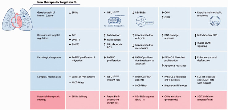
Significant decreases in BMPR2 expression have been found in PAH patients despite an absence of a germline mutation39,40; however, the underlying mechanisms involved in the downregulation of BMPR2 remain poorly understood. Dr. Bisserier’s work built on the recent finding that hypermethylation of the BMPR2 promoter region may impact BMPR2 expression levels41 and investigated the role of switch-independent 3a (SIN3a) in the epigenetic regulation of BMPR2 in PAH. The SIN3 complex, which is composed of the SIN3a and SIN3b isoforms, can either positively or negatively regulate genes through the SIN3/HDAC complex targeting a number of DNA-binding proteins that mediate the deacetylation of histones and the coordination of the methylation status of histone lysines.42 Bisserier and colleagues showed that SIN3a expression is significantly decreased in lungs of PAH patients and rodent models of PH. Overexpression of SIN3a in human pulmonary artery smooth muscle cells (hPASMC) inhibited proliferation and migration, and upregulated BMPR2 expression. Mechanistically, Bisserier and colleagues demonstrated that reduced 10-11 translocase methylcytosine dioxygenase 1/DNA methyltransferase 1 (TET1/DNMT1) ratio regulates the methylation of the promotor region of BMPR2 (Fig. 4). Modulation of the SIN3a expression using an adeno-associated virus encoding human SIN3a (AAV1.hSIN3a) decreased pulmonary artery pressure, vascular remodeling, and Fulton index in rats with MCT-induced PH. Lung-targeted SIN3a delivery also significantly restored BMPR2 expression and reduced the methylation of BMPR2 promoter region by increasing TET1/DNMT1 ratio in lungs of MCT-PAH rats. Together, Dr. Bisserier’s work identified SIN3a gene therapy as a new therapeutic strategy for treating patients with PAH.
Fig. 4.
New therapeutic targets in PH.
SIN3a: switch-independent 3a; TET1: ten-eleven translocase methylcytosine dioxygenase 1; DNMT1: DNA methyltransferase 1; BMPR2: bone morphogenetic protein receptor type 2; PASMCs: pulmonary arterial smooth muscle cells; MCT: monocrotaline; NFU1: mitochondrial iron-sulfur scaffold; FA = fatty acid; ROS: reactive oxygen species; CHK1: checkpoint kinases 1; CHK2: checkpoint kinases 2; sGC β1: soluble guanylate cyclase β1 subunit; cGMP: cyclic 3′5′ guanosine monophosphate; SGLT2: sodium glucose co-transporter 2.
NFU1, iron-sulfur biogenesis, and PH
As increasing number of studies support a link between mitochondrial metabolic reprogramming and progressive tissue dysfunction in the regulation of PH, targeting metabolic dysfunction is an emerging and intriguing therapeutic strategy in patients with PAH.43 At the ATS2020 Conference, James and colleagues investigated targeting the iron-sulfur (Fe-S) scaffold protein NFU1 G206C mutation as a therapeutic target to reverse PH.44 The function of the mitochondrial Fe-S cluster assembly protein NFU1 is largely unknown; however, individuals with mutations in NFU1 develop PH, neurological issue, early death, and multiple mitochondrial dysfunction syndrome (MMDS1).45 NFU1G206C mutant rats have been shown to spontaneously develop PH with increased pulmonary artery thickness, pulmonary pressure, and Fulton index.46 James and colleagues’ work at the ATS2020 Conference examines the impact of impaired Fe-S cluster transfer on PASMC proliferation. The team hypothesized that the NFU1G206C mutation could induce mitochondrial metabolic reprogramming leading to a glycolytic switch and increased proliferation. Using quantitative proteomics, James and colleagues identified 208 mitochondrial proteins which differed in their abundance levels in NFU1G206C rats compared to controls. There was a measurable metabolic shift in the isolated PASMCs with decreased mito-stress and increased glycol-stress and enhanced proliferation of the PASMCs. They measured enhanced fatty acid metabolism with increased fatty acid transport, fatty acid oxidation, and increased mitochondrial reactive oxygen species (ROS) (Fig. 4). Fe-S biology together with corresponding biogenesis genes, including NFU1, iron-sulfur cluster assembly protein (ISC)47 and BolA family member 3 (BOLA3),48 is an emerging area of investigations into the factors contributing to genetic factors predisposing patients to PH development.
Modulation of circadian clock in PAH
The circadian clock plays an essential role in many aspects of behavior and physiology, including sleep/wake cycles, regulations of blood pressure and body temperature, energy metabolism, and inflammation.49,50 Disrupted circadian rhythms of lung function and inflammatory responses have been implicated in pulmonary fibrosis, COPD, asthma, and inflammation51–53; however, the role of the molecular clock and the impact of circadian disruption in the pathogenesis of PAH remain poorly explored. The circadian clock consists of a positive arm – CLOCK and BMAL1 heterodimers, which drive transcription of two inhibitory arms – PER/CRY and REV-ERBα/REV-ERBβ (also known as NR1D1 and NR1D2). As a negative feedback loop, PER and CRY inhibit their own transcription by binding to BMAL and CLOCK/NPAS2. Additionally, REV-ERBα recruits the nuclear receptor co-repressor 1 (NCoR1)/histone deacetylase 3 (HDAC3) complex to inhibit BMAL1/CLOCK heterodimer transactivation function in a negative feedback manner. Dr. Paulin’s work presented at the ATS2020 conference showed a reduced protein expression of REV-ERBα in PASMCs from PAH patients (after 16–20 h of serum shock synchronization) compared to controls. Downregulated REV-ERBα in PASMC was associated with increased proliferation and reduced apoptosis, both of which were inhibited by the treatment of REV-ERBα agonist SR9011 (Fig. 4). The authors carried out RNAseq analyses to identify pathways affected by SR9011 and uncovered 139 downregulated and 101 upregulated genes, and revealed most influenced pathways to related to cell cycle regulation and metabolism, in line with their observed effects on proliferation as well as the effects of ERB proteins observed in human cancers. Using an automatic light system to control the light/dark shift between 6 am and 6 pm, Paulin and colleagues observed that REV-ERBα protein expression was significantly reduced in lungs of MCT rats (at 6 pm) compared to control rats. Moreover, MCT rats left in constant darkness exhibited more frequent and shorter activity–rest cycles compared to their control counterparts under the same conditions. A two-week treatment with SR9011 (at a constant timing of 10 h after light exposure to restore REV-ERBα activity at 6 pm) significantly improved PH phenotypes in MCT rats with decreased PASMC proliferation and increased apoptosis. In summary, Dr. Paulin’s work reveals a role of clock core signaling pathways in PAH etiology and suggests the therapeutic potential of circadian clock modulation for the treatment of PAH.
The role of checkpoint kinases in IPF-PH
Given the limited scope of molecular pathways targeted by current therapeutic strategies coupled with increasingly expanding, and at times contradictory, new mechanistic insights into disease progression of various PH syndromes, cutting-edge efforts have focused on the therapeutic potential of specific molecular targets in different PH groups. In this endeavor, Wu et al. investigated targeting checkpoint kinases in PH-IPF, a progressive fibrotic lung disease with high mortality rate.54,55 PH is a common complication of IPF for which there are no current treatments or approved therapies other than lung transplantation.56–59 In their study, Wu et al. show that checkpoint kinases 1 and 2 (CHK1 and 2) expression and phosphorylation are upregulated in IPF patient lung and distal PAs, localized primarily to PASMCs and fibrotic lesions, as well as in vitro in PASMCs and fibroblasts isolated from IPF patients. CHK1 & 2 are involved in the DNA repair process and regulate cell cycle progression through direct inhibition of CDC25A and activation of p53 and p21.60 They have been implicated in several cancers, and while a number of inhibitors of CHK1 and 2 have not progressed to Phase III clinical trials, in large part owing to toxicity, some recently developed “second-generation” CHK1-selective inhibitors, such as prexasertib, are showing promise.60,61 Evidence by the author’s group and others points to upregulation and dysregulation of the DNA damage response in interstitial fibroblasts and PASMCs in PAH and IPF. This is consistent with the findings of observed resistance to apoptosis and increased proliferation in these conditions.62–64 In a recent publication, the author’s group implicated CHK1 in PAH PASMC upregulation of proliferation and apoptosis resistance via downregulation of DNA repair protein RAD 51 and showed selective CHK1 pharmacological inhibition using MK-8776 improved vascular remodeling and hemodynamics in PAH animal models.65 In the current work presented at the ATS 2020 Conference, Wu et al. also showed an increase in DNA damage assessed by increased phosphorylation of replication protein A 32 (RPA32) and expression of H2A histone family member X and gamma (γH2AX), correlating with increased PA remodeling and fibrosis in IPF patient tissues. Using pharmacological CHK inhibitors (MK-8776 and Prexasertib) in vitro decreased both IPF-fibroblast and PASMC proliferation and increased apoptosis by blocking DNA repair. The authors also showed reduction in profibrotic molecules such as collagen I and MMP2 with prexasertib. In bleomycin-challenged mice, prexasertib reduced lung fibrosis and vascular remodeling, and improved lung function. These findings serve as a promising lead for using CHK inhibitors not only in PAH, as the authors have previously shown, but also in Group 3 PH, as demonstrated by the IPF-PH data presented in this work.
Targeting metabolic syndrome for exercise-induced PH in HFpEF
In addition to PAH and RV failure, Dr. Satoh presented his recent work in EIPH. PH is a common complication of HFpEF, which is associated with high morbidity and mortality. A major symptom of HFpEF patients is exertional dyspnea and exercise intolerance, in large part related to significant increases in pulmonary pressures during exercise. Approximately 50–88% of HFpEF patients develop EIPH, even at low-level exercise, and have an overall worse prognosis with higher risk of right ventricular dysfunction and death.66–68 To date, the pathogenesis and treatment strategy for EIPH remain unclear due to lack of pre-clinical models of EIPH. Since the prevalence of obesity/metabolic phenotype is observed in more than 94% of patients with PH-HFpEF,69,70 the new animal model of EIPH was established based on the previously developed multi-hit rat model of PH-HFpEF, which combines SU5416 (a vascular endothelial growth factor receptor blocker known to induce lung endothelial injury and apoptosis) in rats with multiple features of metabolic syndrome and HFpEF due to double leptin receptors defect (obese ZSF1).71 Consistent with previous findings, SU5416-exposed obese ZSF1 rats (Ob-Su) developed mild PH and higher LV end-diastolic pressure (LVEDP) relative to lean rats at rest. While LV systolic pressure and LVEDP were similarly increased in lean and Ob-Su rats during treadmill exercise, exercise-induced stress significantly exacerbated PH in Ob-Su rats, modeling EIPH. Mechanistically, low plasma levels of cyclic 3ʹ5ʹ guanosine monophosphate (cGMP, which mediates vasodilation) and reduced pulmonary artery expression of soluble guanylate cyclase β1 subunit (sGCβ1, which generates cGMP) were found to be associated with increased ROS production and pulmonary artery dysfunction in rats with EIPH (Fig. 4).
As one of the most recent breakthroughs in cardiology showed that some antidiabetic drugs, such as metformin and sodium glucose co-transporter 2 (SGLT2) inhibitors, are associated with reduction in HF hospitalizations and lower mortality,72–74 Dr. Satoh’s work at the ATS2020 conference also evaluated the effect of SGLT2 inhibitor, empagliflozin, in the newly developed rat model of EIPH. Fourteen-week treatment with empagliflozin (10mg/kg/day) in drinking water significantly improved body weight, HbA1c, and plasma triglyceride in Ob-Su rats at rest. Empagliflozin treatment also significantly reduced exercise-induced changes in PA pressures and LVEDP via restoring sGCβ1-cGMP signaling. Collectively, Dr. Satoh’s work revealed that pulmonary artery dysfunction may contribute to EIPH in metabolic syndrome/HFpEF and that SGLT2 inhibitors may be a promising therapeutic strategy for the treatment of EIPH.
Bonus theme: PH, RV function, and COVID-19
COVID-19 pandemic caused by the SARS-CoV-2 virus continues to inflict significant morbidity and mortality around the globe.75 Recent studies suggest potential pathologic interactions between SARS-CoV-2 infection, RV function, pulmonary vascular damages, and PH.76–81 Lungs of some patients dying with COVID-19 demonstrated pulmonary vascular involvement in the form of severe endothelial injury, cell death, and thrombosis.77,78,80,82 Although the precise mechanisms remain elusive, complement deposition and activation have been suggested as plausible mechanisms for destructive hemorrhagic, capillaritis-like tissue damage, clotting, and hyper-inflammation in COVID-19-associated pulmonary vasculopathy.78
Due to its pathophysiologic relevance, the RV seems to be at higher risk of dysfunction in COVID-19.75,81 In fact, PH and RV dysfunction were associated with increased odds for 30-day inpatient mortality in COVID-19 patients.79 It is postulated that increased RV afterload from acute respiratory distress syndrome and pulmonary thromboembolism, negative inotropic effects of cytokines, and direct angiotensin converting enzyme 2 (ACE2)-mediated cardiac injury from SARS-CoV-2 are potential mechanisms of RV dysfunction in COVID-19.75
The COVID-19 pandemic has presented many unique challenges when caring for patients with PH including clinic and hospital visits, referrals, workup and evaluation, airway management and oxygenation, need for right heart catheterization and enrolment in PAH clinical trials.83,84 Although COVID-19 poses a major threat to the vulnerable PH patient population, based on the available data, the relative number of PH patients with COVID-19-related complications has been low, and most of them have had favorable outcomes.85–87 This has led to the speculation by some that PH patients may be at a lower risk from severe COVID-19 possibly due to a complex interplay between chronic hypoxia exposure together with the beneficial effects with PH therapy on pulmonary vasculature and coagulation profile.87,88 However, this observation needs to be further investigated and validated in the light of the novel SARS-CoV-2 variants.
Summary and discussion from ATS2020 Conference
The ATS2020 Conference highlights the ongoing and future research in respirology. Works presented at the ATS2020 Conference Pulmonary Circulation Assembly mini symposium “What’s New in PAH and RV Signaling: Lessons from the Best Abstracts” grouped in three main topics: (1) understanding and characterization of the mechanisms underlying RV failure, (2) the origin of pro-inflammation phenotype observed in patients, and (3) preclinical models of PH and novel therapeutics targets that emerged from preclinical PH research (Fig. 1).
The RV-focused studies explore vital questions about the pathophysiological changes occurring in RV failure, whether these changes can be blocked or reversed, and if these processes could be harnessed therapeutically or prognostically for PH patients. Ultimately, investigators anticipate that RV-targeted therapies would enhance the ability of the RV to adapt (compensate) to increased afterload, inhibit decompensatory or maladaptive responses, improve the functional capacity of the cardiopulmonary circuit, and enhance quality of life and longevity of patients with PH.5,89 This approach begins with understanding the genetic, epigenetic, molecular, and physiological changes that the RV undergoes during failure paving the way for RV-targeted therapies.
The presentations based on the topic of inflammation identified the contribution of pericytes in the pro-inflammatory PH phenotype. Once activated, those cells produce pro-inflammatory cytokines in LITAF-dependent manner. Two presentations identified TET2 and Uty as new protecting genes against PH, acting by reducing the expression of pro-inflammatory cytokines. Genetic depletion of TET2 and Uty leads to PH development in rodents. Beyond raising the basic PH knowledge, these studies offered new therapeutic targets to tackle inflammation in PH.
Targeting the BMPR2 pathway through the transcriptional regulatory protein SIN3a and mitochondrial metabolism through Fe-S biogenesis have been proposed as attractive and potentially promising therapeutic strategies in PAH treatment. A fascinating discovery of dynamic changes in stability of REV-ERBα protein provides new insights into circadian clock core signaling pathways in the regulation of PAH. This finding also opens a new avenue for the use of selective circadian clock modulators, including REV-ERBα agonists, in the treatment of PAH. Moreover, novel approaches to target checkpoint kinases have shown promise for future therapeutic development for treatment of PAH. Extending checkpoint kinases targeting approaches to other forms of PH, such as Group 3 PH, will not only highlight the importance of these cellular processes in pulmonary vascular dysfunction in general but may also allow development of urgently needed therapies outside of PAH. Furthermore, tackling metabolic syndrome in Group 2 PH using either metformin or SGLT2 inhibitors demonstrated promising preclinical results for EIPH.
Scientific work presented at the ATS2020 Conference also reinforced the ideas around the genetic and epigenetic basis of PH. Mutations of TET2 and NFU1 were associated with the development and progression of PH. Interestingly, work identifying the contribution of two enzymes (both are the members of the TET family) in pulmonary vascular lesions in PH was also presented this year. Both of these enzymes were found to function as regulators of DNA demethylation. These observations suggest a potential contribution of DNA methylation in PAH. However, the role of this epigenetic modification in PAH remains to be investigated fully. Omura and colleagues reported that increased H19 correlates with RV dysfunction and demonstrated the contribution of the first lncRNA in the development of RV failure. These presented works suggest that the development of epigenetic-based therapy may be an appealing approach for future-generation treatments of PAH. PAH is more prevalent in females; however, the disease is more severe in males. Frump and colleagues provide a novel insight into the sex paradox in PAH. They observed that ERα signaling has a protective effect on RV function and that administration of ERα agonist prevents RV dysfunction in the preclinical model of the disease. This observation might provide some insight into the high mortality observed in male PH subjects. Conversely, a study from Cunningham and colleagues demonstrated that the Y-chromosome gene may offer protection against PAH by reducing pro-inflammatory cytokines and endothelial cell death. This observation hints at some molecular mechanisms which may explain the higher female PAH prevalence.
In summary, despite its unusual format, the ATS2020 Conference brought together experts who share their fascinating new work related to basic and translational research in the field of PH, improving our understanding of the disease, raising new questions, and outlining new directions for tomorrow's research.
Acknowledgements
We thank Dr. Sergei Snovida for helpful comments on the manuscript and Elfy Chiang for assistance and production of the figures. The authors also thank the ATS staff, Miriam Rodriguez and Nicole Feijoo, for their help with access to abstracts and presentations.
Footnotes
Authors’ contribution: All authors contributed to writing the article.
Conflict of interest: The author(s) declare that there is no conflict of interest.
Funding: The writing of this review is supported by NIH/NHLBI (R01HL142638) to Y-C. L; NIH/NHLBI (R01HL148712), American Heart Association Innovator grant (18IPA34170257), Gilead Sciences Research Scholars Program in Pulmonary Arterial Hypertension, Vitalant and the Hemophilia Center of Western Pennsylvania to I. AG; AHA Career Development Award (19CDA34660173), ALA Catalyst Award (CA-629145), and Actelion Entelligence Young Investigator Award to A. L. F; lung and heart research institute of Quebec foundation’s grant to F. P; ABRC New Investigator Award (ABRC/ADHS18-198871) and AHA Career Development Award (19CDA34730039) to R.R.V; and NIH/NHLBI (5K08HL141995-03) to S. U.
ORCID iDs: Soban Umar https://orcid.org/0000-0001-8036-3079
Yen-Chun Lai https://orcid.org/0000-0002-4703-5708
References
- 1.Galie N, McLaughlin VV, Rubin LJ, et al. An overview of the 6th World Symposium on Pulmonary Hypertension. Eur Resp J 2019; 53: 1801908. [DOI] [PMC free article] [PubMed] [Google Scholar]
- 2.Simonneau G, Montani D, Celermajer DS, et al. Haemodynamic definitions and updated clinical classification of pulmonary hypertension. Eur Resp J 2019; 53: 1801913. [DOI] [PMC free article] [PubMed] [Google Scholar]
- 3.Vonk Noordegraaf A, Chin KM, Haddad F, et al. Pathophysiology of the right ventricle and of the pulmonary circulation in pulmonary hypertension: an update. Eur Resp J 2019; 53: 1801900. [DOI] [PMC free article] [PubMed] [Google Scholar]
- 4.Vonk Noordegraaf A, Westerhof BE, Westerhof N.The relationship between the right ventricle and its load in pulmonary hypertension. J Am Coll Cardiol 2017; 69: 236–243. [DOI] [PubMed] [Google Scholar]
- 5.Lahm T, Douglas IS, Archer SL, et al. Assessment of right ventricular function in the research setting: knowledge gaps and pathways forward. An Official American Thoracic Society Research Statement. Am J Respir Crit Care Med 2018; 198: e15–e43. [DOI] [PMC free article] [PubMed] [Google Scholar]
- 6.Shimauchi T, Omura J, Bonnet SB, et al. Role of PKM2-PARP1/inflammation/oxidative DNA damage axis in the pathogenesis of right ventricular failure associated with pulmonary arterial hypertension. In: D96 what's new in PAH and RV signaling: lessons from the best abstracts. New York: American Thoracic Society, 2020, pp.A7668–A7668. [Google Scholar]
- 7.Frump AL, Yakubov B, Hester J, et al. Loss of estrogen receptor alpha (ERα) worsens hemodynamic alterations and right ventricular (RV) adaptation in female rats with pulmonary hypertension (PH). In: D96 what's new in PAH and RV signaling: lessons from the best abstracts. New York: American Thoracic Society, 2020, pp.A7673–A7673. [Google Scholar]
- 8.Omura J, Habbout K, Shimauchi T, et al. Long non-coding RNA H19 promotes right ventricular failure in PAH. In: A93 translational research; early clinical findings and omics adventure in pulmonary hypertension: from biomarkers to new therapeutic targets. New York: American Thoracic Society, 2020, pp.A2496–A2496. [Google Scholar]
- 9.Talati M, Hemnes A.Fatty acid metabolism in pulmonary arterial hypertension: role in right ventricular dysfunction and hypertrophy. Pulmon Circ 2015; 5: 269–278. [DOI] [PMC free article] [PubMed] [Google Scholar]
- 10.Archer SL, Fang Y-H, Ryan JJ, et al. Metabolism and bioenergetics in the right ventricle and pulmonary vasculature in pulmonary hypertension. Pulmon Circ 2013; 3: 144–152. [DOI] [PMC free article] [PubMed] [Google Scholar]
- 11.Caruso P, Dunmore BJ, Schlosser K, et al. Identification of microRNA-124 as a major regulator of enhanced endothelial cell glycolysis in pulmonary arterial hypertension via PTBP1 (polypyrimidine tract binding protein) and Pyruvate Kinase M2. Circulation 2017; 136: 2451–2467. [DOI] [PMC free article] [PubMed] [Google Scholar]
- 12.Zhang H, Wang D, Li M, et al. Metabolic and proliferative state of vascular adventitial fibroblasts in pulmonary hypertension is regulated through a MicroRNA-124/PTBP1 (polypyrimidine tract binding protein 1)/pyruvate kinase muscle axis. Circulation 2017; 136: 2468–2485. [DOI] [PMC free article] [PubMed] [Google Scholar]
- 13.Li N, Feng L, Liu H, et al. PARP inhibition suppresses growth of EGFR-mutant cancers by targeting nuclear PKM2. Cell Rep 2016; 15: 843–856. [DOI] [PMC free article] [PubMed] [Google Scholar]
- 14.Tello K, Richter MJ, Yogeswaran A, et al. Sex differences in right ventricular-pulmonary arterial coupling in pulmonary arterial hypertension. Am J Respir Crit Care Med 2020; 202: 1042–1046. [DOI] [PMC free article] [PubMed] [Google Scholar]
- 15.Jacobs W, van de Veerdonk MC, Trip P, et al. The right ventricle explains sex differences in survival in idiopathic pulmonary arterial hypertension. Chest 2014; 145: 1230–1236. [DOI] [PMC free article] [PubMed] [Google Scholar]
- 16.Murphy E.Estrogen signaling and cardiovascular disease. Circ Res 2011; 109: 687–696. [DOI] [PMC free article] [PubMed] [Google Scholar]
- 17.Menazza S, Murphy E.The expanding complexity of estrogen receptor signaling in the cardiovascular system. Circulation research 2016; 118: 994–1007. [DOI] [PMC free article] [PubMed] [Google Scholar]
- 18.Frump AL, Albrecht ME, Yakubov B, et al. 17β-estradiol and estrogen receptor-α protect right ventricular function in pulmonary hypertension via BMPR2 and apelin. J Clin Invest 2021; 13: e129433. [DOI] [PMC free article] [PubMed] [Google Scholar]
- 19.Yao R-W, Wang Y, Chen L-L.Cellular functions of long noncoding RNAs. Nature Cell Biology 2019; 21: 542–551. [DOI] [PubMed] [Google Scholar]
- 20.Keniry A, Oxley D, Monnier P, et al. The H19 lincRNA is a developmental reservoir of miR-675 that suppresses growth and Igf1r. Nature Cell Biol 2012; 14: 659–665. [DOI] [PMC free article] [PubMed] [Google Scholar]
- 21.Luo H, Wang J, Liu D, et al. The lncRNA H19/miR-675 axis regulates myocardial ischemic and reperfusion injury by targeting PPARα. Mol Immunol 2019; 105: 46–54. [DOI] [PubMed] [Google Scholar]
- 22.Ma L, Tian X, Guo H, et al. Long noncoding RNA H19 derived miR-675 regulates cell proliferation by down-regulating E2F-1 in human pancreatic ductal adenocarcinoma. J Cancer 2018; 9: 389–399. [DOI] [PMC free article] [PubMed] [Google Scholar]
- 23.Omura J, Habbout K, Shimauchi T, et al. Identification of long noncoding RNA H19 as a new biomarker and therapeutic target in right ventricular failure in pulmonary arterial hypertension. Circulation 2020; 142: 1464–1484. [DOI] [PubMed] [Google Scholar]
- 24.Prins KW, Archer SL, Pritzker M, et al. Interleukin-6 is independently associated with right ventricular function in pulmonary arterial hypertension. J Heart Lung Transplant 2018; 37: 376–384. [DOI] [PMC free article] [PubMed] [Google Scholar]
- 25.Heresi GA, Aytekin M, Hammel JP, et al. Plasma interleukin-6 adds prognostic information in pulmonary arterial hypertension. Eur Resp J 2014; 43: 912–914. [DOI] [PubMed] [Google Scholar]
- 26.Soon E, Holmes AM, Treacy CM, et al. Elevated levels of inflammatory cytokines predict survival in idiopathic and familial pulmonary arterial hypertension. Circulation 2010; 122: 920–927. [DOI] [PubMed] [Google Scholar]
- 27.Cracowski JL, Chabot F, Labarere J, et al. Proinflammatory cytokine levels are linked to death in pulmonary arterial hypertension. Eur Resp J 2014; 43: 915–917. [DOI] [PubMed] [Google Scholar]
- 28.Yuan K, Shamskhou EA, Orcholski ME, et al. Loss of endothelium-derived Wnt5a is associated with reduced pericyte recruitment and small vessel loss in pulmonary arterial hypertension. Circulation 2019; 139: 1710–1724. [DOI] [PMC free article] [PubMed] [Google Scholar]
- 29.Cong B, Zhang Q, Cao X.The function and regulation of TET2 in innate immunity and inflammation. Protein Cell 2021; 12: 165–173. [DOI] [PMC free article] [PubMed] [Google Scholar]
- 30.Jaiswal S, Natarajan P, Silver AJ, et al. Clonal hematopoiesis and risk of atherosclerotic cardiovascular disease. N Engl J Med 2017; 377: 111–121. [DOI] [PMC free article] [PubMed] [Google Scholar]
- 31.Potus F, Pauciulo MW, Cook EK, et al. Novel mutations and decreased expression of the epigenetic regulator TET2 in pulmonary arterial hypertension. Circulation 2020; 141: 1986–2000. [DOI] [PMC free article] [PubMed] [Google Scholar]
- 32.Umar S, Cunningham CM, Itoh Y, et al. The Y chromosome plays a protective role in experimental hypoxic pulmonary hypertension. Am J Respir Crit Care Med 2018; 197: 952–955. [DOI] [PMC free article] [PubMed] [Google Scholar]
- 33.Galie N, Humbert M, Vachiery JL, et al. 2015 ESC/ERS Guidelines for the diagnosis and treatment of pulmonary hypertension: the Joint Task Force for the Diagnosis and Treatment of Pulmonary Hypertension of the European Society of Cardiology (ESC) and the European Respiratory Society (ERS): Endorsed by: Association for European Paediatric and Congenital Cardiology (AEPC), International Society for Heart and Lung Transplantation (ISHLT). Eur Heart J 2016; 37: 67–119. [DOI] [PubMed] [Google Scholar]
- 34.Nathan SD, Barbera JA, Gaine SP, et al. Pulmonary hypertension in chronic lung disease and hypoxia. Eur Resp J 2019; 53: 1801914. [DOI] [PMC free article] [PubMed] [Google Scholar]
- 35.Han MK, Bach DS, Hagan PG, et al. Sildenafil preserves exercise capacity in patients with idiopathic pulmonary fibrosis and right-sided ventricular dysfunction. Chest 2013; 143: 1699–1708. [DOI] [PMC free article] [PubMed] [Google Scholar]
- 36.Fernandez AI, Yotti R, Gonzalez-Mansilla A, et al. The biological bases of group 2 pulmonary hypertension. Int J Mol Sci 2019; 20: 5884. [DOI] [PMC free article] [PubMed] [Google Scholar]
- 37.Vachiery JL, Tedford RJ, Rosenkranz S, et al. Pulmonary hypertension due to left heart disease. Eur Resp J 2019; 53: 1801897. [Google Scholar]
- 38.Prins KW, Duval S, Markowitz J, et al. Chronic use of PAH-specific therapy in World Health Organization Group III Pulmonary Hypertension: a systematic review and meta-analysis. Pulmon Circ 2017; 7: 145–155. [DOI] [PMC free article] [PubMed] [Google Scholar]
- 39.Soubrier F, Chung WK, Machado R, et al. Genetics and genomics of pulmonary arterial hypertension. J Am Coll Cardiol 2013; 62: D13–D21. [DOI] [PubMed] [Google Scholar]
- 40.Atkinson C, Stewart S, Upton PD, et al. Primary pulmonary hypertension is associated with reduced pulmonary vascular expression of type II bone morphogenetic protein receptor. Circulation 2002; 105: 1672–1678. [DOI] [PubMed] [Google Scholar]
- 41.Liu D, Yan Y, Chen JW, et al. Hypermethylation of BMPR2 promoter occurs in patients with heritable pulmonary arterial hypertension and inhibits BMPR2 expression. Am J Respir Crit Care Med 2017; 196: 925–928. [DOI] [PubMed] [Google Scholar]
- 42.Kadamb R, Mittal S, Bansal N, et al. Sin3: insight into its transcription regulatory functions. Eur J Cell Biol 2013; 92: 237–246. [DOI] [PubMed] [Google Scholar]
- 43.Culley MK, Perk D, Chan SY.NFU1, Iron-sulfur biogenesis, and pulmonary arterial hypertension: a (metabolic) shift in our thinking. Am J Respir Cell Mol Biol 2020; 62: 136–138. [DOI] [PMC free article] [PubMed] [Google Scholar]
- 44.James J, Zemskova M, Eccles CA, et al. Single Mutation in the NFU1 gene metabolically reprograms pulmonary artery smooth muscle cells. Arterioscler Thromb Vasc Biol 2021; 41: 734–754. [DOI] [PMC free article] [PubMed] [Google Scholar]
- 45.Navarro-Sastre A, Tort F, Stehling O, et al. A fatal mitochondrial disease is associated with defective NFU1 function in the maturation of a subset of mitochondrial Fe-S proteins. Am J Hum Genet 2011; 89: 656–667. [DOI] [PMC free article] [PubMed] [Google Scholar]
- 46.Niihori M, Eccles CA, Kurdyukov S, et al. Rats with a human mutation of NFU1 develop pulmonary hypertension. Am J Respir Cell Mol Biol 2020; 62: 231–242. [DOI] [PMC free article] [PubMed] [Google Scholar]
- 47.White K, Lu Y, Annis S, et al. Genetic and hypoxic alterations of the microRNA-210-ISCU1/2 axis promote iron-sulfur deficiency and pulmonary hypertension. EMBO Mol Med 2015; 7: 695–713. [DOI] [PMC free article] [PubMed] [Google Scholar]
- 48.Cameron JM, Janer A, Levandovskiy V, et al. Mutations in iron-sulfur cluster scaffold genes NFU1 and BOLA3 cause a fatal deficiency of multiple respiratory chain and 2-oxoacid dehydrogenase enzymes. Am J Hum Genet 2011; 89: 486–495. [DOI] [PMC free article] [PubMed] [Google Scholar]
- 49.Takahashi JS, Hong HK, Ko CH, et al. The genetics of mammalian circadian order and disorder: implications for physiology and disease. Nat Rev Genet 2008; 9: 764–775. [DOI] [PMC free article] [PubMed] [Google Scholar]
- 50.Bugge A, Feng D, Everett LJ, et al. Rev-erbalpha and Rev-erbbeta coordinately protect the circadian clock and normal metabolic function. Genes Dev 2012; 26: 657–667. [DOI] [PMC free article] [PubMed] [Google Scholar]
- 51.Cunningham PS, Meijer P, Nazgiewicz A, et al. The circadian clock protein REVERBalpha inhibits pulmonary fibrosis development. Proc Natl Acad Sci USA 2020; 117: 1139–1147. [DOI] [PMC free article] [PubMed] [Google Scholar]
- 52.Pariollaud M, Gibbs JE, Hopwood TW, et al. Circadian clock component REV-ERBalpha controls homeostatic regulation of pulmonary inflammation. J Clin Invest 2018; 128: 2281–2296. [DOI] [PMC free article] [PubMed] [Google Scholar]
- 53.Krakowiak K, Durrington HJ.The role of the body clock in asthma and COPD: implication for treatment. Pulm Ther 2018; 4: 29–43. [DOI] [PMC free article] [PubMed] [Google Scholar]
- 54.Travis WD, Costabel U, Hansell DM, et al. An official American Thoracic Society/European Respiratory Society statement: Update of the international multidisciplinary classification of the idiopathic interstitial pneumonias. Am J Respir Crit Care Med 2013; 188: 733–748. [DOI] [PMC free article] [PubMed] [Google Scholar]
- 55.Richeldi L, Collard HR, Jones MG.Idiopathic pulmonary fibrosis. Lancet 2017; 389: 1941–1952. [DOI] [PubMed] [Google Scholar]
- 56.Sakao S, Tanabe N, Tatsumi K.Hypoxic pulmonary vasoconstriction and the diffusing capacity in pulmonary hypertension secondary to idiopathic pulmonary fibrosis. J Am Heart Assoc 2019; 8: e013310. [DOI] [PMC free article] [PubMed] [Google Scholar]
- 57.Raghu G, Nathan SD, Behr J, et al. Pulmonary hypertension in idiopathic pulmonary fibrosis with mild-to-moderate restriction. Eur Resp J 2015; 46: 1370–1377. [DOI] [PubMed] [Google Scholar]
- 58.Shorr AF, Wainright JL, Cors CS, et al. Pulmonary hypertension in patients with pulmonary fibrosis awaiting lung transplant. Eur Resp J 2007; 30: 715–721. [DOI] [PubMed] [Google Scholar]
- 59.Hayes D, Jr, Black SM, Tobias JD, et al. Influence of pulmonary hypertension on patients with idiopathic pulmonary fibrosis awaiting lung transplantation. Ann Thorac Surg 2016; 101: 246–252. [DOI] [PubMed] [Google Scholar]
- 60.Pilie PG, Tang C, Mills GB, et al. State-of-the-art strategies for targeting the DNA damage response in cancer. Nat Rev Clin Oncol 2019; 16: 81–104. [DOI] [PMC free article] [PubMed] [Google Scholar]
- 61.Lee JM, Nair J, Zimmer A, et al. Prexasertib, a cell cycle checkpoint kinase 1 and 2 inhibitor, in BRCA wild-type recurrent high-grade serous ovarian cancer: a first-in-class proof-of-concept phase 2 study. Lancet Oncol 2018; 19: 207–215. [DOI] [PMC free article] [PubMed] [Google Scholar]
- 62.Meloche J, Pflieger A, Vaillancourt M, et al. Role for DNA damage signaling in pulmonary arterial hypertension. Circulation 2014; 129: 786–797. [DOI] [PubMed] [Google Scholar]
- 63.Im J, Lawrence J, Seelig D, et al. FoxM1-dependent RAD51 and BRCA2 signaling protects idiopathic pulmonary fibrosis fibroblasts from radiation-induced cell death. Cell Death Dis 2018; 9: 584. [DOI] [PMC free article] [PubMed] [Google Scholar]
- 64.Sharma S, Aldred MA.DNA damage and repair in pulmonary arterial hypertension. Genes (Basel )2020; 11: 1224. [DOI] [PMC free article] [PubMed] [Google Scholar]
- 65.Bourgeois A, Bonnet S, Breuils-Bonnet S, et al. Inhibition of CHK 1 (Checkpoint Kinase 1) elicits therapeutic effects in pulmonary arterial hypertension. Arterioscler Thromb Vasc Biol 2019; 39: 1667–1681. [DOI] [PMC free article] [PubMed] [Google Scholar]
- 66.Obokata M, Nagata Y, Kado Y, et al. Ventricular-arterial coupling and exercise-induced pulmonary hypertension during low-level exercise in heart failure with preserved or reduced ejection fraction. J Card Fail 2017; 23: 216–220. [DOI] [PubMed] [Google Scholar]
- 67.Borlaug BA, Nishimura RA, Sorajja P, et al. Exercise hemodynamics enhance diagnosis of early heart failure with preserved ejection fraction. Circ Heart Fail 2010; 3: 588–595. [DOI] [PMC free article] [PubMed] [Google Scholar]
- 68.Naeije R, Saggar R, Badesch D, et al. Exercise-induced pulmonary hypertension: translating pathophysiological concepts into clinical practice. Chest 2018; 154: 10–15. [DOI] [PubMed] [Google Scholar]
- 69.Robbins IM, Newman JH, Johnson RF, et al. Association of the metabolic syndrome with pulmonary venous hypertension. Chest 2009; 136: 31–36. [DOI] [PMC free article] [PubMed] [Google Scholar]
- 70.Thenappan T, Shah SJ, Gomberg-Maitland M, et al. Clinical characteristics of pulmonary hypertension in patients with heart failure and preserved ejection fraction. Circ Heart Fail 2011; 4: 257–265. [DOI] [PubMed] [Google Scholar]
- 71.Lai YC, Tabima DM, Dube JJ, et al. SIRT3-AMP-activated protein kinase activation by nitrite and metformin improves hyperglycemia and normalizes pulmonary hypertension associated with heart failure with preserved ejection fraction. Circulation 2016; 133: 717–731. [DOI] [PMC free article] [PubMed] [Google Scholar]
- 72.MacDonald MR, Eurich DT, Majumdar SR, et al. Treatment of type 2 diabetes and outcomes in patients with heart failure: a nested case-control study from the U.K. General Practice Research Database. Diabetes Care 2010; 33: 1213–1218. [DOI] [PMC free article] [PubMed] [Google Scholar]
- 73.Zinman B, Wanner C, Lachin JM, et al. Empagliflozin, cardiovascular outcomes, and mortality in type 2 diabetes. N Engl J Med 2015; 373: 2117–2128. [DOI] [PubMed] [Google Scholar]
- 74.Mahaffey KW, Neal B, Perkovic V, et al. Canagliflozin for primary and secondary prevention of cardiovascular events: results from the CANVAS program (Canagliflozin Cardiovascular Assessment Study). Circulation 2018; 137: 323–334. [DOI] [PMC free article] [PubMed] [Google Scholar]
- 75.Park JF, Banerjee S, Umar S.In the eye of the storm: the right ventricle in COVID-19. Pulmon Circ 2020; 10: 2045894020936660. [DOI] [PMC free article] [PubMed] [Google Scholar]
- 76.Potus F, Mai V, Lebret M, et al. Novel insights on the pulmonary vascular consequences of COVID-19. Am J Physiol Lung Cell Mol Physiol 2020; 319: L277–L288. [DOI] [PMC free article] [PubMed] [Google Scholar]
- 77.Provencher S, Potus F, Bonnet S.COVID-19 and the pulmonary vasculature. Pulmon Circ 2020; 10: 2045894020933088. [DOI] [PMC free article] [PubMed] [Google Scholar]
- 78.Stenmark KR, Frid M, Gerasimovskaya E, et al. Mechanisms of SARS-CoV-2 induced lung vascular disease: potential role of complement. Pulmon Circ 2021; 11: 20458940211015799. [DOI] [PMC free article] [PubMed] [Google Scholar]
- 79.Wats K, Rodriguez D, Prins KW, et al. Association of right ventricular dysfunction and pulmonary hypertension with adverse 30-day outcomes in COVID-19 patients. Pulmon Circ 2021; 11: 20458940211007040. [DOI] [PMC free article] [PubMed] [Google Scholar]
- 80.Low TT, Cherian R, Lim SL, et al. Rethinking COVID-19 'pneumonia' – is this primarily a vaso-occlusive disease, and can early anticoagulation save the ventilator famine? Pulmon Circ 2020; 10: 2045894020931702. [DOI] [PMC free article] [PubMed] [Google Scholar]
- 81.Price LC, Garfield B, Bleakley C, et al. Rescue therapy with thrombolysis in patients with severe COVID-19-associated acute respiratory distress syndrome. Pulmon Circ 2020; 10: 2045894020973906. [DOI] [PMC free article] [PubMed] [Google Scholar]
- 82.Shahin Y, Rajaram S, Parkash V, et al. Patterns of thromboembolic pulmonary vascular disease in COVID-19. Pulmon Circ 2021; 11: 2045894020979198. [DOI] [PMC free article] [PubMed] [Google Scholar]
- 83.Ryan JJ, Melendres-Groves L, Zamanian RT, et al. Care of patients with pulmonary arterial hypertension during the coronavirus (COVID-19) pandemic. Pulmon Circ 2020; 10: 2045894020920153. [DOI] [PMC free article] [PubMed] [Google Scholar]
- 84.Yogeswaran A, Gall H, Tello K, et al. Impact of SARS-CoV-2 pandemic on pulmonary hypertension out-patient clinics in Germany: a multi-centre study. Pulmon Circ 2020; 10: 2045894020941682. [DOI] [PMC free article] [PubMed] [Google Scholar]
- 85.Sahay S, Farber HW.Management of hospitalized patients with pulmonary arterial hypertension and COVID-19 infection. Pulmon Circ 2020; 10: 2045894020933480. [DOI] [PMC free article] [PubMed] [Google Scholar]
- 86.Mandler D, Lichtblau M, Ulrich S.The course of COVID-19 in a 55-year-old patient diagnosed with severe idiopathic pulmonary arterial hypertension. Pulmon Circ 2020; 10: 2045894020936659. [DOI] [PMC free article] [PubMed] [Google Scholar]
- 87.Scuri P, Iacovoni A, Abete R, et al. An unexpected recovery of patients with pulmonary arterial hypertension and SARS-CoV-2 pneumonia: a case series. Pulmon Circ 2020; 10: 2045894020956581. [DOI] [PMC free article] [PubMed] [Google Scholar]
- 88.Horn EM, Chakinala M, Oudiz R, et al. Could pulmonary arterial hypertension patients be at a lower risk from severe COVID-19? Pulmon Circ 2020; 10: 2045894020922799. [DOI] [PMC free article] [PubMed] [Google Scholar]
- 89.Frump AL, Bonnet S, de Jesus Perez VA, et al. Emerging role of angiogenesis in adaptive and maladaptive right ventricular remodeling in pulmonary hypertension. Am J Physiol Lung Cell Mol Physiol 2018; 314: L443–l460. [DOI] [PMC free article] [PubMed] [Google Scholar]


