Abstract
We discuss a case of secondary syphilis with pulmonary involvement in a 45-year-old man who tested positive for HIV. He presented with dyspnoea, chest pain and a rash on his limbs and torso. A CT showed multiple bilateral necrotic lung nodules. A diagnosis of pulmonary syphilis was made due to his respiratory symptoms and imaging, his serological, histopathology findings, and the resolution of symptoms on treatment with benzathine penicillin.
Keywords: HIV / AIDS, syphilis
Background
Syphilis is known to manifest with a variety of clinical presentations. Early disease (primary and secondary) presents with a typical ulcerative lesion at the site of inoculation followed, a number of weeks later, by dissemination of the infection leading to widespread cutaneous, mucosal and systemic manifestations.1 Most new cases, particularly in the UK, are sexually acquired with the rates of infection increasing dramatically over the last 10 years.2 HIV coinfection may alter the presentation of secondary syphilis by suppressing the immune system leading to atypical presentation.3
Due to the wide range of possible clinical presentations, syphilis can pose diagnostic challenges and often needs to be considered high up in the differentials.4 Resolution of signs and symptoms after treatment can help to differentiate syphilis as the causative pathogen.3 Here, we describe a case of lung nodules and respiratory symptoms in a patient with HIV diagnosed with secondary syphilis.
Case presentation
A 45-year-old man of Mexican origin, living in the UK for the last 20 years, presented in July 2019 with a 4-week history of malaise, with associated myalgia and arthralgia. His symptoms had deteriorated over the 4 days prior to presentation at the Accident and Emergency (A&E) department with dyspnoea and left-sided, anterior, upper chest pain. On that occasion, his observations were normal as were his chest X-ray, urine dip, electrocardiography and troponin. He was known to have HIV which was well controlled on Truvada (emtricitabine/tenofovir disoproxil fumarate) and raltegravir. His HIV viral load (VL) was undetectable (<20 cp/mL) at the time of presentation and his most recent CD4 count was 677 cells/mm3 (6 months prior). He worked in an office, had no pets and was a non-smoker with no documented alcohol or recreational drug use.
Of relevance in his medical history was that he had been diagnosed with HIV 1 year ago (VL 286 000 cp/mL and CD4+ 263 cells/mm3), with a concomitant diagnosis of secondary neurosyphilis (presented with severe rash, fever, headaches, deranged liver function tests (LFTs) and a rapid plasma reagin (RPR) of 1:128) which was treated at the time with doxycycline 200 mg two times per day for 28 days. He initially refused first-line treatment of benzylpenicillin due to a possible childhood allergy to penicillin.
Two days later, he was reviewed and had a new non-productive cough, worsening chest pain that had spread to the left lower posterior side of his chest, night sweats, frontal headache and some erythematous papules on his left hand and forearm, scalp and neck which had been occurring over the preceding 5 weeks.
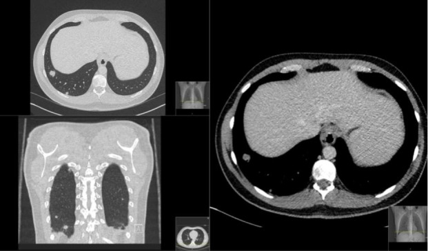
He was noted to have deranged LFTs (alanine aminotransferase 81, alkaline phosphatase 241), and because of this, he underwent further investigation with a liver ultrasound which showed fatty infiltration and an ill-defined, echogenic lesion in the left lower lobe, prompting further imaging with CT abdomen and pelvis (CT AP). The CT AP showed non-alcoholic, fatty liver disease but an otherwise unremarkable abdomen and pelvis. The higher cuts of the CT showed, however, the presence of lower lobe lung nodules. These were further investigated with a CT chest showing several bilateral lower lobe, necrotic, nodules. No lymphadenopathy was seen in the thorax (figure 1).
Figure 1.

Transverse, coronal and sagittal planes of a CT chest showing pulmonary nodules in the right lower lobe.
A CT-guided fine needle aspiration of the nodules showed necrotising granulomatous inflammation. Staining showed no signs of fungi and the acid fast bacilli test for tuberculosis (TB) was negative. Paenibacillus species was isolated from the biopsy, which was thought to be non-clinically significant in this context. He continued to have ongoing symptoms and his regular male partner had also started to have symptoms of fatigue and a mild fever.
Differential diagnosis
He was referred to the TB clinic and HIV team for further management due to the pulmonary nodules seen, and at review, a syphilis serology was requested and was positive with an RPR of 1:256. Of note, he had a previous RPR done in February 2019 which was 1:2, showing good treatment response to his secondary syphilis diagnosed and treated in October 2018.
As such, a diagnosis of secondary syphilis was reached.
Treatment
Following these results, both he and his partner were treated for secondary syphilis with a single dose of benzathine penicillin 2.4 million units after his penicillin allergy had been investigated and ruled out.
Outcome and follow-up
This patient was reviewed 6 weeks post treatment for secondary syphilis, at which point his symptoms had completely resolved. A repeat CT chest showed resolution of his lung nodules and a repeat of his serological RPR test showed an adequate treatment response, with a decrease to 1:64. He had received no other treatment at any point since his initial presentation to the A&E department 2 months prior.
Discussion
Pulmonary involvement has been well documented in cases of tertiary or congenital syphilis; however, it is a rare manifestation of secondary syphilis.5 The clinical presentation can vary greatly from being asymptomatic to causing cough, haemoptysis, dyspnoea and chest pain in association with typical symptoms of secondary syphilis such as rash, fever and lymphadenopathy.3 5 Radiological signs can also vary with case reports of both single and multiple lesions in the upper, middle and lower zones.5–8
Coleman et al proposed five clinical criteria for the diagnosis of pulmonary involvement of secondary syphilis9:
History and physical findings of typical secondary syphilis.
Serological test results positive for syphilis.
Pulmonary abnormalities seen on radiographs with or without associated pulmonary symptoms or signs.
Exclusion of other forms of pulmonary disease when possible by findings of serological tests, sputum smears and cultures, and cytological examination of sputum.
Therapeutic response to antisyphilitic treatment visible on radiographs.
In this case, our patient complied with all criteria. He presented with a widespread macularpapular rash, a positive syphilis serology as well as respiratory signs with pulmonary abnormalities seen on CT. According to the literature data, lung lesions respond well and rapidly to treatment,3 as was shown in this case with symptoms and lung nodules resolving shortly after treatment for secondary syphilis.
In relation to criteria 4, Paenibacillus species was found on the culture of the lung biopsy. Paenibacillus species can be isolated from a wide range of sources including soil and water. They are opportunistic microorganisms and tend to be found in immunocompromised hosts, although with different extents of pathogenicity.10 Our patient was on stable antiretroviral treatment, had a good T CD4+ lymphocyte count (677 cells/mm3) and an undetectable VL at the time of presentation; it would therefore be very unlikely for the Paenibacillus to be causing or contributing to his clinical presentation. Moreover, most species of Paenibacillus would be unlikely to cause necrotising granulomas on FNA and show resistance to penicillin, making it even less likely that total resolution of signs and symptoms would occur after treatment with penicillin only.11
Pulmonary manifestations of secondary syphilis are treated in the same way as other manifestations of secondary syphilis with a single dose of benzathine penicillin 2.4 million units or doxycycline 100 mg two times daily for 14 days. It does not require more intensive treatment (such as intravenous penicillin or doxycycline 200 mg two times per day) which is normally reserved for neuro, ocular or oto syphilis.
Although pulmonary syphilis is rare, syphilis itself is not. This patient was seen by both the emergency medicine and the medical teams over the course of five visits and a differential diagnosis of syphilis had not been considered prior to him being reviewed by the HIV team, despite symptoms of rash, fever, myalgia and deranged LFTs, which are common symptoms of secondary syphilis.1 Consequently, he underwent possibly unnecessary investigations, including an invasive procedure (FNA of lung nodules), as well as suffered from a prolonged period of morbidity.
Learning points.
The presence of pulmonary nodules caused by secondary syphilis is a rare diagnosis, however, syphilis is not.
In any patient presenting with symptoms including myalgia, deranged liver function tests and a rash, a high index of suspicion should be maintained for an underlying diagnosis of syphilis, particularly in patients that are within a high-risk population.
Efforts should be made to improve awareness in the Accident and Emergency department and general medicine physicians of syphilis risk factors and arrays of clinical presentations.
Specialist advice should be sought early so that unnecessary investigations and patient morbidity can be avoided.
Footnotes
Contributors: NB: Complied and written the report. MB: Reviewed and treated the patient and reviewed and modified the report. AP: Reviewed and treated the patient and reviewed and modified the report.
Funding: The authors have not declared a specific grant for this research from any funding agency in the public, commercial or not-for-profit sectors.
Competing interests: None declared.
Provenance and peer review: Not commissioned; externally peer reviewed.
Ethics statements
Patient consent for publication
Obtained.
References
- 1.Hook EW, Peeling RW. Syphilis control — a continuing challenge. N Engl J Med Overseas Ed 2004;351:122–4. 10.1056/NEJMp048126 [DOI] [PubMed] [Google Scholar]
- 2.Amin R. Sexually transmitted infections and screening for Chlamydia in England, 2019: annual official statistics, data to end of December 2019. public health England. Available: https://assets.publishing.service.gov.uk/government/uploads/system/uploads/attachment_data/file/914184/STI_NCSP_report_2019.pdf
- 3.Florencio KBV, Costa ADda, Viana TCdeM, et al. Secondary syphilis with pulmonary involvement mimicking lymphoma: a case report. Rev Soc Bras Med Trop 2019;52:e20190044. 10.1590/0037-8682-0044-2019 [DOI] [PubMed] [Google Scholar]
- 4.Bissessor M, Chen M. Syphilis, the great mimicker, is back. Aust Fam Physician 2009;38:384-7–7. [PubMed] [Google Scholar]
- 5.McCready JB, Skrastins R, Downey JF, et al. Necrotic pulmonary nodules in secondary syphilis. CMAJ 2011;183:E163–6. 10.1503/cmaj.091479 [DOI] [PMC free article] [PubMed] [Google Scholar]
- 6.Kim SJ, Lee J-H, Lee E-S, et al. A case of secondary syphilis presenting as multiple pulmonary nodules. Korean J Intern Med 2013;28:231–5. 10.3904/kjim.2013.28.2.231 [DOI] [PMC free article] [PubMed] [Google Scholar]
- 7.Futami S, Takimoto T, Nakagami F, et al. A lung abscess caused by secondary syphilis - the utility of polymerase chain reaction techniques in transbronchial biopsy: a case report. BMC Infect Dis 2019;19:598. 10.1186/s12879-019-4236-4 [DOI] [PMC free article] [PubMed] [Google Scholar]
- 8.Cholankeril JV, Greenberg AL, Matari HM, et al. Solitary pulmonary nodule in secondary syphilis. Clin Imaging 1992;16:125–8. 10.1016/0899-7071(92)90126-T [DOI] [PubMed] [Google Scholar]
- 9.Coleman DL, McPhee SJ, Ross TF, et al. Secondary syphilis with pulmonary involvement. West J Med 1983;138:875–8. [PMC free article] [PubMed] [Google Scholar]
- 10.Grady EN, MacDonald J, Liu L, et al. Current knowledge and perspectives of Paenibacillus: a review. Microb Cell Fact 2016;15:203. 10.1186/s12934-016-0603-7 [DOI] [PMC free article] [PubMed] [Google Scholar]
- 11.Celandroni F, Salvetti S, Gueye SA, et al. Identification and Pathogenic Potential of Clinical Bacillus and Paenibacillus Isolates. PLoS One 2016;11:e0152831. 10.1371/journal.pone.0152831 [DOI] [PMC free article] [PubMed] [Google Scholar]


