

A 30-year-old woman diagnosed with a neuroendocrine tumour of cecum–appendix (grade 2; Ki67 5%) was treated surgically through an appendectomy and right hemicolectomy 3 years ago. In the follow-up studies with thoracoabdominal computed tomography (CT) (Figure 1), there was no evidence of disease. Due to an elevation of serum chromogranin A, 3 years after the surgery, a Edotreotide Gallium Ga-68 (DOTATOC) positron emission tomography-computed tomography (PET/CT) scan was performed revealing multiple DOTATOC-avid lymphadenopathies (Figure 2) with an intense uptake in the right axillary and supraclavicular region and lower uptake in the laterocervical, left axillary and bilateral ilioinguinal region, with no other 68Ga-DOTATOC-avid lesions in the rest of the study.
Figure 1.
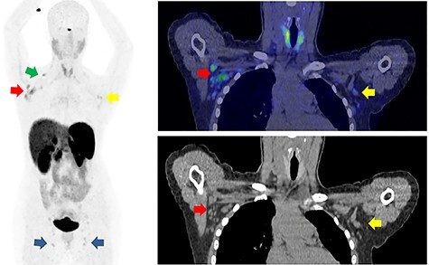
On the left, maximum intensity projection (MIP) image showing multiple 68Ga-DOTATOC-avid lymph nodes with an intense uptake in the right axillary (red arrow) and supraclavicular (green arrow) region and lower uptake in the laterocervical, left axillary (yellow arrow) and bilateral ilioinguinal region (blue arrows). On the right, a coronal view located at the level of axillary lymphadenopathies showing an intense uptake in the right (red arrow) and lower uptake in the left (yellow arrow) lymph nodes.
Figure 2.
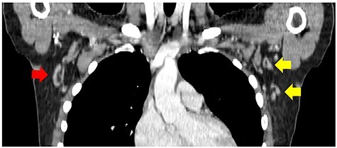
In the follow-up studies with contrast-enhanced TACT, before the elevation of serum chromogranin A, there was no evidence of axillary disease. In the image, performed 2 years after the surgery, few lymph nodes were observed in both axilas without suspicious thickening. TACT, thoracoabdominal computed tomography.
An ultrasound of the right axillary lymphadenopathies showed normal morphology with central hilum and thin cortex. A surgical resection of one of these right axillary nodes revealed a benign reactive lymph node with CD10+ B cell populations identified by flow cytometry. A blood test with complete blood count did not show any alterations and serology was negative for infectious diseases.
The medical history revealed that the patient received two doses of coronavirus disease 2019 (COVID-19) mRNA vaccine Pfizer BioNTech at the right upper arm deltoid muscle at 40 and 62 days prior to PET/CT examination, with posterior pain and swelling on the arm that went away within a few days. The histology, cytology and PET findings suggest immune system activation due to a recent vaccination.
Reactive nodes are a common pitfall of 68Ga-DOTATOC PET/CT and the uptake is explained by the expression of somatostatin receptor 2 (SSTR2) on the leukocytes and macrophages in the node.
Contributor Information
Michal Pudis, Nuclear Medicine - PET department (IDI), Bellvitge University Hospital - IDIBELL, L’Hospitalet de Llobregat, Barcelona, Spain.
José Luis Vercher Conejero, Nuclear Medicine - PET department (IDI), Bellvitge University Hospital - IDIBELL, L’Hospitalet de Llobregat, Barcelona, Spain.
Juan José Martín Marcuartu, Nuclear Medicine - PET department (IDI), Bellvitge University Hospital - IDIBELL, L’Hospitalet de Llobregat, Barcelona, Spain.
Montserrat Cortés Romera, Nuclear Medicine - PET department (IDI), Bellvitge University Hospital - IDIBELL, L’Hospitalet de Llobregat, Barcelona, Spain.