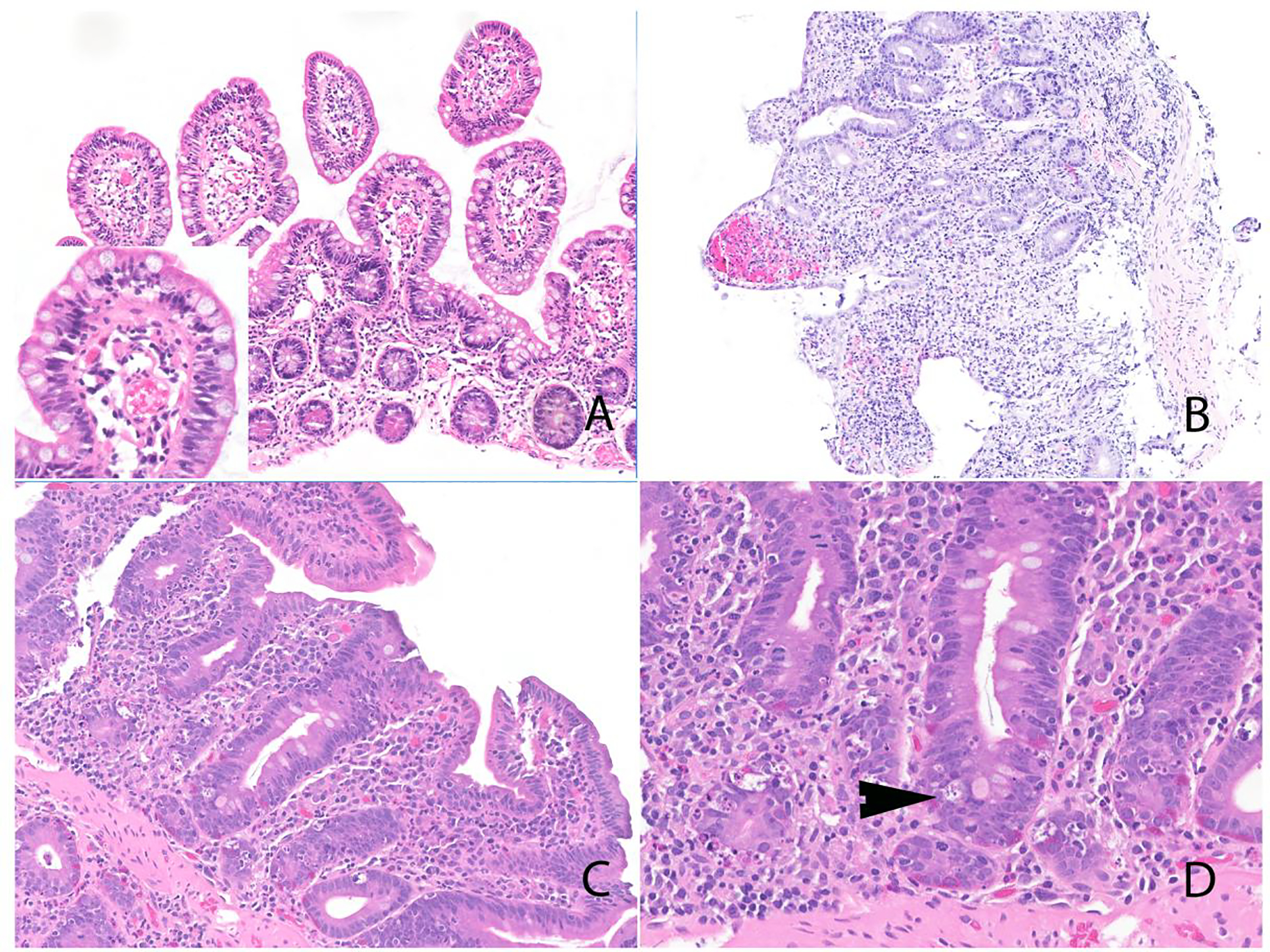
Figure 2:

Histologic features of ICI-associated duodenitis. A) Duodenal mucosa with intact villus:crypt ratio and intraepithelial lymphocytosis; inset shows high-power image. B) Duodenum with villous blunting, diffuse epithelial injury, and increase in mucosal lymphocytes and plasma cells. Notably, there was no increase in intraepithelial lymphocytes. C) Duodenal mucosa with villous blunting and expansion of the mucosa by lymphoid infiltrate. D) Prominent increase in apoptotic activity (arrowhead).
