

Abstract
We present an endovascular repair of aortic transection at distal thoracic level due to traumatic burst fracture. The association of blunt aortic transections and thoracic burst fractures is very rare. Contemporary preferred treatment approach is endovascular aortic repair, because of low mortality rates. The aortic repair procedure should be performed before spinal stabilization surgery. In this case report, we present a 49-year-old male patient with blunt traumatic descending thoracic aortic transection, treated by endovascular aortic repair. In conclusion, the emergent endovascular repair is a preferable method to treat the traumatic distal thoracic aortic transection.
Keywords: Aortic transection, Thoracic burst fracture, Endovascular repair
Introduction
Blunt traumatic aortic injury (BTAI), associated with thoracic spine fracture, is a very rare condition, for which thoracic endovascular aortic repair (TEVAR) is an appropriate treatment choice [1, 2]. Herein, we present a case of thoracic spine fracture, leading to aortic transection, which was treated by endovascular aortic repair.
Case report
A 49-year-old male patient was admitted to the emergency department with multiple injuries after falling down from height. He had no confusion on admission. Physical examination showed a blood pressure of 131/78 mmHg and a pulse of 88 bpm. The patient suffered from abdominal and lower extremities pain. Fecal incontinence was observed as a neurological deficit. Bilateral femoral arteries had weak pulses on physical examination. Immediately, whole-body computed tomography (CT) scan with intravenous iodinated contrast media was obtained because of high-energy trauma. CT scan showed grade-I hepatic laceration, grade-III splenic laceration, bilateral hemothorax, multiple rib fractures, and lumbar transverse process fractures. There was a vertebral burst fracture at T11 level, resulting in aortic transection as grade-IV traumatic aortic injury and periaortic hematoma at the same level (Fig. 1a, b). After an emergent multidisciplinary evaluation, aortic injury was accepted as first ranked life-threatening condition. The patient was transferred to an interventional radiology suite for the TEVAR procedure without loss of time. Under general anesthesia, transfemoral access was secured via 6-French sheath. The origin of the celiac axis was detected by the aid of thoracic angiography, to exclude it while deploying stent graft. Distal landing zone was determined as 3 cm distal to the injury, and just before the celiac axis. In preoperative CT, the proximal diameter of the aorta was measured as 25 mm in the trauma region. 28 mm × 28 mm × 150 mm Valiant thoracic stent graft (Medtronic, Minneapolis, MN) was placed throughout the injury level. Afterwards, balloon dilatation was performed carefully to smooth out the graft. Overall endovascular procedure time was 28 min and 60 ml of contrast agent was used. No complications or endoleaks were found in the angiographic control (Fig. 2). The day after, thoracolumbar spine fixation was carried out from T9 to L, with placement of rods in prone position. Transpedicular listhesis screws were placed at T9–10 and L1 bilaterally, and on the left side at T12 level. Posterolateral fusion with allograft bone was performed from T9 to L1. Postoperative CT scan showed no evidence of endoleak after spinal fixation (Fig. 3). Fecal incontinence resolved after surgery. Prophylactic antibiotics were administered after spine surgery and prophylaxis for deep venous thrombosis was initiated, due to immobilization. Blood pressure, pulse rate, and hemoglobin levels were followed intermittently. For hemothorax, the chest tube was placed on the admission day, and was removed after 10 days. The patient recovered completely and was discharged to home on postoperative day 15, with an appropriate physical rehabilitation program.
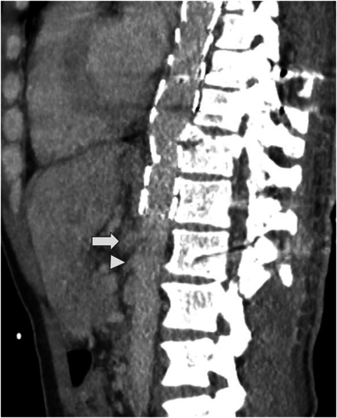
Fig. 1.

Reformatted CT angiography showing distal thoracic aortic transection (arrow) adjacent to vertebral burst fracture (arrowhead) at T11 level (a). Axial image in the same CT angiography reveals aortic transection (arrow) and periaortic hematoma (asterisks) (b)
Fig. 2.

Completion angiogram showing adequate stent-graft placement with no endoleak or kink
Fig. 3.
Postoperative reformatted CT scan shows the aortic stent graft with no endoleak or kink. Note that the celiac artery (arrow) and the superior mesenteric artery (arrowhead) are patent
Discussion
The most common location of aortic injury with blunt trauma is at the isthmus distal to the left subclavian artery. BTAIs of the distal, compared with proximal, thoracic aorta were associated with more thoracic spine injuries, and had a higher mortality owing to associated non-aortic injuries [2]. Only 2% of blunt aortic injuries were reported to be located near the diaphragm [3]. Motor vehicle accidents are the traumatic mechanism most frequently associated with BTAI; however, our patient was admitted to the emergency department after fall [2]. Proximal thoracic aortic endovascular repairs are more complicated and need different techniques including chimney graft or periscope graft [4]. In aortic injuries evaluation, grade-I BTAI may be managed conservatively, grade-II treatment is medical or endovascular repair, and recommendation is endovascular repair for grade III to IV injuries [2]. Endovascular repair was preferred as treatment of choice, because the current case had grade-IV traumatic aortic injury. Sizing for TEVAR in BTAI is still a controversial subject. The size selection of the stent graft was recommended as 10–20% oversizing in relation to the aortic diameter at the attachment site [5]. As sometimes, the patient may be hypovolemic, we may underestimate the diameter, also an oversizing in a weak aorta can be dangerous. However, our case had normal blood pressure. Also, there was a mismatch among the proximal (25 mm) and distal (20 mm) landing zones. Therefore, the size selection of the stent graft was performed as 12% oversizing, based on the proximal diameter of the aorta, to avoid oversizing at the distal site. If the distal landing zone above the celiac axis is inadequate, several endovascular approaches are possible: hybrid procedures with TEVAR and open by-pass to the celiac artery, custom made stent graft with scallop or fenestration for the celiac artery, or intentional coverage of the celiac artery [6]. However, the distal landing zone was sufficient in this case. Vascular damage, recognized and classified by CT and angiography, should be treated before spine stabilization, to avoid uncontrolled bleeding, as in our case. Traction of the aorta and its vessels is realized by dislocated vertebral fractures. Most common vascular damage is the rupture, followed by intimal tear and pseudoaneurysm [7]. Endovascular approach, which has a high success rate, may be an alternative to surgical therapy in suitable lesions in the treatment of traumatic vascular injuries [8]. TEVAR has largely replaced open aortic repair, since its approval by the Food and Drug Administration (FDA), resulting in a 50% reduction in mortality from BTAI [1]. Distal descending aortic transections, related with thoracic spine fractures and treated by endovascular approach, are extremely rare. As an example, Chock et al. presented a case of thoracic fracture resulting in aortic transection; however, that patient was paraplegic after a motor vehicle accident and had aortic pseudoaneurysm [9].
Our patient had aortic transection with fecal incontinence as a neurological deficit. Immediately, successful TEVAR procedure, followed by spinal surgery, was performed. The patient has recovered satisfactorily; however, strict long-term follow-up is necessary.
Conclusion
Aortic injuries caused by vertebral burst fractures at distal thoracic level are very rare. Emergent endovascular repair is a preferable method to treat traumatic distal thoracic aortic transection.
Funding
No funds, grants, or other support was received.
Compliance with ethical standards
Conflict of interest
The authors have no conflicts of interest to declare that are relevant to the content of this article.
Statement of human and animal rights
This case report does not involve animals.
Consent to participate
“Exceptions where it is not necessary to obtain consent: Images such as x rays, laparoscopic images, ultrasound images, brain scans, pathology slides unless there is a concern about identifying information in which case, authors should ensure that consent is obtained.” This case report involves only computed tomography and angiography images in accordance with this rule.
Footnotes
Publisher’s note
Springer Nature remains neutral with regard to jurisdictional claims in published maps and institutional affiliations.
References
- 1.Scalea TM, Feliciano DV, DuBose JJ, Ottochian M, O’Connor JV, Morrison JJ. Blunt thoracic aortic injury: endovascular repair is now the standard. J Am Coll Surg. 2019;228:605–610. doi: 10.1016/j.jamcollsurg.2018.12.022. [DOI] [PubMed] [Google Scholar]
- 2.Sabra MJ, Dennis JW, Allmon JC, Gautam S, Habib J. Identification of unique characteristics and the management of blunt traumatic aortic injuries occurring at unusual locations in the descending thoracic aorta. J Vasc Surg. 2019;69:40–46. doi: 10.1016/j.jvs.2018.06.208. [DOI] [PubMed] [Google Scholar]
- 3.Burkhart HM, Gomez GA, Jacobson LE, Pless JE, Broadie TA. Fatal blunt aortic injuries: a review of 242 autopsy cases. J Trauma. 2001;50:113–115. doi: 10.1097/00005373-200101000-00020. [DOI] [PubMed] [Google Scholar]
- 4.Canyiğit M, Küçüker A, Hıdıroğlu M, Çam A, Şener E. Endovascular aortic repair with periscope graft technique in traumatic aortic transection. Turk Gogus Kalp Dama. 2015;23:129–133. doi: 10.5606/tgkdc.dergisi.2015.10221. [DOI] [Google Scholar]
- 5.Zipfel B, Chiesa R, Kahlberg A, Marone EM, Rousseau H, Kaskarelis I, Riambau V, Coppi G, Ferro C, Sassi C, Esteban C, Mangialardi N, Tealdi DG, Nano G, Schoder M, Funovics M, Buz S, Hetzer R, RESTORE Investigators Endovascular repair of traumatic thoracic aortic injury: final results from the relay endovascular registry for thoracic disease. Ann Thorac Surg. 2014;97:774–780. doi: 10.1016/j.athoracsur.2013.09.034. [DOI] [PubMed] [Google Scholar]
- 6.Falkenberg M, Lönn L, Schroeder T, Delle M. TEVAR and covering the celiac artery. Is it safe or not? J Cardiovasc Surg. 2010;51:177–182. [PubMed] [Google Scholar]
- 7.Santoro G, Ramieri A, Chiarella V, Vigliotta M, Domenicucci M. Thoraco-lumbar fractures with blunt traumatic aortic injury in adult patients: correlations and management. Eur Spine J. 2018;27:248–257. doi: 10.1007/s00586-018-5601-5. [DOI] [PubMed] [Google Scholar]
- 8.Şahin S, Parıldar M, Okbay AM, et al. Endovascular therapeutic approaches and short term results in traumatic vascular injuries. Turk Gogus Kalp Dama. 2006;14:141–145. [Google Scholar]
- 9.Chock MM, Aho J, Naik N, Clarke M, Heller S, Oderich GS. Endovascular treatment of distal thoracic aortic transection associated with severe thoracolumbar spinal fracture. Vascular. 2015;23:550–552. doi: 10.1177/1708538114560458. [DOI] [PubMed] [Google Scholar]