Abstract
Structural failure of mechanical heart valve was a known feature when it was evolving in the 1960s and 1970s. With the advent of pyrolytic carbon and a better design, it is a rare entity with present valves. We report a case of disc fracture leading to acute mitral regurgitation in TTK Chitra heart valve prosthesis (CHVP) (TTK Healthcare Limited, India) heart valve, 6 years after its implantation in mitral position.
Keywords: TTK Chitra heart valve prosthesis, Disc fracture, Structural valve dysfunction
Introduction
Valve replacement with mechanical prosthesis is being done since decades. Late mechanical prosthetic valve dysfunction usually occurs due to non-structural valve failure, generally precipitated by infection, thrombosis, or in-growth of pannus. The incidence of structural valve dysfunction in mechanical valve caused by material wear and tear is very rare, and when they occur, it often results in catastrophic events. Björk-Shiley convexo-concave valves have had a high incidence of structural dysfunction [1]. Ball fracture and cloth wear and tear have been reported with Starr-Edwards ball and cage valve, leading to valve regurgitation in its early models, until the introduction of models 1260 and 6120 [2, 3]. Chitra heart valve prosthesis (CHVP), a tilting disc valve developed in India, is being implanted since 1990. Our experience with CHVP dates back to 1992, when we first implanted a CHVP in the mitral position. We report a unique case of disc fracture of mitral CHVP, 6 years after implantation.
Case report
A 32-year-old female underwent mitral valve replacement (MVR) with 29-mm CHVP for rheumatic mitral re-stenosis in 2012 (status post-balloon mitral valvotomy in 1997). Her pre-discharge echocardiogram showed normal prosthetic valve function with no prosthetic valve regurgitation and she was discharged home on oral coumadin. She was on regular follow-up and in New York Heart Association (NYHA) class I. Routine annual echocardiography done at subsequent follow-ups showed normal prosthetic valve function.
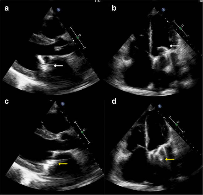
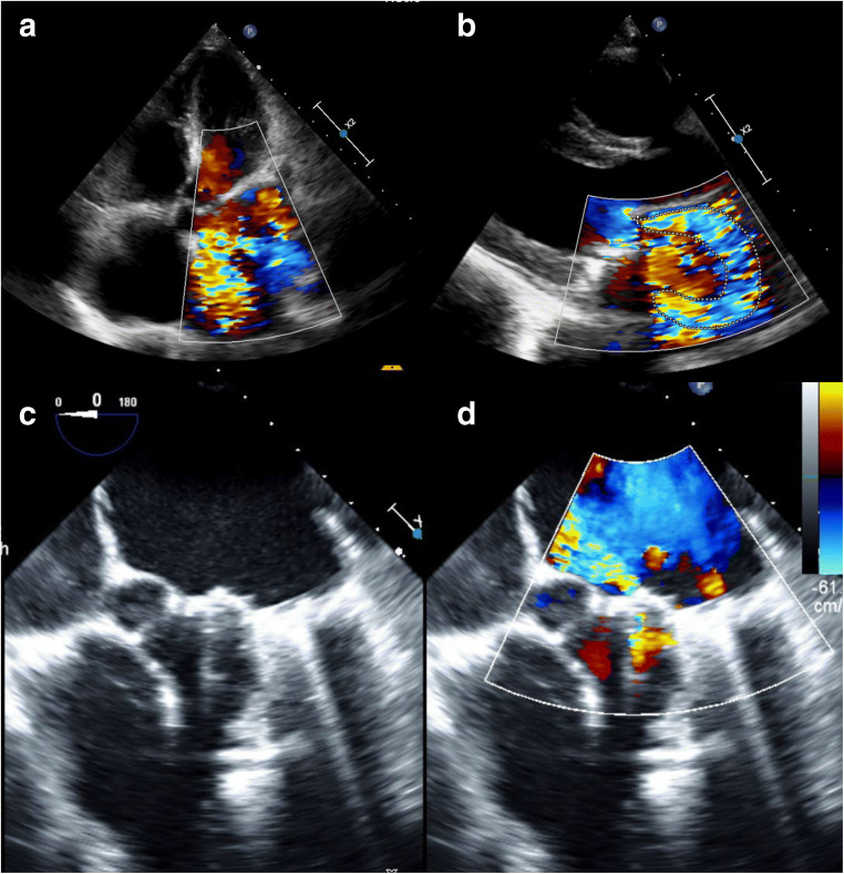
In July 2018, she presented to us with complaints of dyspnoea (NYHA class III) for 10 days duration. On general clinical examination, she had features of congestive cardiac failure (CCF). On auscultation, prosthetic valve click was audible, however, with a grade 3/6 pan-systolic murmur. Transthoracic echocardiography (TTE) demonstrated normal disc movement with severe prosthetic valve regurgitation, severe tricuspid regurgitation and severe pulmonary arterial hypertension (right ventricular systolic pressure: 87 mm of Hg). The TTE also showed an unusual component of the mitral prosthesis protruding into the left atrium (Figs. 1 and 2). Initially, it was suspected to be severe para-valvular regurgitation. As she was symptomatic and worsened to NYHA-IV, she was taken up for an emergency redo MVR. Intraoperative transesophageal echocardiogram (TEE), before going on cardiopulmonary bypass (CPB), showed severe intra-valvular regurgitation with suspicion of structural valve dysfunction.
Fig. 1.
Parasternal long axis view (a) and apical 4 chamber view (b) showing complete opening of the disc with no restriction of movement (white arrow). Parasternal long axis view (c) and apical 4 chamber view (d) showing an unusual component of the mitral prosthesis protruding into the left atrium (yellow arrow)
Fig. 2.
a, b Pre-operative echocardiogram showing severe prosthetic valve regurgitation. c Intraoperative transesophageal echocardiogram showing mitral valve prosthesis in systole with a component of the mitral prosthesis protruding into the left atrium. d Intraoperative transesophageal echocardiogram showing severe prosthetic valve regurgitation, possibly intra-valvular
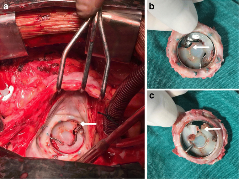
After an uneventful redo-sternotomy, CPB was instituted with standard aorto-bi-caval cannulation; left atriotomy was performed and the prosthetic valve inspected. The CHVP mitral mechanical prosthesis was in situ, with well-endothelialised sewing rim. There was a transverse fracture of the CHVP disc, resulting in a severe intra-valvular leak, thereby confirming the TEE findings (Fig. 3 a–c). The valve was explanted in toto and examined. Although the disc was fractured, no part of the disc was missing. A new 31-mm CHVP was implanted. Teflon felt reduction annuloplasty was performed for severe tricuspid regurgitation through a right atriotomy. The patient was weaned off CPB in sinus rhythm. Intraoperative TEE, post CPB, showed normal prosthetic valve movement, with no intra- or para-valvular regurgitation with trivial to mild tricuspid regurgitation. She made an uneventful recovery and was discharged home in a stable condition. She is doing well at 2-year follow-up and is in NYHA functional class I. TTE performed at 2 years post redo-surgery revealed normally functioning mitral mechanical prosthesis.
Fig. 3.
Intraoperative images. a The TTK Chitra mitral prosthesis in situ, with well endothelialisedsewing rim. b Superior surface of the explanted valve. c Inferior surface of the explanted valve. The white arrow in each image shows the transverse fracture of the disc occluder
Discussion
The invention and implantation of a valve prosthesis for valvular heart disease were a game changer in the early 1960s. However, the research for an ideal prosthetic valve continues until date.
The first mechanical prosthesis for the heart valve was the ball and cage valve, developed and implanted by Albert Starr and Lowell Edwards on September 21, 1960 [4], which evolved over time to mono-leaflet valves and eventually to the currently used bi-leaflet valves. Although we do not have the ideal mechanical prosthesis, as they need lifelong anticoagulation, mechanical heart valves are primarily indicated in patients less than 50 years of age [5]. Currently, CHVP is the only tilting mono-disc valve that is being used.
Mechanical prosthetic valve regurgitation can be either intra-valvular or para-valvular. Intra-valvular regurgitation in a mechanical valve can be either immediate or delayed. In case of an immediate presentation after replacement, regurgitation may result due to incomplete disc closure, as a result of sub-valvular chordae or suture hindering the occluder. However, delayed regurgitation is usually due to a thrombus or pannus resulting in restricted leaflet movement. Infective endocarditis can also cause delayed intra-valvular regurgitation. However, these should not be mistaken for the signature ‘washing jets’, which prevents stagnation of blood or clot formation on the disc and struts of the valve [2].
Structural damage to the valve can be caused by material wear and tear. It may be due to (a) disc dislodgement, (b) disc fracture or (c) strut fracture. Mechanical damage to the implanted valve is rare, but when it occurs, it causes several complications including risk to life. Bjork-Shiley convexo-concave valve model had several incidences of transverse disc fracture, and also strut fracture, leading to embolism and hence was removed from the market in 1986 [6, 7].
The patients may present varyingly from no symptoms to cardiac failure and even with sudden cardiac arrest. Though TTE is most commonly used for the diagnosis, TEE is more sensitive. The exact mechanism and cause of structural failure are often difficult to identify, but may be thrombus versus pannus or intra- versus para-valvular regurgitation.
CHVP is a single leaflet valve with a housing made of Haynes-25 alloy (cobalt-nickel-chromium-tungsten alloy), ultra-high molecular weight (UHMW) polyethylene rotatable disc occluder, and a sewing ring made of polyester. No structural valve dysfunction has been reported for CHVP until date.
In our case, the patient had been on regular follow-up. The TTE done during her previous (5 years post-implant) review also showed normal prosthetic valve—both structurally and functionally. However, she came back a year later (6 years post-implant) with features of CCF and was diagnosed to have severe prosthetic valve regurgitation. Initial suspicion was in favour of infective endocarditis or restricted valve movements due to thrombus or pannus. As she had no fever, no vegetations in TTE, and no laboratory parameters suggestive of infection, prosthetic valve endocarditis was ruled out.
Multiple factors have been hypothesised to cause leaflet fracture:
Inadequate compliance of the sewing ring causing reduced shock absorption as the leaflet moves.
Surgical mishandling during implantation.
The pressure difference in the blood flow, leading to unstable air bubble formation, releases energy on collapsing (high frequency pressure fluctuations) leading to the formation of microcracks in the disc—cavitation [8, 9].
The UHMW polyethylene disc is manufactured in such a way so that the disc rotates on its own axis, and is also able to slide in its plane, thus distributing the stress all over the plane while in operation [10]. We are unable to explain the mechanism which caused the disc fracture in our case. We presume that there must have been some thrombus impairing the rotation of the disc on its axis, which in turn led to disc fracture; however, this presumption was not evident on examination of the valve.
Fracture of the leaflet is a dreaded complication as the fractured bit can migrate into the systemic circulation. The clinical signs and symptoms of structural failure are exactly similar to those of acute valvular regurgitation, which includes features of CCF. Leaflet fracture can be differentiated from valve thrombosis by abrupt symptom onset. Immediate medical attention and emergency surgical replacement of the prosthesis are essential. In our case, fortunately, there was no disc embolization, and the patient presented with features of acute mitral regurgitation with heart failure and hence was taken up for emergency redo mitral valve replacement.
Conclusion
Structural failure had been a reported problem with the older generation of prosthetic heart valves. However, it is a very rare occurrence with the current generation of mechanical prosthetic valves. There are no reports of a disc fracture in the CHVP prosthesis. In a patient with a mechanical prosthesis, who presents with sudden decompensation and acute onset of symptoms; although rare, the serious complication of structural valve failure must be suspected, besides infective endocarditis and prosthetic valve thrombosis. Early diagnosis and appropriate treatment is the key to success in such rare cases.
Funding
None.
Compliance with ethical standards
Conflict of interest
The authors declare that there is no conflict of interest.
Statement of human and animal rights
This article does not contain any studies with animals performed by any of the authors.
Informed consent
Informed consent was obtained from the patient.
Footnotes
Publisher’s note
Springer Nature remains neutral with regard to jurisdictional claims in published maps and institutional affiliations.
References
- 1.Gott VL, Alejo DE, Cameron DE. Mechanical heart valves: 50 years of evolution. Ann Thorac Surg. 2003;76:S2230–S2239. doi: 10.1016/j.athoracsur.2003.09.002. [DOI] [PubMed] [Google Scholar]
- 2.Misawa Y. Valve-related complications after mechanical heart valve implantation. Surg Today. 2015;45:1205–1209. doi: 10.1007/s00595-014-1104-0. [DOI] [PMC free article] [PubMed] [Google Scholar]
- 3.Miller DC, Oyer PE, Mitchell RS, et al. Performance characteristics of the Starr-Edwards Model 1260 aortic valve prosthesis beyond ten years. J Thorac Cardiovasc Surg. 1984;88:193–207. doi: 10.1016/S0022-5223(19)38353-9. [DOI] [PubMed] [Google Scholar]
- 4.Russo M, Taramasso M, Guidotti A, et al. The evolution of surgical valves. Cardiovasc Med. 2017;20:285–292. doi: 10.4414/cvm.2017.00532. [DOI] [Google Scholar]
- 5.Nishimura RA, Otto CM, Bonow RO, Carabello BA, Erwin JP, III, Fleisher LA, Jneid H, Mack MJ, McLeod CJ, O’Gara PT, Rigolin VH, Sundt TM, III, Thompson A. 2017 AHA/ACC focused Update of the 2014 AHA/ACC Guideline for the Management of patients with valvular heart disease: a report of the American College of Cardiology/American Heart Association Task Force on Clinical Practice Guidelines. JAm Coll Cardiol. 2017;70:252–289. doi: 10.1016/j.jacc.2017.03.011. [DOI] [PubMed] [Google Scholar]
- 6.González-Santos JM, Arnáiz-García ME, Dalmau-Sorlí MJ, et al. Complete transversal disc fracture in a Björk-Shiley delrin mitral valve prosthesis 43 years after implantation. Ann Thorac Surg. 2016;102:e283–5. [DOI] [PubMed]
- 7.Davis PK, Myers JL, Pennock JL, Thiele BL. Strut fracture and disc embolization in Björk-Shiley mitral valve prostheses: diagnosis and management. Ann Thorac Surg. 1985;40:65–8. [DOI] [PubMed]
- 8.van Steenbergen GGJ, Tsang QHY, van der Heide SM, Verkroost MWA, Li WWL, Morshuis WJ. Spontaneous leaflet fracture resulting in embolization from mechanical valve prostheses. J Card Surg. 2019;34:124–130. doi: 10.1111/jocs.13975. [DOI] [PMC free article] [PubMed] [Google Scholar]
- 9.Lee WY, Hong JM, Bae J-W. A Case of fatal mechanical mitral valve leaflet fracture embolization - a case report. Korean J Crit Care Med. 2011;26:101.
- 10.Vayalappil MC, Bhuvaneswar GS. The saga of Chitra heart valve prosthesis. 2005. 10.13140/2.1.4281.2488.


