Abstract
Objectives:
To investigate the predictability of arch expansion using Invisalign.
Materials and Methods:
Sixty-four adult white patients were selected to be part of this retrospective study. Pre- and posttreatment digital models created from an iTero scan were obtained from a single orthodontist practitioner. Digital models from Clincheck were also obtained from Align Technology. Linear values of upper and lower arch widths were measured for canines, premolars, and first molars at two different points: lingual gingival margins and cusp tips. A paired t-test was used to compare expansion planned on Clincheck with the posttreatment measurements. Variance ratio tests were used to determine if a larger change planned was associated with larger error.
Results:
For every maxillary measurement, there was a statistically significant difference between Clincheck and final outcome (P < .05), with prediction worsening toward the posterior region of the arch. For the lower arch measurements at the gingival margin, there was a statistically significant difference between the Clincheck planned expansion and the final outcome (P < .05). Points measured at the cusp tips of the lower arch teeth showed nonstatistically significant differences between Clincheck prediction and the final outcome (P > .05). Variance ratios for upper and lower arches were significant (P < .05).
Conclusions:
The mean accuracy of expansion planned with Invisalign for the maxilla was 72.8%. The lower arch presented an overall accuracy of 87.7%. Clincheck overestimates expansion by body movement; more tipping is observed. Overcorrection of expansion in the posterior region of the maxillary arch seems appropriate.
Keywords: Invisalign, Predictability
INTRODUCTION
Invisalign involves a series of plastic aligners that move the teeth. The aligners are removable and are made of 0.75-mm-thick polyurethane.1,2 Patients are to wear an aligner for a period of 1–2 weeks and then change to the next one. Each aligner is programmed to produce a precise movement on a tooth of about 0.15–0.25 mm.3 The stereolithographic technology is used to fabricate custom aligners from an impression or an intraoral digital image scanned in the dental office. Patient compliance is mandatory to achieve good results with Invisalign. It is important for patients to wear their aligners 22 hours a day or more.4
Arch expansion is possible with Invisalign and may be required as a perceived need to improve the esthetics of the smile by broadening the dental arches5 or as a mechanism to create space for resolution of crowding.6,7 It can also be used as a way of correcting dentoalveolar posterior crossbites.8
In their 2001 publication on treatment of complex malocclusion using Invisalign, Boyd and Vlaskalic3 reported that buccal expansion can be achieved to alleviate crowding or to modify the arch form. The range of expansion would be 2–4 mm. In an article by Ali et al.2 in 2012, it was stated that dentoalveolar expansion is possible with Invisalign and can be an alternative to interproximal reduction. According to the same authors, expansion of the dental arches should be limited to 2–3 mm of arch width per quadrant to minimize the risk of relapse and gingival recession. Malik et al.4 in 2013 reported that expansion is an indication to use Invisalign when having to resolve 1–5 mm of crowding. In the same article, dental expansion using Invisalign was also recommended for blocked out teeth.
There are limited data on the amount of discrepancy between predicted and actual achieved movements with Invisalign.9 In a prospective clinical study by Kravitz et al.10 in 2009, the mean accuracy of tooth movement in the anterior region was found to be 41% with Invisalign. An internal study from Align Technology found that one should expect about 80% of tooth movement seen on Clincheck.11
Given the scarcity of data in the literature, especially regarding posterior transverse changes, the present study aims at comparing Clincheck transverse measurements with the actual clinical outcome. Knowing the accuracy of the software at predicting changes could help the practitioner to anticipate the need of overcorrection, thereby reducing refinements, midcourse corrections, and treatment time.
MATERIALS AND METHODS
An ethics approval certificate was issued by the Bannatyne Campus Research Ethics Boards at the University of Manitoba on May 14, 2014, before the beginning of this retrospective study.
An assumption was made that 70% of the expansion predicted would be achieved with a margin of error of 10%. Sample size calculation was based on a power of 0.8 and a confidence interval of 95%, and estimated it to be of 64 patients. The sample was obtained from a single specialist at an orthodontic practice in Adelaide, Australia. Patient age and gender as well as three .stl files required (pretreatment, predicted treatment, and posttreatment) were recorded. The sample was composed of 41 women and 23 men with a mean age of 31.2 years and a range from 18 to 61 years. Twenty of these patients had a dentoalveolar crossbite involving at least one tooth, mainly premolars. All patients had both arches treated with Invisalign only. The mean treatment duration was 56 weeks. The study looked only at the first round of aligners. No refinement was included.
Records were randomly obtained for patients who met the inclusion/exclusion criteria. Patients whose growth was complete (older than 18 years), treated with nonextraction, and without any auxiliaries other than Invisalign attachments were included in the study. Patients had to wear each aligner for 2 weeks. Compliance was assessed by the practitioner. Those with missing teeth or who had interproximal reduction prescribed distal to the canines were excluded from the study. Any patient needing midcourse correction or treated after the introduction of the Smart track material was also excluded.
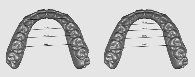
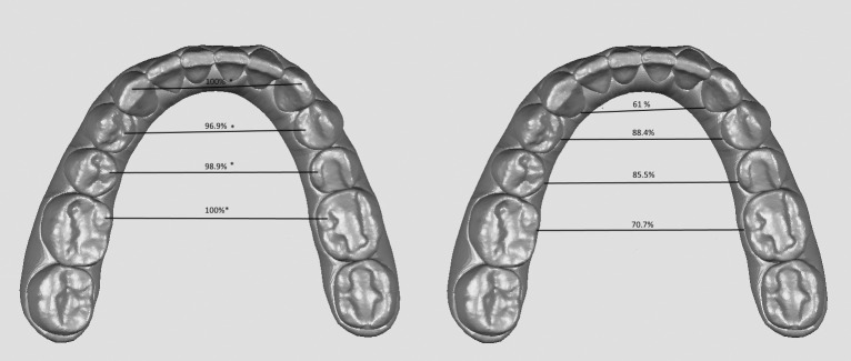
Pre- and posttreatment digital models (.stl files), created from an iTero scan, were obtained from the 64 patients in the study. Digital models from Clincheck were also obtained from Align Technology to measure the planning accuracy. Patient confidential data were deidentified by an assistant who assigned a unique number for each patient. The digital .stl files from the iTero scanner and Clincheck were uploaded in Geomagic Qualify (Geomagic, Morissville, NC) software. Arch width measurements using the software's digital caliper were recorded by the primary investigator. Linear values of upper and lower arch widths were measured at the cusp tip and most lingual points at the gingival margin of canines, premolars, and first molars according to the illustrations in Figures 1 and 2.
Figure 1.
Landmarks and accuracy of transverse measurements in the maxillary arch. (Left) Cusp tip. (Right) Cervical margins.
Figure 2.
Landmarks and accuracy of transverse measurements in the maxillary arch. (Left) Cusp tip. (Right) Cervical margins.
Interdental width linear measurements were recorded pretreatment, based on the Clincheck plan and posttreatment (or before refinement). In cases of wear facets, an estimation of the cusp tip point was used.12
Digital models were measured with Geomagic Qualify by the principal investigator. To test the intra- and interexaminer reliability, 20% of the sample size was randomly chosen to be measured again 2 weeks after the first assessment.13 The reliability of the measures was assessed by the mean of an interclass correlation coefficient (ICC) with Shrout-Fleiss derivation. SPSS (Statistical Package for the Social Sciences), version 18.0 (IBM Corp, Chicago, Ill) was the chosen software to analyze the data. Normality of distribution was assessed using the QQ-plots method. A paired t-test was selected to compare the Clincheck prediction with the posttreatment measurements. The level of significance was set at 5%. The amount of change achieved was also calculated in percentage10: percentage of accuracy = 100% − [(lpredicted – achievedl)/lachievedl] × 100%. This equation ensures that calculated values do not exceed 100%. The variance ratio test was used to determine if larger planned changes were correlated with larger inaccuracies.14 This test was chosen to compare the amount of expansion requested (Clincheck pretreatment) to the amount of expansion obtained (Clincheck posttreatment). A simple correlation could not be used because Clincheck measurements were present in both equations.
RESULTS
The ICC test showed almost perfect agreement with regard to interrater reliability with a score of .97. The value for the intrarater reliability also showed almost perfect agreement with a value of .98.15 A mean of planned transverse changes was calculated as well as the mean difference between the amount of change planned and the final outcome (Tables 1 and 2).
Table 1.
Predictability of Changes for Maxillary Measurements
| Tooth Type |
Predicted Change as per Clincheck, mm |
SD |
95% CI |
Mean Difference (Posttreatment, Clincheck) |
SD |
95% CI |
Clincheck vs Posttreatment P Value |
Accuracy of Change, % |
| Canine tip | 1.92 | 2.05 | 1.42–2.42 | 0.22 | 0.74 | 0.03–0.40 | .0225* | 88.7 |
| Canine gingival | 1.85 | 1.76 | 1.41–2.28 | 0.6 | 1.02 | 0.34–0.85 | <.001* | 67.8 |
| First premolar tip | 3.77 | 2.34 | 3.20–4.35 | 0.58 | 1.14 | 0.03–0.58 | .001* | 84.7 |
| First premolar gingival | 3.36 | 2.04 | 2.86–3.86 | 1.09 | 1.22 | 0.78–1.39 | <.001* | 67.6 |
| Second premolar tip | 4.11 | 3.06 | 3.36–4.85 | 0.75 | 1.54 | 0.37–1.13 | <.001* | 81.7 |
| Second premolar gingival | 3.45 | 2.56 | 2.83–4.08 | 1.3 | 1.61 | 0.90–1.7 | <.001* | 62.3 |
| First molar tip | 3.28 | 3.13 | 2.51–4.04 | 0.77 | 1.84 | 0.31–1.23 | .001* | 76.6 |
| First molar gingival | 3.02 | 2.58 | 2.39–3.65 | 1.42 | 1.9 | 0.95–1.90 | .001* | 52.9 |
P < .05.
Table 2.
Predictability of Changes for Mandibular Measurements
| Tooth Type |
Predicted Change as per Clincheck, mm |
SD |
95%CI |
Mean Difference (Posttreatment, Clincheck) |
SD |
95% CI |
Clincheck vs. Posttreatment P Value |
Accuracy of Change, % |
| Canine tip | 1.39 | 1.84 | 0.93 to 1.84 | −0.08 | 0.81 | −0.28 to 0.12 | .430 | 100 |
| Canine gingival | 1.66 | 2.12 | 1.14 to 2.18 | 0.65 | 1.01 | 0.39 to 0.90 | <.001* | 61 |
| First premolar tip | 2.47 | 2.88 | 1.77 to 3.18 | 0.07 | 0.96 | −0.16 to 0.32 | .520 | 96.9 |
| First premolar gingival | 2.30 | 2.64 | 1.65 to 2.94 | 0.27 | 1.00 | 0.02 to 0.52 | .037* | 88.4 |
| Second premolar tip | 3.07 | 3.12 | 2.30 to 3.83 | 0.07 | 1.15 | −0.25 to 0.32 | .810 | 98.9 |
| Second premolar gingival | 2.58 | 2.51 | 1.96 to 3.20 | 0.38 | 1.16 | 0.09 to 0.66 | .012* | 85.5 |
| First molar tip | 2.14 | 2.38 | 1.56 to 2.72 | 0.03 | 1.33 | −0.36 to 0.3- | .849 | 100 |
| First molar gingival | 1.84 | 1.99 | 1.36 to 2.33 | 0.54 | 1.34 | 0.21 to 0.87 | .002* | 70.7 |
P < .05.
Table 1 shows that for every maxillary measurement, there was a statistically significant difference between Clincheck and the final outcome. The lingual gingival margin at the first molar was the area with less accuracy (52.9%) corresponding to a mean difference of 1.42 ± 1.90 mm (Figure 1). The most reliable area to predict transverse changes in the maxilla was the canine cusp tip with 88.9% of the change achieved, a mean difference of 0.22 ± 0.74 mm. Table 2 shows that for every lower arch measurement at the gingival margin, there was a statistically significant difference between the Clincheck plan and the final outcome. Prediction accuracy ranged from 61.0% (canine) to 88.4% (first premolar; Figure 2). The mean difference between Clincheck and the final outcome on these teeth ranged from 0.27 ± 1.00 mm (first premolar) to 0.65 ± 1.01 mm (canine). Measurements at the cusp tips in the lower arch showed nonstatistically significant differences between Clincheck and the final outcome. Variance was not equal for any of the measurements done either upper or lower, meaning that the amount of change planned was not associated with prediction error. All of the P values were recorded as significant for the variance ratio tests (Tables 3 and 4).
Table 3.
Variance Ratios for Maxillary Measurements
| Tooth Type |
Variance Ratio |
P Value |
| Canine tip | 7.66 | ≤.001* |
| Canine gingival | 2.96 | ≤.001* |
| First premolar tip | 4.25 | ≤.001* |
| First premolar gingival | 2.82 | ≤.001* |
| Second premolar tip | 3.96 | ≤.001* |
| Second premolar gingival | 2.52 | ≤.001* |
| First molar tip | 2.91 | ≤.001* |
| First molar gingival | 1.85 | .02* |
P < .05.
Table 4.
Variance Ratios for Mandibular Measurements
| Tooth Type |
Variance Ratio |
P Value |
| Canine tip | 5.12 | ≤.001* |
| Canine gingival | 4.43 | ≤.001* |
| First premolar tip | 8.91 | ≤.001* |
| First premolar gingival | 6.96 | ≤.001* |
| Second premolar tip | 7.32 | ≤.001* |
| Second premolar gingival | 4.72 | ≤.001* |
| First molar tip | 3.2 | ≤.001* |
| First molar gingival | 2.22 | .002* |
P < .05.
DISCUSSION
The purpose of this study was to assess the reliability of Invisalign Clincheck when planning for transverse changes. There are only a limited number of studies comparing Clincheck to the final outcome of orthodontic treatment.10–12 From these studies, only two assessed expansion.10,11 To our knowledge, as of July 2015, no study evaluated the accuracy of posterior transverse changes with Invisalign.
An adult population was chosen to participate in this study to avoid bias due to normal transverse growth of the jaws. As reported by Bishara et al.,16 the practitioner should not expect arch width change when the eruption of the permanent dentition has already been completed.
Our results showed large variability depending on the tooth studied, the point of measurement, and the arch studied. Transverse changes in the upper arch were found to be 72.8% accurate overall, 82.9% at the cusp tips, and 62.7% at the gingival margins. The most accurate prediction was for canine cusp tip, with an accuracy of 88.7%, meaning that 0.22 mm of the 1.92-mm expansion requested was not achieved (Table 1). This difference was statistically significant but may not be clinically relevant. On the other hand, at the first molar gingival margin, the accuracy was 52.9%. Almost half of the changes planned at the gingival margin of the upper first molars did not occur (1.42 mm of 3.02 mm planned). At the cusp tip of the same tooth, 0.77 mm of the 3.28 mm planned were not achieved with the aligners, an accuracy of 76.6%. Overall, the upper first molars were the teeth with the lowest tracking accuracy. In fact, there was a trend observed in the upper arch showing that Clincheck accuracy decreases when moving posteriorly into the arch. This difference is most likely the result of root anatomy, cortical plate thickness, higher mastication loading, and greater soft tissue resistance from the cheeks in the posterior region.
The lower arch presented an overall accuracy of 87.7%, 98.9% at the cusp tips and 76.4% at the gingival margins. Cusp tip posttreatment measurements were all found to be close to 100% accuracy (Table 2). In terms of gingival margin measurements, the lower canines were found to be the area with the biggest prediction error with 62% accuracy, followed by the molars (70.7%), the second premolar (85.5%), and the first premolar (88.4%). These differences were all found to be statistically significant. Results were different at the cusp tips, where all the differences between Clincheck and the clinical outcome were not statistically significant. This means that the software was able to predict accurately the changes that occurred. This better result may be explained by the fact that the amount of change requested in the lower arch is usually less than in the upper arch. Also, resistance is reduced given the fact that the upper arch is being expanded simultaneously. A 2015 systematic review concluded that clear aligners are able to control posterior buccolingual inclination, although the evidence found was considered weak.17
In 2009, Kravitz et al.10 reported 40.5% accuracy of tooth movement for labial expansion of the anterior teeth. One of their recommendations was to treat cases with severe lower crowding mostly by interproximal reduction (IPR) instead of dentoalveolar expansion. This recommendation comes from the finding that retraction is more accurate than dentoalveolar expansion of the lower anterior teeth. The higher accuracy with Clincheck in our study may be explained by the fact that the database is coming from a single well-experienced practitioner who has been working with the Invisalign system for many years. Also, this study was completed a few years later than the study mentioned above. New versions of the software, changes in the algorithm, and improvements to the technique may also explain why the accuracy of Clincheck was found to be higher in our study. A new study looking at the accuracy of anterior teeth expansion would help validate this assumption.
It was also found that greater expansion planned with Clincheck was not associated with less accuracy. In other words, the tracking will not necessarily be better if the amount of expansion requested is less. This finding needs to be interpreted with caution as the amount of expansion in this study was small overall. The largest amount of expansion was of 4.11 mm at the upper second premolar cusp tip. Lack of association may also derive from the experience of the clinician. Knowing the limitations of the appliance helps to minimize errors. According to Phan and Ling,18 the greater success is obtained with Invisalign when treating nonskeletally constricted arches by tipping movement, which is usually in the range between 0.1 and 5.0 mm.18 Also, the lowest expansion requested was 1.39 mm at the cusp tip of the lower canine. This correlates with recommendations from Schulof et al.,19 who reported that expansion of the mandibular intercanine width poses the greatest risk of relapse following treatment.
Overcorrection aligners did not lead to bias in this study because every digital model requested to Align Technology was obtained before the overcorrection trays. No further expansion during overcorrection was planned for any of the patients included in the study. The clinician may have planned for expansion overcorrection during Clincheck knowing that all of it could not be obtained as requested. This was one of the recommendations mentioned by Tuncay in 200611 study to think of overcorrection and even refinement to achieve the best result.
Geomagic software was selected to make the linear measurements required for this study. The accuracy of Geomagic to make linear measurements was demonstrated by Sousa et al.20 in 2012. Because of the very high values of ICC tests, it was possible to ascertain that linear measurements to assess transverse changes in digital dental arches is reproducible using Geomagic. The abilities of the software include the possibility to rotate and magnify the digitals models, which may explain such high values for the ICC test.
Two landmarks (cusp tip and gingival margin) were selected to represent transverse tipping and bodily movement, respectively. Our data suggest that Clincheck is predicting more bodily movement than Invisalign actually can achieve. This conclusion is similar to that of Pavoni et al.21 in 2011, who compared dentoalveolar expansion between Invisalign and the Damon system.
It has been reported that as much as 70% to 80% of the patients treated with Invisalign would require a midcourse correction or a refinement.10 These numbers suggest that the accuracy of treatment planning with Clincheck is low. Midcourse corrections or refinements have some consequences: longer treatment time for the patient, increased chair time and costs for the orthodontist, and higher manufacturing demand for Align Technology. Some of these inaccuracies can derive from the practitioner inexperience with the technique, the software, or the low level of patient compliance. Studies on the accuracy of Clincheck may help to reduce the rate of midcourse corrections and refinements.
Pretreatment arch form can possibly be used as a predictor of how much dentoalveolar expansion can be achieved during treatment. Lingually tipped teeth might offer more magnitude of expansion. This assumption would have to be verified in a future study.
When it comes to planning expansion on Clincheck, overcorrection needs to be considered, especially in the posterior region of the maxillary arch. Auxiliaries such as crossbite elastics may be used to improve the transverse relationship of the teeth.17 Conventional expansion appliances prescribed before Invisalign are another treatment modality that can be used.
CONCLUSIONS
When dentoalveolar expansion is planned with Invisalign, the mean accuracy for the maxilla is 72.8%: 82.9% at the cusp tips and 62.7% at the gingival margins.
Invisalign becomes less accurate going from the anterior to the posterior region.
When dentoalveolar expansion is planned, the lower arch presented an overall accuracy of 87.7%: 98.9% for the cusp tips and 76.4% for the gingival margins. All cusp tip posttreatment measurements were found to have a nonstatistically significant difference when compared with Clincheck.
Clincheck prediction of expansion involves more bodily movement of the teeth than can be seen clinically. More dental tipping was observed.
Careful planning with overcorrection and other auxiliary methods of expansion may help reduce the rate of midcourse corrections and refinements, especially in the posterior region of the maxilla.
ACKNOWLEDGMENT
A sincere thank you to Dr Grant Duncan and his staff at Duncan Orthodontics in Adelaide, Australia, for their assistance during this investigation.
REFERENCES
- 1.Lagravere MO, Flores-Mir C. The treatment effects of Invisalign orthodontic aligners: a systematic review. J Am Dent Assoc. 2005;136:1724–1729. doi: 10.14219/jada.archive.2005.0117. [DOI] [PubMed] [Google Scholar]
- 2.Ali SA, Miethke HR. Invisalign, an innovative invisible orthodontic appliance to correct malocclusions: advantages and limitations. Dent Update. 2012;39:254–256. doi: 10.12968/denu.2012.39.4.254. 258–260. [DOI] [PubMed] [Google Scholar]
- 3.Vlaskalic V, Boyd R. Orthodontic treatment of a mildly crowded malocclusion using the Invisalign system. Aust Orthod J. 2001;17:41–46. [PubMed] [Google Scholar]
- 4.Malik OH, McMullin A, Waring DT. Invisible orthodontics part 1: Invisalign. Dent Update. 2013;207–210:213–215. doi: 10.12968/denu.2013.40.3.203. 40:203–204, [DOI] [PubMed] [Google Scholar]
- 5.Krishnan V, Daniel ST, Lazar D, Asok A. Characterization of posed smile by using visual analog scale, smile arc, buccal corridor measures, and modified smile index. Am J Orthod Dentofacial Orthop. 2008;133:515–523. doi: 10.1016/j.ajodo.2006.04.046. [DOI] [PubMed] [Google Scholar]
- 6.Lee RT. Arch width and form: a review. Am J Orthod Dentofacial Orthop. 1999;115:305–313. doi: 10.1016/S0889-5406(99)70334-3. [DOI] [PubMed] [Google Scholar]
- 7.Womack WR, Ahn JH, Ammari Z, Castillo A. A new approach to correction of crowding. Am J Orthod Dentofacial Orthop. 2002;122:310–316. doi: 10.1067/mod.2002.127477. [DOI] [PubMed] [Google Scholar]
- 8.Giancotti A, Mampieri G. Unilateral canine crossbite correction in adults using the Invisalign method: a case report. Orthodontics (Chic.) 2012;13:122–127. [PubMed] [Google Scholar]
- 9.Krieger E, Seiferth J, Marinello I, et al. Invisalign® treatment in the anterior region: were the predicted tooth movements achieved? J Orofac Orthop. 2012;73:365–376. doi: 10.1007/s00056-012-0097-9. [DOI] [PubMed] [Google Scholar]
- 10.Kravitz ND, Kusnoto B, BeGole E, Obrez A, Agran B. How well does Invisalign work? A prospective clinical study evaluating the efficacy of tooth movement with Invisalign. Am J Orthod Dentofacial Orthop. 2009;135:27–35. doi: 10.1016/j.ajodo.2007.05.018. [DOI] [PubMed] [Google Scholar]
- 11.Tuncay OC. The Invisalign System. Chicago, Ill: Quintessence; 2006. [Google Scholar]
- 12.Taner TU, Ciger S, El H, Germeç D, Es A. Evaluation of dental arch width and form changes after orthodontic treatment and retention with a new computerized method. Am J Orthod Dentofacial Orthop. 2004;126:464–475. doi: 10.1016/j.ajodo.2003.08.033. [DOI] [PubMed] [Google Scholar]
- 13.Houston WJ. A comparison of the reliability of measurement of cephalometric radiographs by tracings and direct digitization. Swed Dent J Suppl. 1982;15:99–103. [PubMed] [Google Scholar]
- 14.Tu YK, Baelum V, Gilthorpe MS. The problem of analysing the relationship between change and initial value in oral health research. Eur J Oral Sci. 2005;113:271–278. doi: 10.1111/j.1600-0722.2005.00228.x. [DOI] [PubMed] [Google Scholar]
- 15.Cicchetti DVB, James N, Nelson LD. Guidelines, criteria, and rules of thumb for evaluating normed and standardized assessment instruments in psychology. Psychol Assess. 1994;6:284–290. [Google Scholar]
- 16.Bishara SE, Jakobsen JR, Treder J, Nowak A. Arch width changes from 6 weeks to 45 years of age. Am J Orthod Dentofacial Orthop. 1997;111:401–409. doi: 10.1016/s0889-5406(97)80022-4. [DOI] [PubMed] [Google Scholar]
- 17.Rossini G, Parrini S, Castroflorio T, Deregibus A, Debernardi CL. Efficacy of clear aligners in controlling orthodontic tooth movement: a systematic review. Angle Orthod. 2015;85:881–889. doi: 10.2319/061614-436.1. [DOI] [PMC free article] [PubMed] [Google Scholar]
- 18.Phan X, Ling PH. Clinical limitations of Invisalign. J Can Dent Assoc. 2007;73:263–266. [PubMed] [Google Scholar]
- 19.Schulhof RJ, Lestrel PE, Walters R, Schuler R. The mandibular dental arch: part III. Buccal expansion. Angle Orthod. 1978;48:303–310. doi: 10.1043/0003-3219(1978)048<0303:TMDAPI>2.0.CO;2. [DOI] [PubMed] [Google Scholar]
- 20.Sousa MV, Vasconcelos EC, Janson G, Garib D, Pinzan A. Accuracy and reproducibility of 3-dimensional digital model measurements. Am J Orthod Dentofacial Orthop. 2012;142:269–273. doi: 10.1016/j.ajodo.2011.12.028. [DOI] [PubMed] [Google Scholar]
- 21.Pavoni C, Lione R, Laganà G, Cozza P. Self-ligating versus Invisalign: analysis of dento-alveolar effects. Ann Stomatol (Roma) 2011;2:23–27. [PMC free article] [PubMed] [Google Scholar]


