Abstract
Objectives:
To evaluate the maxillary sinus volumes in unilaterally impacted canine patients and to compare the volumetric changes that occur after the eruption of canines to the dental arch using cone beam computed tomography (CBCT).
Materials and Methods:
Pre- (T0) and posttreatment (T1) CBCT records of 30 patients were used to calculate maxillary sinus volumes between the impacted and erupted canine sides. The InVivoDental 5.0 program was used to measure the volume of the maxillary sinuses. The distance from impacted canine cusp tip to the target point on the palatal plane was also measured.
Results:
Right maxillary sinus volume was statistically significantly smaller compared to that of the left maxillary sinus when the canine was impacted on the right side at T0. According to the T1 measurements there was no significant difference between the mean volumes of the impaction side and the contralateral side. The distance from the canine tip to its target point on the palatal plane were 17.17 mm, and the distance from the tip to the target point was 15.14 mm for the left- and right-side impacted canines, respectively, and there was a significant difference between the mean amount of change of both sides of maxillary sinuses after treatment of impacted canines.
Conclusions:
Orthodontic treatment of impacted canines created a significant increase in maxillary sinus volume when the impacted canines were closer with respect to the maxillary sinus.
Keywords: Maxillary sinus, Impacted canine, CBCT, Sinus volume, Upper airway
INTRODUCTION
The maxillary sinus is a bilateral air-filled cavity that is located in the maxillary complex and is the largest of the four paranasal sinuses.1,2 The paranasal sinuses have various functions, including moisturizing the air, equilibrating air pressure changes, assisting with resonance, expanding the olfactory mucosa area, decreasing the weight of the cranium, and playing an important role in facial development.3,4 Maxillary sinuses are surrounded by the uncinate process of the ethmoid bone from the superior, the ethmoidal process of the inferior nasal concha from the inferior, the vertical part of the palatine from the posterior, and the lacrimal bone from the superior and anterior.4 The floor is formed by the alveolar process of the maxilla, and when the sinuses are above average size, the premolar and molar roots are in close relationship to each other. Roots are generally separated with a compact bone, but yet more the floor can also be perforated by the apices of these teeth. Occasionally, even in a normally aligned dental arch, canine roots also interfere with the inferior wall of the maxillary sinus.5 Apart from being in normal alignment, canines move into an even closer relationship with the maxillary sinuses when they are in an impacted position.6
It is known that a wide array of factors, such as pathological findings,7 could affect the maxillary sinus volume; most of these factors could be asymptomatic. Apart from pathological findings, as a result of the proximity of the premolars and canines to the maxillary sinuses, it is known that sinus pneumatization and/or sinus expansion can also occur after the extraction of these teeth.8 However, to date, none of the applicable studies have evaluated three-dimensional volumetric changes in maxillary sinuses when impacted canines are erupted by orthodontic treatment.
The maxillary sinuses have been measured with conventional two-dimesional visualization methods in previous studies.2,9 Endo et al.2 investigated the maxillary sinus size in different malocclusion groups on lateral cephalometric radiographs. Oktay9 also investigated the size of the maxillary sinus area, but this study's measurements were made on orthopantomographs. However, the results of these studies were limited in terms of defining a complex three-dimensional anatomic structure. With recent advances in medical imaging, computerized tomography (CT) scanning has become a widely used imaging modality for evaluating the paranasal sinus volumes. This method allows for proper assessment with the acquisition of axial, sagittal, and coronal sections.10–12 In the orthodontic literature,10,13 maxillary sinus volumes have been a consideration of research designed mainly to investigate the volumetric changes before and after rapid maxillary expansion.
In recent years, cone beam computed tomography (CBCT) has also become a golden standard with which to widely assess the position of the impacted canines. Most of the studies14,15 that have been performed using CBCT discuss the location of impaction, diagnostic methods, or possible root resorptions that were observed in adjacent teeth. No studies have been published relating the maxillary impacted canine to the maxillary sinuses. Therefore, the aim of this study was to investigate the maxillary sinus volume in unilaterally impacted canine patients and to compare the maxillary sinus volumes after eruption of impacted canines with orthodontic treatment.
MATERIALS AND METHODS
The experimental protocol used in this study was approved by the Institutional Review Board of Ondokuz Mayıs University in Samsun, Turkey. CBCT scans of 30 skeletal Class I individuals (14 males, 16 females; aged 13.1–18.2 years) who had a unilaterally impacted canine, 17 of whom had records that had been previously used in a different study, were included.16 Of these 30 patients, 15 had impacted canines on the right side and 15 on the left side. For the purposes of this study, 120 maxillary sinus regions from a total of 60 pretreatment (T0) and posttreatment (T1) CBCT records were evaluated. Patients having (1) a normal breathing pattern, (2) unilaterally impacted maxillary canine, and (3) enough space for traction of the impacted canine with nonextraction orthodontic treatment were included to the study. Patients having congenital craniofacial deformities (cleft lip and palate, maxillary hypoplasia, etc), history of mouth breathing, nasal obstruction, snoring, adenoidectomy, detectable pharyngeal pathology through inspection of the images, and pathologies relating to the maxillary sinuses were not included to the study.
A nonextraction treatment plan was performed for all of the patients. The teeth were bonded with 0.018 × 0.025–inch preadjusted brackets (Dentsply GAC, Bohemia, NY). Each impacted canine was exposed surgically by removing the palatal flap, and a gold chain was bonded. The orthodontic traction was applied via ballista spring during the eruption of the impacted teeth into the dental arch. When the canine was visible, elastomeric thread was applied to align the teeth. The average orthodontic treatment time was 21.1 ± 4.5 months.
CBCT images were taken with the Iluma Cone Beam CT Scanner (3M Imtec, Ardmore, Okla) at settings of 3.8 mA, 120 kV, and 19 × 24–cm field of view (FOV). The main reason for this large FOV was the necessity to obtain lateral cephalometric, anteroposterior, and submentovertex radiographs in order to evaluate the skeletal and dental characteristics of the recruited patients and pretreatment and posttreatment positional changes of the canines in the previously mentioned study.16 Furthermore, the images that were obtained from the existing database of Case Western Reserve University School of Dental Medicine are a routine part of the initial diagnostic records for orthodontic patients. Each patient's image data consisted of 385 slices, with a slice thickness of 0.3 mm.
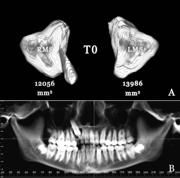
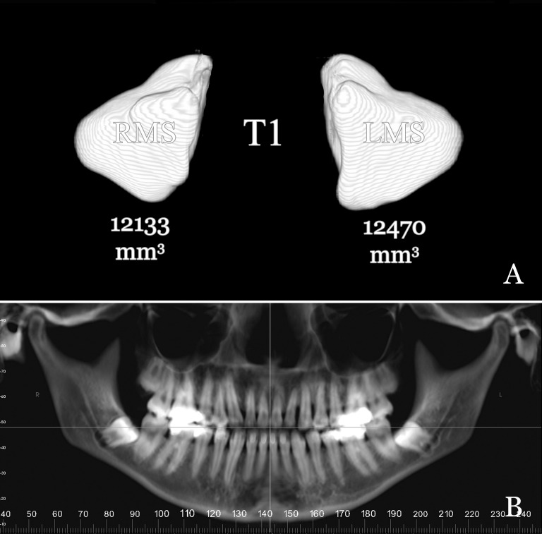
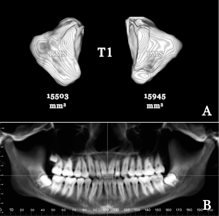
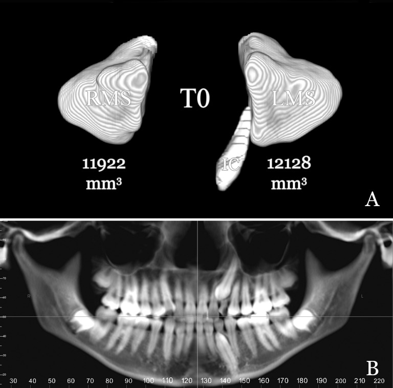
Volumetric measurements of the maxillary sinuses were performed twice at an interval of 15 days using the InVivoDental 5.3 (Anatomage Inc, San Jose, Calif) software program; an experienced orthodontist in the field of airway visualization obtained the measurements. Images were oriented in three spatial planes. The axial slice was adjusted to represent the Frankfort horizontal plane, the sagittal slice was adjusted to represent the midsagittal plane, and the coronal slice was adjusted to pass from the furcation of the upper first molar roots. The oriented image was then transferred to the volume render tool and inverted. While in the inversed mode, the opacity was decreased until the airway became visible. Using the coronal, sagittal, and axial view tools, the maxillary sinuses were cut out from the rest of the image, and opacity was increased until the sinuses appeared as solid structures. The right and left sinuses were separated and measured individually. Eventually, the volume measurement tool was used to calculate the volume of the sinuses. The volume of the right/left maxillary sinus on the impaction side was compared with the contralateral side for both the T0 and T1 time intervals (Figures 1–4). The comparison of the amount of change from the T0 to T1 time interval was also calculated. Furthermore, in order to determine the level of impaction, a line from the canine tip perpendicular to the palatal plane was drawn and the distance was measured. It was determined that the lower the distance, the higher the canine was impacted.
Figure 1.
Volumetric measurements of right maxillary sinus (RMS) and left maxillary sinus (LMS) (A) and panoramic view (B) of a representative patient with right maxillary impacted canine at T0.
Figure 4.
Volumetric measurements of right maxillary sinus (RMS) and left maxillary sinus (LMS) (A) and panoramic view (B) of same patient as in Figure 3 after orthodontic treatment (T1).
The SPSS Statistics 17.0 (SPSS Inc, Chicago, Ill) program was used for all statistical analysis. The Kruskal-Wallis test was used to check the normality of the data. As a result of the nonnormality of the distribution of the data, nonparametric Wilcoxon signed rank tests were used to compare the changes in the maxillary sinus volume. Intraoperator reliability for each measurement was estimated using the intraclass correlation coefficient (ICC).
RESULTS
According to a post hoc power analysis, the computed achieved power of the study was 75%, largely due to the sample size. The ICC for all measured parameters showed high reliability and reproducibility of measurements (r > 0.95).
The maxillary sinus volumes of the impaction and nonimpaction sides at T0 and T1 time intervals are given at Tables 1 and 2. Right maxillary sinus volume was statistically significantly smaller compared to the left maxillary sinus when the canine was impacted on the right side at T0 (P = .015). No such difference was observed between the maxillary sinuses when the canine was impacted on the left side at T0 (P = .211) (Table 1). There was no statistically significant difference between right and left maxillary sinus volumes regardless of the impaction side for the T1 time interval (Table 2).
Table 1.
Descriptive Demographics and the Comparison of Right and Left Maxillary Sinuses at Pretreatment (T0) Time Interval
|
Impacted Canine Side |
Mean ± SD, mm3 |
Median |
Minimum |
Maximum |
P |
| Left (n = 15) | |||||
| Right maxillary sinus (T0) | 12,945.53 ± 3531.33 | 11,594.00 | 8911.00 | 21,199.00 | .211 |
| Left maxillary sinus (T0) | 12,643.33 ± 3189.14 | 11,657.00 | 8482.00 | 19,731.00 | |
| Right (n = 15) | |||||
| Right maxillary sinus (T0) | 11,532.13 ± 3296.65 | 10,433.00 | 7898.00 | 20,820.00 | .015* |
| Left maxillary sinus (T0) | 12,319.93 ± 3509.15 | 12,151.00 | 8112.00 | 22,881.00 | |
indicates statistically significant (P < .05).
Table 2.
Descriptive Demographics at Posttreatment (T1) Time Interval and the Comparison of Right and Left Maxillary Sinuses at T1 Time Interval
|
Impacted Canine Side |
Mean ± SD, mm3 |
Median |
Minimum |
Maximum |
P |
| Left (n = 15) | |||||
| Right maxillary sinus (T1) | 14,481.67 ± 3195.38 | 13,890.00 | 10,272.00 | 21,401.00 | .281 |
| Left maxillary sinus (T1) | 14,779.53 ± 3315.04 | 14,770.00 | 9848.00 | 22,280.00 | |
| Right (n = 15) | |||||
| Right maxillary sinus (T1) | 13,781.93 ± 2933.74 | 12,857.00 | 10,236.00 | 21,819.00 | .733 |
| Left maxillary sinus (T1) | 13,886.80 ± 2925.90 | 14,002.00 | 10,307.00 | 22,046.00 | |
There was a statistically significant increase for all of the maxillary sinus volumes from T0 to T1 (Table 3). All maxillary sinuses showed a significant volumetric increase at the end of orthodontic treatment. When the impacted canine was on the right side, the mean amount of change was 2249.80 mm3 and 1566.87 mm3 for the right and left maxillary sinuses, respectively, where a statistically significant difference was observed (P = .015) (Table 3; Figures 1 and 2). When the maxillary impacted canine was on the left side, the mean amount of change was 1536.13 mm3 and 2136.20 mm3 for the right and left sides, respectively (P = .017) (Table 3; Figures 3 and 4). The mean distance from the canine tip to the palatal plane was 17.17 ± 2.29 mm and 15.14 ± 2.75 mm for the left- and right-side impacted canines, respectively, meaning that the right side impactions were located significantly higher (P = .037) compared to the left side (Table 4).
Table 3.
Comparisons of the Mean Amount of Change from Pretreatment (T0) to Posttreatment (T1) Time Intervala
|
Impacted Canine Side |
Mean Amount of Change, mm3 (T1–T0) |
P |
P′ |
| Left (n = 15) | |||
| Right maxillary sinus (T1) − right maxillary sinus (T0) | 1536.13 | .001 | .017 |
| Left maxillary sinus (T1) − left maxillary sinus (T0) | 2136.20 | .001 | |
| Right (n = 15) | |||
| Right maxillary sinus (T1) − right maxillary sinus (T0) | 2249.80 | .001 | .015 |
| Left maxillary sinus (T1) − left maxillary sinus (T0) | 1566.87 | .001 | |
P-values represent the asymptotic significance (two-tailed) for the same region but at different time intervals. P′ values represent the asymptotic significance (two-tailed) of the comparison of amount of change.
Figure 2.
Volumetric measurements of right maxillary sinus (RMS) and left maxillary sinus (LMS) (A) and panoramic view (B) of same patient as in Figure 1 after orthodontic treatment (T1).
Figure 3.
Volumetric measurements of right maxillary sinus (RMS) and left maxillary sinus (LMS) (A) and panoramic view (B) of a representative patient with left maxillary impacted canine at T0.
Table 4.
Comparison of the Distance of Canine Incisal Tip to Palatal Plane With Respect to Impaction Side
|
Impaction Side |
Mean ± SD, mm |
Median |
Minimum |
Maximum |
P |
| Left impacted canine (n = 15) | 17.17 ± 2.29 | 17.40 | 12.60 | 20.20 | .037* |
| Right impacted canine (n = 15) | 15.14 ± 2.75 | 15.70 | 9.80 | 18.60 |
indicates statistically significant (P < .05).
DISCUSSION
Maxillary sinus volume and relationship between the maxillary sinus and several maxillary posterior teeth have been investigated by several authors.8,17,18 According to the results of these previous studies, especially maxillary molar teeth roots were in close relationship or even inside the maxillary sinuses or maxillary sinus floor. Authors18 also concluded that the shape and size of the maxillary sinuses were affected by the proximity of the roots. Therefore, it is not erroneous to think that maxillary sinus shape and, as a result, volume may be affected by a close neighboring structure, such as an impacted canine. However, none of the previous studies took into account whether erupting impacted canines with orthodontic treatment have any effect on the maxillary sinus volume using a three-dimensional imaging techniques.
Some recent studies using CT exhibited a high prevalence of incidental findings concerning the paranasal sinuses without any clinical symptoms. These findings included acute and chronic inflammatory disease, primary and secondary neoplastic disease, malformation, and bone dysplasia among others.19 Pazera et al.19 showed a prevalence of 46.8%, while Havas et al.20 found 42.5% of incidental findings for the paranasal sinuses. It has also been well documented that such findings cause a volumetric change in the paranasal sinuses. Therefore, all patient CBCTs were closely monitored, and in the event of such findings the patients were excluded from the study in order to investigate the possible extent of the effect of impacted canines on maxillary sinus volume.
Growth of the maxillary sinuses begins during intrauterine life, and the sinuses are present at birth as rudimentary air cells. They present an accelerated growth pattern for 3 years following birth. Subsequent to this rapid growth period, a decrease in growth rate and a quiescence period takes place until 7 years of age. Growth of the maxillary sinuses reaccelerates between the ages of 7 to 12 years and approximates the adult volume.21 All of the patients that were included to the study were over 13 years of age, so we did not expect any maxillary sinus volume changes during the fixed orthodontic treatment that would be due to the patient's growth. But both impacted and contralateral sides showed a significant amount of volumetric change between the two intervals (T0–T1). According to the results of this study, it may be concluded that the maxillary sinus continues to grow and exhibits a volumetric increase after 12 years of age.
The maxillary sinus is reported to be the largest sinus among the paranasal sinuses, and the volume averages a wide range of 8.6 to 24.9 cm3.22 In the present study, the volumetric values that we encountered at the T0 time interval fall within the specified limits. Shahbazian et al.23 and Ariji et al.24 indicated that most of their patients revealed symmetric maxillary sinus morphology, and they did not observe a statistically significant morphologic and volumetric difference between males and females. However, Barghout et al.25 investigated three-dimensional sizes of the paranasal sinuses with magnetic resonance imaging, and they reported volumetric differences between the left and right maxillary sinuses. The aforementioned studies were performed using subjects without any impacted teeth.
The ICC for all measured parameters showed high reliability and reproducibility of measurements (r > 0.95). However, automatic segmentation programs may be affected by noise, artifacts, and CBCT exposure settings and may not present very accurate results. Therefore, the volumetric measurements that are presented here may not be reflective of the real-life volumes.
In the current study, there was a significant amount of maxillary sinus volume change between the impaction and contralateral side on both left and right sides (P = .017 for the left side, P = .015 for the right side). It was observed that the volumetric measurements at the end of the active treatment phase were symmetric (Table 2) where the impaction was corrected and all teeth were in their respective positions. However, right maxillary sinus volume was significantly lower compared to that of the contralateral side when the canine was impacted on the right side at T0. When the canine teeth were impacted on the right side, the mean distance from the canine tip to its target point on the palatal plane was decreased significantly compared to measurements involving the left-side impactions. Therefore, it may be interpreted that the maxillary canines that were impacted on the right side were more superiorly positioned compared to the left-impacted canines. This situation may have caused a more significant change in the maxillary sinus shape and thereby the volume at T0. When the canine teeth were erupted into the oral cavity, this significant difference seems to diminish as a result of the adaptability of the maxillary sinuses, as mentioned previously.
Between T0 and T1 there was a statistically significant increase for both maxillary sinuses. However, although not significant, impacted canine-side maxillary sinus volumes increased more compared to the contralateral side after the eruption of the canine to the dental arch and approached a value that was very similar to that of the nonimpaction side. Rusu et al.5 presented a case report that included bilateral dissection of the maxilla of a human adult cadaver head. According to this case report, an oblique impacted canine was evidenced, and its root apex was tangent to the sinus floor, and the root was in a close relationship within the floor of the expanded inferior nasal meatus. In the current study, we also found that the relationships of the right impacted maxillary canines were closer with the maxillary sinuses, and this relationship could explain the significant increase of right maxillary sinus volume after orthodontic treatment of impacted canine.
Clinicians should be wary of the results, since the calculated achieved power of the study is below 80%. Further studies with larger sample sizes are necessary in order to confirm the findings that are presented here.
CONCLUSION
Impacted canines have no significant impact on the size of the maxillary sinuses unless the canine is deeply impacted, with closer relationship to the maxillary sinuses. In such a case, when the impacted canines are erupted to the dental arch, a volumetric increase in the maxillary sinuses may be expected.
REFERENCES
- 1.Emirzeoglu M, Sahin B, Bilgic S, Celebi M, Uzun A. Volumetric evaluation of the paranasal sinuses in normal subjects using computer tomography images: a stereological study. Auris Nasus Larynx. 2007;34:191–195. doi: 10.1016/j.anl.2006.09.003. [DOI] [PubMed] [Google Scholar]
- 2.Endo T, Abe R, Kuroki H, Kojima K, Oka K, Shimooka S. Cephalometric evaluation of maxillary sinus sizes in different malocclusion classes. Odontology. 2010;98:65–72. doi: 10.1007/s10266-009-0108-5. [DOI] [PubMed] [Google Scholar]
- 3.Rennie CE, Hood CM, Blenke EJ, et al. Physical and computational modeling of ventilation of the maxillary sinus. Otolaryngol Head Neck Surg. 2011;145:165–170. doi: 10.1177/0194599811401202. [DOI] [PubMed] [Google Scholar]
- 4.Fehrenbach MJ, Herring SW, Thomas P. Philadelphia, Penn: WB Saunders; 1996. Illustrated Anatomy of the Head and Neck. [Google Scholar]
- 5.Rusu MC, Comes CA, Stanciu D, et al. Altered anatomy in a case with a buccally impacted maxillary canine tooth. Rom J Morphol Embryol. 2010;51:783–786. [PubMed] [Google Scholar]
- 6.Kwak HH, Park HD, Yoon HR, Kang MK, Koh KS, Kim HJ. Topographic anatomy of the inferior wall of the maxillary sinus in Koreans. Int J Oral Maxillofac Surg. 2004;33:382–388. doi: 10.1016/j.ijom.2003.10.012. [DOI] [PubMed] [Google Scholar]
- 7.Stephens JC, Saleh HA. Evaluation and treatment of isolated maxillary sinus disease. Curr Opin Otolaryngol Head Neck Surg. 2013;21:50–57. doi: 10.1097/MOO.0b013e32835af905. [DOI] [PubMed] [Google Scholar]
- 8.Sharan A, Madjar D. Maxillary sinus pneumatization following extractions: a radiographic study. Int J Oral Maxillofac Implants. 2008;23:48–56. [PubMed] [Google Scholar]
- 9.Oktay H. The study of the maxillary sinus areas in different orthodontic malocclusions. Am J Orthod Dentofacial Orthop. 1992;102:143–145. doi: 10.1016/0889-5406(92)70026-7. [DOI] [PubMed] [Google Scholar]
- 10.Pangrazio-Kulbersh V, Wine P, Haughey M, Pajtas B, Kaczynski R. Cone beam computed tomography evaluation of changes in the naso-maxillary complex associated with two types of maxillary expanders. Angle Orthod. 2011;82:448–457. doi: 10.2319/072211-464.1. [DOI] [PMC free article] [PubMed] [Google Scholar]
- 11.El H, Palomo JM. Airway volume for different dentofacial skeletal patterns. Am J Orthod Dentofacial Orthop. 2011;139:e511–e521. doi: 10.1016/j.ajodo.2011.02.015. [DOI] [PubMed] [Google Scholar]
- 12.El H, Palomo JM. Measuring the airway in 3 dimensions: a reliability and accuracy study. Am J Orthod Dentofacial Orthop. 2010;137:S50.e1–S50.e9. doi: 10.1016/j.ajodo.2010.01.014. [DOI] [PubMed] [Google Scholar]
- 13.Darsey DM, English JD, Kau CH, Ellis RK, Akyalcin S. Does hyrax expansion therapy affect maxillary sinus volume? A cone-beam computed tomography report. Imaging Sci Dent. 2012;42:83–88. doi: 10.5624/isd.2012.42.2.83. [DOI] [PMC free article] [PubMed] [Google Scholar]
- 14.Oberoi S, Knueppel S. Three-dimensional assessment of impacted canines and root resorption using cone beam computed tomography. Oral Surg Oral Med Oral Pathol Oral Radiol. 2012;113:260–267. doi: 10.1016/j.tripleo.2011.03.035. [DOI] [PubMed] [Google Scholar]
- 15.Guerrero ME, Shahbazian M, Elsiena Bekkering G, Nackaerts O, Jacobs R, Horner K. The diagnostic efficacy of cone beam CT for impacted teeth and associated features: a systematic review. J Oral Rehabil. 2011;38:208–216. doi: 10.1111/j.1365-2842.2010.02141.x. [DOI] [PubMed] [Google Scholar]
- 16.Oz AZ. Evaluation of Diagnosis and Orthodontic Treatment of Impacted Maxillary Canines with 3 Dimensional Imaging Method [PhD Thesis] Ankara, Turkey: Institute of Health Sciences, Hacettepe University; 2012. [Google Scholar]
- 17.Eberhardt JA, Torabinejad M, Christiansen EL. A computed tomographic study of the distances between the maxillary sinus floor and the apices of the maxillary posterior teeth. Oral Surg Oral Med Oral Pathol. 1992;73:345–346. doi: 10.1016/0030-4220(92)90133-b. [DOI] [PubMed] [Google Scholar]
- 18.Lana JP, Carneiro PM, Machado Vde C, de Souza PE, Manzi FR, Horta MC. Anatomic variations and lesions of the maxillary sinus detected in cone beam computed tomography for dental implants. Clin Oral Implants Res. 2012;23:1398–1403. doi: 10.1111/j.1600-0501.2011.02321.x. [DOI] [PubMed] [Google Scholar]
- 19.Pazera P, Bornstein MM, Pazera A, Sendi P, Katsaros C. Incidental maxillary sinus findings in orthodontic patients: a radiographic analysis using cone-beam computed tomography (CBCT) Orthod Craniofac Res. 2011;14:17–24. doi: 10.1111/j.1601-6343.2010.01502.x. [DOI] [PubMed] [Google Scholar]
- 20.Havas TE, Motbey JA, Gullane PJ. Prevalence of incidental abnormalities on computed tomographic scans of the paranasal sinuses. Arch Otolaryngol Head Neck Surg. 1988;114:856–859. doi: 10.1001/archotol.1988.01860200040012. [DOI] [PubMed] [Google Scholar]
- 21.Lawson W, Patel ZM, Lin FY. The development and pathologic processes that influence maxillary sinus pneumatization. Anat Rec (Hoboken) 2008;291:1554–1563. doi: 10.1002/ar.20774. [DOI] [PubMed] [Google Scholar]
- 22.Fernandez JS, Escuredo JA, Del Rey AS, Montoya FS. Morphometric study of the paranasal sinuses in normal and pathological conditions. Acta Otolaryngol. 2000;120:273–278. doi: 10.1080/000164800750001080. [DOI] [PubMed] [Google Scholar]
- 23.Shahbazian M, Xue D, Hu Y, van Cleynenbreugel J, Jacobs R. Spiral computed tomography based maxillary sinus imaging in relation to tooth loss, implant placement and potential grafting procedure. J Oral Maxillofac Res. 2010;1:e7. doi: 10.5037/jomr.2010.1107. [DOI] [PMC free article] [PubMed] [Google Scholar]
- 24.Ariji Y, Kuroki T, Moriguchi S, Ariji E, Kanda S. Age changes in the volume of the human maxillary sinus: a study using computed tomography. Dentomaxillofac Radiol. 1994;23:163–168. doi: 10.1259/dmfr.23.3.7835518. [DOI] [PubMed] [Google Scholar]
- 25.Barghouth G, Prior JO, Lepori D, Duvoisin B, Schnyder P, Gudinchet F. Paranasal sinuses in children: size evaluation of maxillary, sphenoid, and frontal sinuses by magnetic resonance imaging and proposal of volume index percentile curves. Eur Radiol. 2002;12:1451–1458. doi: 10.1007/s00330-001-1218-9. [DOI] [PubMed] [Google Scholar]


