Abstract
Early B-cell factor (EBF) is a transcription factor suggested as essential for early B-lymphocyte development by findings in mice where the coding gene has been inactivated by homologous disruption. This makes the identification of genetic targets for this transcription factor pertinent for the understanding of early B-cell development. The lack of B29 transcripts, coding for the β subunit of the B-cell receptor complex, in pro-B cells from EBF-deficient mice suggested that B29 might be a genetic target for EBF. We here present data suggesting that EBF interacts with three independent sites within the mouse B29 promoter. Furthermore, ectopic expression of EBF in HeLa cells activated a B29 promoter-controlled reporter construct 13-fold and induced a low level of expression from the endogenous B29 gene. Finally, mutations in the EBF binding sites diminished B29 promoter activity in pre-B cells while the same mutations did not have as striking an effect on the promoter function in B-cell lines of later differentiation stages. These data suggest that the B29 gene is a genetic target for EBF in early B-cell development.
B-lymphocyte development is a complex differentiation process that generates antibody-secreting cells. One key issue for the understanding of this process is how transcription is controlled in order to achieve lineage- and stage-specific expression of selected genes. Transcriptional control has been extensively studied in B cells, and several transcription factors have been shown to be essential for the development of the B-cell lineage (3, 5). One of these transcription factors is early B-cell factor (EBF) expressed in all stages of B-cell development with the exception of the plasma cell stage (7, 9). EBF is B-cell restricted but also expressed in adipocytes and in olfactory neurons (7, 35). The factor binds to the DNA core sequence CCCNNGGG as a homodimer by a large DNA binding domain containing a zinc coordination motif (7, 8, 34). The dimerization is mediated by two α-helixes with homology to helix two of the helix-loop-helix (HLH) domain found in transcription factors of the basic HLH family (7, 8). EBF has been shown to contain two independent transactivation domains, one located within the DNA binding domain and another in the carboxy-terminal part of the protein (8). EBF interacts functionally with transcription control elements regulating a number of B-cell-specific genes (for a review, see reference 28). The first identified genetic target was the mb-1 gene (9), which encodes the α-subunit of the B-cell receptor complex (13, 14). Here, EBF was shown to interact with an element important for full activity of the promoter (9). However, the mb-1 gene can be expressed in cell lines in the absence of EBF as well, demonstrating that this promoter is not completely dependent on EBF in transformed cells (29). The pre-B-cell-specific surrogate light-chain genes λ5 and VpreB (for a review, see reference 22) have also been suggested to be direct targets for EBF since both contain EBF binding sites important for promoter function (21, 26, 29). This was further supported by the observation that ectopic expression of EBF in the immature hematopoietic cell line Ba/F3 resulted in activation of the endogenous λ5 and VpreB genes (29). EBF has also been demonstrated to interact with immunoglobulin (Ig) light-chain promoters (30) and to functionally modulate the Ig heavy-chain intron-enhancer (1).
Homologous disruption of the EBF coding gene resulted in mice lacking IgM expressing B cells, showing the necessity for EBF in the development of mature B lymphocytes (19). The developmental block of B-cell differentiation occurred at an early stage, before the onset of Ig recombination but after the initiation of expression of sterile Ig transcripts (Iμ and μ0) and terminal deoxynucleotide transferase. Flow cytometric analysis showed that the bone marrow of EBF−/− mice contained cells expressing B220 and CD43 but not BP1 or heat-stable antigen, suggesting that these cells are arrested in the transition from fraction A (pro-B) to fraction B (pre-B) cells (10). The interleukin-7 receptor was expressed, but no detectable mRNA for Rag-1, Ig α-chain, VpreB, λ5, or Igβ-chain (Igβ) were detected (19). The absence of Igβ (B29) transcripts was striking since mRNA coding for Igβ is present in normal fraction A pro-B cells (17). Furthermore, Igβ transcripts can be detected in mice homozygous for an inactivated E2A gene (38), which have a similar developmental block of B-cell development. These observations suggested to us that the B29 promoter might be a genetic target for EBF.
The B29 gene encodes the β component of the B-cell receptor complex and acts as a signal transducer molecule after antigen recognition of surface Ig (for reviews, see references 2, 13, and 14). The gene has been shown to be essential for B-cell development, since mice homozygous for a mutation in the B29 gene display an early arrest of B-cell development, after the onset of D-J but before VDJ recombination of the Ig heavy-chain locus (6). B29 is expressed in cell lines representing all B-cell differentiation stages (11); it was proposed that there is a partial but not complete overlap of EBF and B29 expression. The B29 promoter has been suggested to interact with IKAROS, OCT, and Ets proteins (12, 25). The importance of these factors has also been displayed by mutagenesis experiments where the Ets and the OCT protein binding sites were shown to be important for the function of the B29 promoter (25). The B29 gene has also been shown to be under the control of repressor elements located 5′ of the promoter (20).
Here we show data suggesting that EBF binds to the B29 promoter and has the ability to activate the promoter in HeLa cells. Furthermore, we present data indicating that the EBF binding sites are of great importance in the function of the promoter in pre-B-cell lines while they have less impact on promoter function in B-cell lines representing later developmental stages. These experiments suggest that the B29 gene is a direct genetic target for EBF and would explain the absence of B29 transcripts in pro-B cells in EBF−/− mice.
MATERIALS AND METHODS
Tissue culture conditions.
All cells were grown in RPMI medium supplemented with 7.5% fetal calf serum, 10 mM HEPES, 2 mM pyruvate, 50 μM 2-mercaptoethanol, and 50 μg of gentamicin per ml (all purchased from Life Technologies AB, Täby, Sweden) at 37°C and 5% CO2.
Transient transfections and luciferase assays.
A total of 250,000 HeLa cells were grown overnight in 1 ml of RPMI medium in a 24-well plate. The cells were washed once with serum-free medium (OPTIMEM; Life Technologies), and 800 μl of the serum-free medium was added for transfection. Five microliters of Lipofectin (Life Technologies) was diluted in 100 μl of serum-free medium, incubated for 45 min at room temperature, and mixed with the DNA diluted in 100 μl of serum-free medium. The mixture was incubated for 25 min, and the combined volume of 200 μl was added to the HeLa cells. The cells were then incubated in a CO2 incubator at 37°C for 6 h, after which the transfection medium was removed and replaced by RPMI medium supplemented with 10% fetal calf serum. The cells were harvested after 40 h, and protein extracts were prepared by using 100 μl of cell lysis buffer (Promega, Scandinavian Diagnostic Services AB, Falkenberg, Sweden). The luciferase assay was then conducted with 15 μl of the obtained extracts and 200 μl of luciferase assay reagent (Promega). To induce expression of endogenous B29 expression in HeLa cells, 2 million cells were transfected with 2 μg of EBF expression plasmid by a scaled-up Lipofectin protocol as described above.
All other cell lines were washed twice in Tris-buffered saline (TBS; 140 mM NaCl, 5 mM KCl, 25 mM Tris [pH 7.4], 0.6 mM phosphate, 0.5 mM MgCl2, 0.7 mM CaCl2), and 2.5 × 10⁶ cells were transfected with 2 μg of reporter gene construct in 0.65 ml of TBS with 0.7 mg of DEAE-dextran (Pharmacia, Uppsala, Sweden) per ml for 30 min at 20°C. After a single wash in TBS, the transfected cells were cultured in 5 ml of complete RPMI medium in six-well plates for 48 h. Preparation of protein extracts and luciferase assays were performed with a luciferase assay kit (Promega) by using 20% of the protein extract.
Protein extracts and electrophoretic mobility shift assay (EMSA).
Nuclear extracts were prepared as described by Schreiber et al. (27). DNA probes were labeled with [γ-32P]ATP by incubation with T4 polynucleotide kinase (Life Technologies), annealed, and purified on a 5% polyacrylamide Tris-borate-EDTA (TBE) gel. The B29 probe used in Fig. 5 was generated by PCR amplification of the B29 promoter cloned in pGem 3Z (see below) by using the B29 EBF-3 antisense oligonucleotide together with an SP6 primer (Promega). This PCR product includes all three EBF binding sites (positions −106 to −152) but lacks OCT, Ets, and SP1 binding sites. The probe was digested with HindIII and fill-in labeled with [α-32P]dCTP by incubation with Klenow fragment. The B29 wild-type promoter used as the probe in the experiment shown in Fig. 6 was generated by PCR using an SP6 primer (Promega) together with the B29 antisense primer and pGem B29 as the template. The resulting full-length B29 promoter was digested with HindIII and HaeIII and fill-in labeled with [α-32P]dCTP by incubation with Klenow fragment, which generates a truncated (−88 to −152) probe. The mutated B29 promoters used in the experiment shown in Fig. 6 were also generated by PCR using RVprimer3 (Promega) together with the B29 antisense primer and the respective B29 promoter mutants cloned in pGL3 basic (Promega) as templates (see below). The resulting full-length B29 promoter mutants were digested with MluI and HaeIII and fill-in labeled with [α-32P]dCTP by incubation with Klenow fragment, which generates truncated (−88 to −152) probes. All PCR-generated probes were purified on 5% polyacrylamide TBE gels. Nuclear extract or in vitro-transcribed-translated protein was incubated with labeled probe (20,000 cpm; 3 fmol) for 30 min at room temperature in binding buffer (10 mM HEPES [pH 7.9], 70 mM KCl, 1 mM dithiothreitol, 1 mM EDTA, 2.5 mM MgCl2, 0.05% Nonidet P-40) with 0.75 μg poly(dI/dC) (Pharmacia). DNA competitors were added 10 min before the addition of the DNA probe. The samples were separated on a 6% polyacrylamide TBE gel, which was dried and subjected to autoradiography. Competitors based on synthetic oligonucleotides were added at the molar excesses indicated in the respective figures. Full-length B29 promoters (+1 to −152) were generated by PCR (see below) and were added at the molar excesses indicated in the respective figures. λ5 full-length promoter and actin control competitors were also generated by PCR (for details, see reference 29) and were added at the molar excesses indicated in the respective figures
FIG. 5.
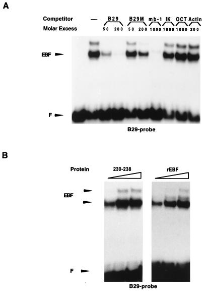
The B29 promoter interacts with EBF in 230–238 pre-B cells and has the ability to interact with multiple EBF homodimers simultaneously. (A) EMSA using an end-labeled truncated B29 promoter spanning nucleotides −105 to −152 and nuclear extracts from 230–238 pre-B cells. The obtained band shifts were competed for by the addition of the PCR-amplified full-length (+1 to −152) wild-type (B29) or EBF-mutated (B29M) B29 promoter; the mb-1 EBF, IKAROS consensus, or OCT protein binding site; and an actin PCR fragment as indicated. (B) EMSA using the B29 −105 to −152 probe and increasing amounts of either 230–238 pre-B-cell nuclear extracts or in vitro-translated recombinant EBF (rEBF). F indicates the free probe.
FIG. 6.
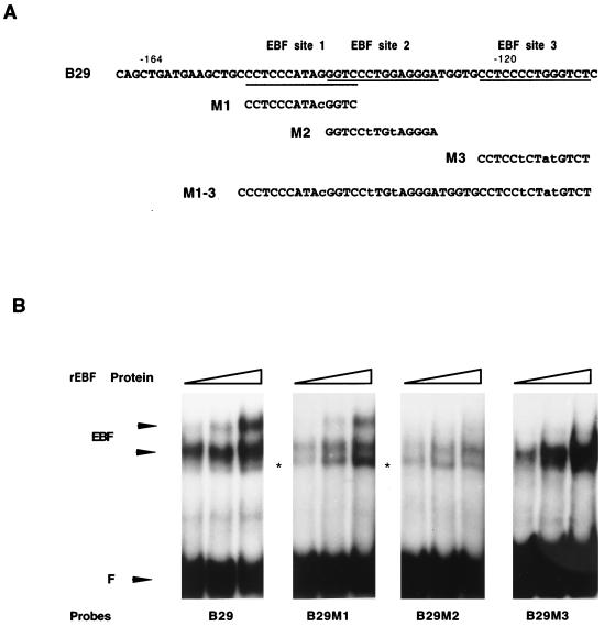
EBF binding sites 2 and 3 are essential for the formation of the low-mobility complex. (A) Schematic drawing of the B29 promoter fragments that were used as binding sites for EBF in the EMSA. Introduced mutations are indicated by lowercase letters. (B) EMSA analysis with increasing amounts (0.5, 2, and 4 μl) of recombinant EBF and 20,000 cpm of the indicated end-labeled B29 promoter variants as binding sites. The band indicated by an asterisk was not competed for by an mb-1 promoter EBF binding site. F indicates free probe.
Oligonucleotides used for EMSAs were as follows: mb-1 EBF sense (5′-GAGAGAGACTCAAGGGAATTGTGG), mb-1 EBF antisense (5′-CCACAATTCCCTTGAGTCTCTCTC), B29 EBF-1 sense (5′-TAGGAGCTCCCATAGGGTCCCTGTAGGGAT), B29 EBF-1 antisense (5′-ATCCCTACAGGGACCCTATGGGAGCTCCTA), B29 EBF-2 sense (5′-TAGGAGCTCCCATACGGTCCCTGGAGGGATGGT), B29 EBF-2 antisense (5′-ACCATCCCTCCAGGGACCGTATGGGAGCTCCTA), B29 EBF-3 sense (5′-GAGGGATGGTGCCTCCCCTGGGTCTCAATT), B29 EBF-3 antisense (5′-AATTGAGACCCAGGGGAGGCACCATCCCTC), SP6κ EBF sense (5′-ATGGATTACTTTGCATAGATCCCTAGAGGCCAGCACAGCTGCT), SP6κ EBF antisense (5′-AGCAGCTGTGCTGGCCTCTAGGGATCTATGCAAAGTAATCCAT), OCT sense (5′-TTCATTGATTTGCATCGCATGAGACGCTAACATCGTACGTTC), OCT antisense (5′-GAACGTACGATGTTAGCGTCTCATGCGATGCAAATCAATGAA), IKAROS consensus sense (5′-TAGAATTCTTGGGAATACCGATCTT), and IKAROS consensus antisense (5′-AAGATCGGTATTCCCAAGAATTCTA).
Plasmids and constructs.
The EBF expression plasmid (29) was based on the eukaryotic expression vector cDNA3 (Invitrogen BV, NV Leek, The Netherlands), which places the inserted cDNA under the control of a cytomegalovirus promoter. The full-length B29 promoter (+1 to −152) was PCR amplified by using B29 sense and antisense primers with genomic mouse DNA as the template. The resulting PCR product was cloned in pGem 3Z (Promega) and verified by sequencing. Luciferase reporter constructs were based on the pGL3 basic reporter vector (Promega). The wild-type B29 promoter was isolated as a BglII-SacI fragment from pGem 3Z and subcloned in the pGL3 basic reporter vector. B29 promoters with mutated EBF sites (+1 to −152) were constructed by using PCR primer B29M1, B29M2, B29M3, B29M, or B29M1–3 (see Fig. 3A and 6A) together with the B29 antisense primer. The B29-containing pGem 3Z vector was used as the template. The resulting B29 promoter mutants were cloned blunt in pGL3 basic reporter vector. All constructs were verified by sequencing.
FIG. 3.
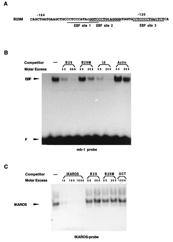
Mutations in the three defined EBF binding sites impair the ability of the B29 promoter to bind EBF. (A) Schematic drawing indicating the mutations introduced into the B29 promoter by PCR using an oligonucleotide with inserted mutations. Lowercase letters indicate the mutations. (B) EMSA in which the binding of in vitro-translated recombinant EBF to the mb-1 promoter EBF binding site was competed for by the PCR-amplified (+1 to −152) wild-type B29 (B29), the mutated B29 (B29M), or the λ5 promoter. An actin PCR fragment served as a negative control. (C) EMSA using nuclear extracts from 230–238 pre-B cells and a consensus IKAROS binding site (3). The obtained band shift was competed for by an IKAROS consensus binding site, by the PCR-amplified wild-type (B29) or EBF mutant B29 (B29M) promoter, and by an octamer-containing oligonucleotide (OCT) as indicated. F indicates the free probe.
Oligonucleotides used for B29 promoter constructs included the following: B29 sense (5′-TAGGAGCTCCCATAGGGTCCCTGGAGGGAT), B29 antisense (5′-CAGAGATCTGGTCACTGCTCTGTCTCTGAGCCC), B29M (5′-TAGGAGCTCCCATACGGTCCCTGTAGGGATGGTGCCTCCCCTATGT CTCAATTTGC), B29M1 (5′-TAGGAGCTCCCATACGGTCCCTGGAGGGATGGT), B29M2 (5′-TAGGAGCTCCCATAGGGTCCTTGTAGGGATGGTGCCTCCCCTGGGTCTCAA), B29M3 (5′-TAGGAGCTCCCATAGGGTCCCTGGAGGGATGGTGCCTCCTCTATGTCTCAA), and B29M1–3 (5′-TAGGAGCTCCCATACGGTCCTTGTAGGGATGGTGCCTCCTCTATGTCTCAA).
In vitro transcription and translation.
Recombinant proteins were generated by coupled in vitro transcription-translation by using a reticulocyte lysate kit (Promega). A total of 0.5 to 2 μl of a 25-μl reaction mix was used for EMSAs.
RT and PCRs.
RNA was prepared from cells by using RNAzol B (Tel-Test Inc., Friendswood, Tex.). cDNA was generated by annealing 1 μg of total RNA and 0.5 μg of dT18 oligonucleotide in 10 μl of diethylpyrocarbonate-treated water. Reverse transcriptase (RT) reactions were performed with 200 U of SuperScript RT (Life Technologies) in the manufacturer’s buffer supplemented with 0.5 mM deoxynucleoside triphosphate, 10 mM dithiothreitol, and 20 U of RNase inhibitor (Boehringer Mannheim, Bromma, Sweden), in a total volume of 20 μl at 37°C for 1 h. One-twentieth of the RT reaction mix was used for the PCR assays.
PCRs were performed with 1 U of Taq polymerase (Life Technologies) in the manufacturer’s buffer supplemented with 0.2 mM deoxynucleoside triphosphate, in a total volume of 25 μl. The conditions of each PCR cycle were 94°C for 30 s, 60°C for 30 s, and 72°C for 30 s. Primers were added to a final concentration of 1 mM. Twenty-five cycles were used for all sets.
PCR products were blotted onto Zeta-Probe GT nylon membranes (Bio-Rad Laboratories AB, Sundbyberg, Sweden) by the vacuum transfer method. Membranes were prehybridized in 0.5 M NaHPO4 (pH 7.5)–7% sodium dodecyl sulfate (SDS) at 57°C for 10 min and hybridized with γ-32P-labeled B29 oligonucleotide for 12 h at 57°C in the same solution. Membranes were washed at room temperature three times in 40 mM NaHPO4 (pH 7.5)–1% SDS for 15 min.
Oligonucleotides used for RT-PCR included the following: B29 cDNA sense, exon 3 (5′-TCTGGCAGAGCCCACGTTTCATA); B29 cDNA antisense, exon 6 (5′-CTCATAGGTGGCTGTCTGGTCAAT); B29 cDNA hybridization, exon 3 (5′-TGAAGCTGGAAAAGGGCCGCAT); GADPH sense (5′-CCACCCATGGCAAATTCCATGGCA); and GADPH antisense (5′-TCTAGACGGCAGGTCAGGTCCACC).
RESULTS
EBF interacts with three independent sites in the B29 promoter.
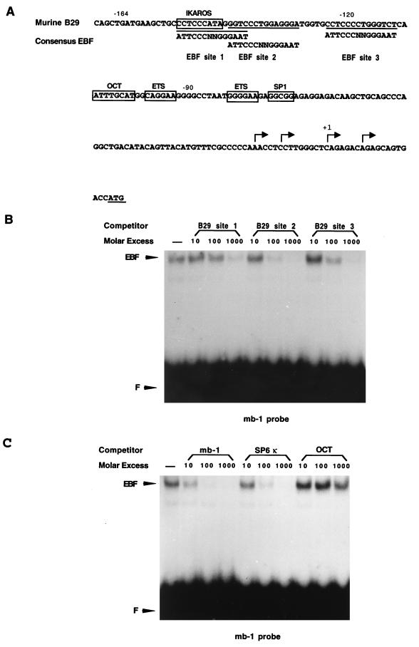
We initially examined the mouse B29 promoter for potential EBF binding sites and observed three putative recognition sequences in a region spanning from position −160 to the major transcription initiation site. Figure 1 shows the sequence of the B29 promoter (25) with the three potential EBF binding sites indicated. The potential EBF binding sites were all located between bases −150 and −110 in a region known to be important for promoter function (25). These sites were also conserved between the mouse and the human B29 promoter, even though there were single base substitutions within the sites (1a, 31). To determine whether these sites bound to EBF, we performed an EMSA using double-stranded oligonucleotides, spanning these sites, as competitors for binding of recombinant in vitro-translated EBF to the EBF binding site from the mb-1 promoter. To compare the binding efficiency of the B29 sites to that of other characterized EBF sites, we also included the mb-1 and the SP6κ promoter EBF binding sites as competitors. An OCT binding site was used as a specificity control. The results shown in Fig. 1 suggest that EBF indeed interacted with all three B29 promoter sites, although sites 1 and 3 appeared to be of low affinity. Site 2 bound EBF with a similar efficiency as the EBF binding site in the mb-1 promoter. Thus, we conclude that the B29 promoter contains three EBF binding sites.
FIG. 1.
The B29 promoter contains three independent EBF binding sites. (A) Schematic drawing of the B29 minimal promoter as defined by Omori and Wall (25). The potential EBF binding sites are underlined, and a consensus EBF binding site (34) is shown. (B) EMSA in which the binding of in vitro-translated recombinant EBF to the mb-1 promoter binding sites was competed for by potential EBF binding sites from the B29 promoter. (C) EMSA in which the binding of in vitro-translated recombinant EBF to the mb-1 promoter binding site was competed for by EBF binding sites from the mb-1 and SP6κ promoters and a control octamer-containing oligonucleotide (OCT). F indicates the free probe.
EBF can activate the B29 promoter in an HeLa cell.
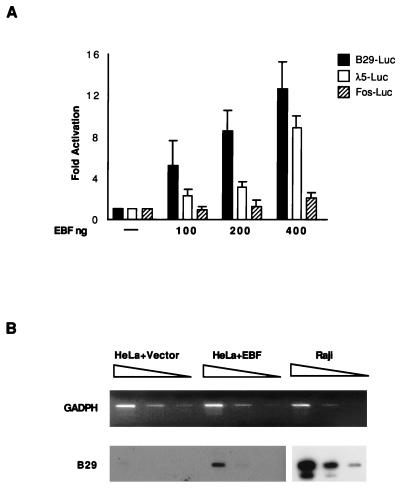
Knowing that EBF bound to the B29 promoter, we wanted to determine whether EBF was able to directly activate transcription from the B29 promoter in a non-B cell. To study this, we cloned the B29 promoter (+1 to −152) in front of a luciferase reporter gene (pGL3) and transfected this construct together with an expression plasmid for EBF into HeLa cells. Figure 2A shows a diagram of the resulting luciferase activity from this experiment. The activity of the B29 promoter was induced up to 13-fold upon cotransfection with increasing amounts of EBF expression plasmid. The λ5 promoter, previously identified as an EBF target, was induced to a similar level, while a fos promoter was unaffected by the inclusion of an EBF expression vector. We next investigated whether endogenous B29 expression was also induced by ectopic expression of EBF. The results when cDNA from HeLa cells transiently transfected either with empty or EBF-encoding expression vector were analyzed for endogenous B29 expression by RT-PCR analysis are shown in Fig. 2B. Transcripts were detected in the EBF-transfected cells, but the RT-PCR analysis suggests that the B29 levels were low (less then 1%) in comparison to the levels found in the Raji B-cell line. Longer exposures of the filters revealed a low level of B29 transcripts also in HeLa cells transfected with the empty expression plasmid. We conclude from this analysis that overexpression of EBF in a non-B cell activates some transcription from the B29 promoter either in the form of a naked template or in the chromatin context of an endogenous gene.
FIG. 2.
The B29 promoter is induced by EBF in HeLa cells. (A) Luciferase activity assay results when a fos, a λ5, or a B29 promoter (+1 to −152) construct was transiently transfected into HeLa cells together with increasing amounts of EBF expression plasmid. The DNA content in each transfection was equalized by the addition of empty expression plasmid (cDNA3). The data represent four transfections from two independent experiments. (B) RT-PCR analysis of HeLa cells transiently transfected with EBF expression plasmid. The upper panel shows an ethidium bromide-stained agarose gel with the resulting amount of GADPH (glyceraldehyde-3-phosphate dehydrogenase) PCR products obtained from HeLa cells transfected with either an empty or an EBF-containing cDNA3 expression plasmid and from the human B-cell line Raji. The data show a serial dilution using 1, 1/5, 1/25 dilutions of the template cDNA after 25 cycles of PCR. The lower panel shows an autoradiogram from RT-PCR analysis indicating the amount of B29 transcripts in the same cDNA preparations after 25 cycles of PCR. The PCR products were blotted onto a nylon membrane and hybridized to an internal B29 oligonucleotide. The filters with PCR product from transfected HeLa cells were exposed for 24 h, while the corresponding filters for Raji cells were exposed for 4 h.
EBF binding sites are important for full functional activity of the B29 promoter in pre-B-cell lines.
Knowing that EBF has the ability to functionally interact with the B29 promoter, we wanted to investigate the impact of mutations in the EBF binding sites on the function of the B29 promoter in B cells at different differentiation stages. To this end, mutations were introduced in the three EBF binding sites. The mutations were introduced to avoid disruption of other overlapping binding sites such as those for IKAROS or transcription factor E3 (Fig. 3A). To confirm that these mutations resulted in a B29 promoter with impaired ability to interact with EBF, we performed an EMSA analysis in which the PCR-amplified full-length (+1 to −152) wild-type or EBF-mutated promoter fragment was used to compete for binding of in vitro-translated EBF to the mb-1 promoter EBF site. Figure 3B shows that the wild-type B29 promoter competed at least as efficiently as the λ5 promoter (+1 to −415) while the mutated promoter competed with low efficiency. No competition was observed when an actin PCR fragment was used as the control. The residual EBF binding capacity of the mutated B29 promoter was probably due to the fact that we introduced only subtle mutations to avoid disruption of other overlapping binding sites. To examine whether the mutations affected the binding of factors to the IKAROS consensus site in a pre-B cell, we performed an EMSA using nuclear extract from cell line 230–238 and a duplex consensus IKAROS binding site as the probe. This resulted in two minor complexes and one major complex, but none of these were competed for by either the wild-type or the mutated B29 promoter fragment (Fig. 3C). Thus, we conclude that proteins in 230–238 cells that bind to an IKAROS consensus binding site do not interact efficiently with the B29 promoter and that this interaction was not modified by the mutations introduced in the EBF sites while binding of EBF was impaired.
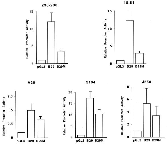
To investigate the functional role of the EBF binding sites in B cells, luciferase reporter constructs under the control of either the wild-type or the EBF-mutated B29 promoter (positions +1 to −152) were transiently transfected into B-cell lines representing different developmental stages. As shown in Fig. 4, transfection of the wild-type B29 promoter fragment into the pre-B-cell line 230–238 or 18.81 resulted in a 13-fold induction of luciferase activity in comparison to that resulting from transfection with the basic reporter vector (pGL3). When the EBF mutant B29 promoter was used, the corresponding induction was 3.5-fold, a 4-fold reduction in functional activity. In contrast, when the same promoter constructs were transfected into cells from the B-cell lymphoma A20 or the plasmacytomas J558 and S194, the EBF mutations resulted in less than a twofold reduction in the B29 promoter activity. This suggests that EBF is an important regulator of B29 transcription in early B-cell development while other factors control the B29 gene in later stages of B-cell differentiation.
FIG. 4.
The EBF binding sites are important for the function of the B29 promoter in pre-B cells. The resulting activities of wild-type (B29) and EBF-mutated (B29M) B29 promoter constructs relative to that of a basic luciferase vector (pGL3) after transient transfections into transformed mouse cells of the B-cell lineage are shown. The data are collected from four transfections and two independent experiments from the following cell lines: 230–238 pre-B cells, 18.81 pre-B cells, A20 mature B cells, S194 plasma cells, and J558 plasma cells.
EBF interacts with B29 EBF sites 2 and 3 to form a low-mobility complex.
To further examine the proteins interacting with the region from −105 to −152 of the B29 promoter in pre-B cells, we used this region as a probe in an EMSA with nuclear extracts from the 230-238 pre-B-cell line. Two complexes were detected (Fig. 5A), one major and one minor with slower mobility. Both complexes were competed for by the B29 promoter fragment and the mb-1 EBF binding site, while neither of the complexes was efficiently competed for by an EBF mutant B29 promoter, an IKAROS consensus binding site, or an OCT binding site. We interpret these data as suggesting that EBF is the major factor interacting with this region of the B29 promoter in pre-B cells. The presence of two complexes that could be competed for by the mb-1 EBF binding site indicated that the high-mobility complex represented a B29 promoter occupied by more than one EBF homodimer. To examine this more carefully, we used the B29 promoter −105 to −152 fragment as a probe and used increasing amounts of 230–238 cell nuclear extract or in vitro-translated EBF in an EMSA (Fig. 5B). This experiment suggested that with increasing amounts of recombinant protein, a second high-mobility complex, migrating with the same mobility as that of the complex found in nuclear extracts, was formed. These data supported the idea that multiple EBF homodimers bind simultaneously to the B29 promoter but do not discriminate between double occupancy due to interaction with two sites or due to formation of a ternary complex on one site (7). To address this, we introduced mutations in the individual EBF binding sites by PCR and used the mutated promoters as binding sites in EMSAs with increasing amounts of recombinant EBF. Figure 6A shows the promoter mutants used, with the introduced mutations indicated. It is important to note that these promoter fragments span a larger part (−152 to −88) of the promoter than the probe used in Fig. 5A. When the wild-type promoter or the promoter with a mutation introduced in site 1 was used as a probe, formation of the low-mobility complex occurred (Fig. 6B). This complex was competed for by the addition of an mb-1 promoter, while the complex indicated by an asterisk in Fig. 6B was not (data not shown). In contrast, the promoters carrying mutations in site 2 or 3 did not form any low-mobility EBF complex. The promoter carrying a mutation in site 2 also appeared to interact with EBF with a reduced affinity, suggesting that site 2 is the major EBF binding site in the promoter. These experiments lead us to suggest that the low-mobility complex consists of two EBF homodimers bound to sites 2 and 3.
B29 EBF sites 2 and 3 are important for the full function of the B29 promoter.
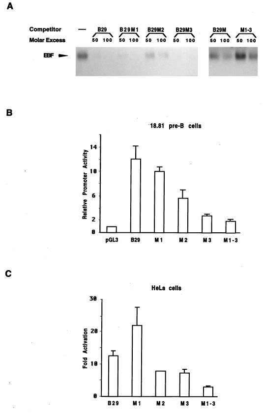
Having established that EBF interacts with functionally important sites in the B29 promoter, we wanted to examine the individual contributions of the sites to promoter function. To this end, we introduced point mutations in site 1, 2, or 3 or in all three binding sites. The contribution of the individual sites to the total EBF affinity of the promoter was examined in competition experiments using recombinant EBF bound to the mb-1 promoter EBF binding site. This suggested that a B29 promoter with a mutation in EBF site 1 still competed efficiently for binding (Fig. 7A). A mutation in site 2 resulted in a decrease in EBF binding, while a mutation in site 3 reduced it but to a lesser extent. This suggests that EBF site 2 is critical for the high affinity of the B29 promoter for EBF. The point mutations were more drastic than those used in our initial analysis (Fig. 3A), and the EMSA experiment illustrated in Fig. 7A shows that the promoter with all three mutations (B29M1–3) did not compete for the binding of recombinant EBF to the mb-1 EBF binding site to the same extent as the previously used promoter mutant (B29M) did (Fig. 7A).
FIG. 7.
EBF binding sites 2 and 3 are important for high-affinity binding and full functional activity of the B29 promoter. (A) Results of an EMSA in which the binding of 0.5 μl of recombinant in vitro-translated EBF to an excess of the mb-1 promoter EBF binding site was competed for by increasing amounts (50 and 100 times molar excess) of wild-type (B29) or EBF-mutated full-length B29 promoters (B29M1, B29M2, B29M3, M1-3, and B29M). The autoradiogram is cut to show only the EBF-DNA complex and not the free probe present in all lanes. (B) Luciferase activity resulting from DEAE-mediated transient transfection of luciferase reporter constructs controlled by wild-type (B29) or EBF site-mutated B29 promoters (M1, M2, M3, and M1-3) into 18–81 pre-B cells. Data represent the results of four experiments. (C) Resulting induction of activity of 250 ng of B29 promoter-controlled luciferase reporter gene in the presence of 400 ng of EBF expression plasmid after Lipofectin-mediated transient transfections into HeLa cells. The DNA content in each transfection was normalized with empty expression plasmid (cDNA3), and the data are taken from three independent transfections. Induction was calculated based on the activity of the different promoter constructs when transfected together with 400 ng of empty expression plasmid.
To examine the functional relevance of the individual sites, we transiently transfected reporter constructs under the control of the mutated promoters into 18–81 pre-B cells. A mutation in site 1 did not affect the function of the promoter, while a mutation in site 2 resulted in a 50% decrease in promoter activity. A mutation in site 3 resulted in a 75% reduction, while a mutation in all the sites resulted in an 80% reduction in promoter activity. The same observation was made in 230–238 pre-B cells (data not shown). The relatively low impact of the mutation on site 2 may be explained by the fact that this region has been suggested to interact with a repressor in B cells. The loss of affinity for the repressor may then compensate for loss of function of the EBF site. To examine this more carefully, we used the same reporter constructs in transient transfections in HeLa cells and assayed the induction of promoter activity in the presence of either 400 ng of empty or EBF-encoding expression plasmid. A mutation in site 1 did not impair induction, while a mutation in either site 2 or 3 resulted in a 50% reduction in reporter gene activation. Mutations in all three sites resulted in an 85% reduction of induced activity. Thus, we conclude that sites 2 and 3 are the binding sites of greatest importance for B29 promoter function.
DISCUSSION
The finding that pro-B cells from mice carrying a mutation in the gene encoding EBF did not express the B29 gene suggested to us that B29 may be a genetic target for EBF. Examination of the promoter sequence revealed three potential EBF binding sites within 160 bp upstream of the main transcription initiation site. All three sites were shown to interact with EBF, although affinities varied from high to low among the sites. Furthermore, we showed that EBF can interact functionally with the B29 promoter since ectopic expression of EBF in HeLa cells resulted in induction of promoter activity. EMSAs showed that EBF bound to the B29 promoter in pre-B-cell extracts and transient transfections showed that the binding sites were important for promoter activity in these cells. Thus, we suggest that EBF plays a critical role in the transcriptional control of the B29 gene in pre-B cells.
EBF is not a unique activator of B29 transcription, since it has been shown that B29 can be expressed in cell lines where EBF is not detected. Thus, B29 transcripts have been detected in both the bone marrow-derived cell line Ba/F3 that lacks EBF (29) and in plasma cell lines that should be devoid of EBF (11). This probably reflects the fact that other DNA binding factors like OCT and Ets family proteins can interact with, and activate, the promoter (25, 31). This resembles the situation found in the mb-1 promoter, where EBF has been shown to be one of several transcription factors with functional importance (4, 9, 33). The mutation of the EBF binding sites also resulted in a slight reduction of promoter activity in plasma cells, suggesting that the EBF binding sites overlap with another factor present in these cells. One such candidate would be the factor defined as BF/FA (18, 30), which has been shown to be involved in octamer-dependent activation of the SP6κ promoter. Even though B29 can be expressed in the absence of EBF, the role of EBF in the activation of B29 may still be pivotal in normal early B-cell development since EBF-deficient mice lack B29 transcripts in the pro-B-cell compartment (19).
The presence of multiple EBF binding sites within the promoter and the fact that several of these sites can be occupied simultaneously even with a large excess of binding sites indicate that EBF may bind cooperatively to sites 2 and 3 in the B29 promoter. This is, however, weakly supported by the functional analysis of the individual sites since only the mutation in site 3 resulted in a significant reduction of promoter activity in pre-B cells. The explanation for this may come from experiments by Omori and Wall (25) that suggested the presence of a repressor activity overlapping with site 2. A mutation in EBF site 2 would then result in the neutralization of both EBF and repressor activities, to result in only a modest loss of function. Functional cooperation also could not be supported by induction experiments with HeLa cells, where a mutation in either site 2 or 3 resulted in only a modest reduction of activity. This could depend on the fact that the EBF levels in these experiments were high, which may compensate for the loss of affinity due to loss of one of the binding sites. The contribution to the function of the independent sites and the ability of EBF to bind cooperatively to target genes are therefore still unclear. However, the large number of EBF target genes carrying multiple EBF binding sites in their promoters suggests that cooperative binding and activation may be a common theme in promoter activation by EBF.
The B29 gene is the fourth important target gene defined for EBF in pre-B cells. It has previously been shown that EBF is an activator of mb-1 (7, 9), as well as of the λ5 and VpreB genes (29). This would suggest that EBF is a pleiotropic activator of early B-cell-specific genes, similar to the role of MyoD in myocyte development (for reviews, see references 36 and 37). In contrast to MyoD, however, there are no data supporting the possibility that EBF would be a master regulator of B-cell development, able to initiate the full developmental program. On the contrary, it appears as if B-cell fate determination is initiated at a stage before EBF expression since EBF−/− mice still develop fraction A pro-B cells. Furthermore, ectopic expression of EBF in the immature hematopoietic cell line Ba/F3 resulted only in activation of certain pre-B-cell-specific genes (29), which would suggest that EBF controls only a subset of genes important for B-cell differentiation. Neither does EBF, in contrast to MyoD, appear to regulate its own transcription (30a). A positive autoregulation may be an important feature for a fate-determining factor to maintain its own activity and drive differentiation processes. An alternative possibility would be that EBF acts together with another transcription factor to initiate the full differentiation program. Such a synergy has been shown in adipogenesis where PPARγ2 and C/EBPα cooperate to drive a fibroblast into an adipocyte (32) and in myogenesis where coexpression of myeloid HLH proteins with MEF2 induces myogenesis (23). One such cooperation partner for EBF is the E2A-encoded HLH transcription factor E47 (for reviews, see references 15 and 24). Mice deficient in a functional E2A gene display a B-cell differentiation block with similarities to that observed in EBF-deficient mice (1b, 19, 38). Furthermore, EBF and E47 have been shown to act in synergy to stimulate transcription of the B-cell-specific genes λ5 and VpreB (29) but, again, they only induced expression of some early B-cell markers in stably transfected Ba/F3 cells. The apparent lack of a master gene activity for B-cell development may reflect an inherent difference in the nature of the developmental process from a fibroblast to a myocyte, as compared to the more complex B-lymphoid differentiation process.
Even though both λ5 and B29 have been shown to be essential for early B-cell development (6, 16), their absence cannot explain the early B-cell differentiation block in EBF-deficient mice. This makes further identification of genetic targets for EBF an important issue in the effort to understand the earliest stages of B-cell development.
ACKNOWLEDGMENTS
We thank R. Grosschedl for the kind gift of the EBF expression plasmid.
This work was funded by the Swedish Medical Research Council, American Cancer Society, The Swedish Cancer Society, and The Crafoord, Österlunds, and Kocks Foundations.
REFERENCES
- 1.Åkerblad P, Sigvardsson M, Leanderson T. Early B cell factor (EBF) down regulates immunoglobulin heavy chain intron enhancer function in a plasmacytoma cell line. Scand J Immunol. 1996;44:145–149. doi: 10.1046/j.1365-3083.1996.d01-283.x. [DOI] [PubMed] [Google Scholar]
- 1a.Åkerblad, P. Unpublished data.
- 1b.Bain G, Robanus Maandag E, Izon D, Amsen D, Kruisbeek A, Weintraub B, Krop I, Schlissel M, Feeney A, van Roon M, van der Valk M, te Riele H, Berns A, Murre C. E2A proteins are required for proper B cell development and initiation of immunoglobulin gene rearrangements. Cell. 1994;79:885–892. doi: 10.1016/0092-8674(94)90077-9. [DOI] [PubMed] [Google Scholar]
- 2.Borst J, Jacobs H, Brouns G. Composition and function of T cell receptor and B cell receptor complexes on precursor lymphocytes. Curr Opin Immunol. 1996;8:181–190. doi: 10.1016/s0952-7915(96)80056-2. [DOI] [PubMed] [Google Scholar]
- 3.Clevers H, Grosschedl R. Transcriptional control of lymphoid development: lessons from gene targeting. Immunol Today. 1996;17:336–343. doi: 10.1016/0167-5699(96)10019-0. [DOI] [PubMed] [Google Scholar]
- 4.Fitzsimmons D, Hodsdon W, Wheat W, Maira S-M, Wasylyk B, Hagman J. Pax-5 (BSAP) recruits Ets proto-oncogene family proteins to form functional ternary complexes on a B-cell-specific promoter. Genes Dev. 1996;10:2198–2211. doi: 10.1101/gad.10.17.2198. [DOI] [PubMed] [Google Scholar]
- 5.Georgopoulos K. Transcription factors required for lymphoid lineage commitment. Curr Opin Immunol. 1997;9:222–227. doi: 10.1016/s0952-7915(97)80139-2. [DOI] [PubMed] [Google Scholar]
- 6.Gong S, Nussenzweig M. Regulation of an early developmental checkpoint in the B cell pathway by Igβ. Science. 1996;272:411–414. doi: 10.1126/science.272.5260.411. [DOI] [PubMed] [Google Scholar]
- 7.Hagman J, Belanger C, Travis A, Turck C, Grosschedl R. Cloning and functional characterization of early B-cell factor, a regulator of lymphocyte-specific gene expression. Genes Dev. 1993;7:760–773. doi: 10.1101/gad.7.5.760. [DOI] [PubMed] [Google Scholar]
- 8.Hagman J, Gutch M, Lin H, Grosschedl R. EBF contains a novel zinc coordination motif and multiple dimerization and transcriptional activation domains. EMBO J. 1995;14:2907–2916. doi: 10.1002/j.1460-2075.1995.tb07290.x. [DOI] [PMC free article] [PubMed] [Google Scholar]
- 9.Hagman J, Travis A, Grosschedl R. A novel lineage-specific nuclear factor regulates mb-1 gene transcription at the early stages of B cell differentiation. EMBO J. 1991;10:3409–3417. doi: 10.1002/j.1460-2075.1991.tb04905.x. [DOI] [PMC free article] [PubMed] [Google Scholar]
- 10.Hardy R, Carmack C, Shinton S, Kemp J, Hayakawa K. Resolution and characterization of pro-B and pre-pro-B cell stages in normal mouse bone marrow. J Exp Med. 1991;173:1213–1225. doi: 10.1084/jem.173.5.1213. [DOI] [PMC free article] [PubMed] [Google Scholar]
- 11.Hermanson G, Eisenberg D, Kincade P, Wall R. B29 a member of the immunoglobulin gene superfamily exclusively expressed on beta lineage cells. Proc Natl Acad Sci USA. 1988;85:6890–6894. doi: 10.1073/pnas.85.18.6890. [DOI] [PMC free article] [PubMed] [Google Scholar]
- 12.Hermanson G, Briskin M, Sigman D, Wall R. Immunoglobulin enhancer and promoter motifs 5′ of the B29 B-cell specific gene. Proc Natl Acad Sci USA. 1989;86:7341–7345. doi: 10.1073/pnas.86.19.7341. [DOI] [PMC free article] [PubMed] [Google Scholar]
- 13.Hombach J, Lottspeich F, Reth M. Identification of the genes encoding the Ig-α and Ig-β components of the IgM antigen receptor complex by aminoterminal sequencing. Eur J Immunol. 1990;20:2795–2799. doi: 10.1002/eji.1830201239. [DOI] [PubMed] [Google Scholar]
- 14.Hombach J, Tsubata T, Leclercq L, Stappert H, Reth M. Molecular components of the B-cell antigen receptor complex of the IgM class. Nature. 1990;343:760–762. doi: 10.1038/343760a0. [DOI] [PubMed] [Google Scholar]
- 15.Kadesch T. Helix-loop-helix proteins in the regulation of immunoglobulin gene transcription. Immunol Today. 1992;13:31–36. doi: 10.1016/0167-5699(92)90201-h. [DOI] [PubMed] [Google Scholar]
- 16.Kitamura D, Kudo A, Schaal S, Muller W, Melchers F, Rajewsky K. A critical role of λ5 protein in B cell development. Cell. 1992;69:823–831. doi: 10.1016/0092-8674(92)90293-l. [DOI] [PubMed] [Google Scholar]
- 17.Li Y-S, Wasserman R, Hayakawa K, Hardy R. Identification of the earliest B lineage stage in mouse bone marrow. Immunity. 1996;5:527–535. doi: 10.1016/s1074-7613(00)80268-x. [DOI] [PubMed] [Google Scholar]
- 18.Liberg D, Sigvardsson M, Bemark M, Leanderson T. Differentiation specific, octamer dependent costimulation of κ transcription. J Immunol. 1998;160:3899–3907. [PubMed] [Google Scholar]
- 19.Lin H, Grosschedl R. Failure of B-cell differentiation in mice lacking the transcription factor EBF. Nature. 1995;376:263–267. doi: 10.1038/376263a0. [DOI] [PubMed] [Google Scholar]
- 20.Malone C, Omori S, Wall R. Silencer elements controlling the B29 (Igβ) promoter are neither promoter nor cell-type-specific. Proc Natl Acad Sci USA. 1997;94:12314–12319. doi: 10.1073/pnas.94.23.12314. [DOI] [PMC free article] [PubMed] [Google Scholar]
- 21.Mårtensson A, Mårtensson I-L. Early B cell factor binds to a site critical for lambda5 core enhancer activity. Eur J Immunol. 1997;27:315–320. doi: 10.1002/eji.1830270145. [DOI] [PubMed] [Google Scholar]
- 22.Melchers F, Karasuyama H, Haasner D, Bauer S, Kudo A, Sakaguchi N, Jameson B, Rolink A. The surrogate light chain in B-cell development. Immunol Today. 1993;14:60–68. doi: 10.1016/0167-5699(93)90060-X. [DOI] [PubMed] [Google Scholar]
- 23.Molkentin J, Black B, Martin J, Olson E. Cooperative activation of muscle gene expression by MEF" and myogenic bHLH proteins. Cell. 1995;83:1125–1136. doi: 10.1016/0092-8674(95)90139-6. [DOI] [PubMed] [Google Scholar]
- 24.Murre C, Bain G, van Dijk M, Engel I, Furnari B, Massari M, Matthews J, Quong M, Rivera R, Stuiver M. Structure and function of helix-loop-helix proteins. Biochim Biophys Acta. 1994;1218:129–135. doi: 10.1016/0167-4781(94)90001-9. [DOI] [PubMed] [Google Scholar]
- 25.Omori S, Wall R. Multiple motifs regulate the B cell specific promoter of the B29 gene. Proc Natl Acad Sci USA. 1993;90:11732–11727. doi: 10.1073/pnas.90.24.11723. [DOI] [PMC free article] [PubMed] [Google Scholar]
- 26.Persson C, Måartensson A, Måartensson I-L. Identification of a tissue and differentiation stage specific enhancer of the VpreB1 gene. Eur J Immunol. 1998;28:787–798. doi: 10.1002/(SICI)1521-4141(199803)28:03<787::AID-IMMU787>3.0.CO;2-H. [DOI] [PubMed] [Google Scholar]
- 27.Schreiber E, Matthias P, Muller M, Schaffner W. Rapid detection of octamer binding proteins with “mini-extracts” prepared from a small number of cells. Nucleic Acids Res. 1989;17:6419. doi: 10.1093/nar/17.15.6419. [DOI] [PMC free article] [PubMed] [Google Scholar]
- 28.Sigvardsson M, Grosschedl R. Transcriptional control of B cell differentiation by EBF and E47. In: Monroe J G, Rothenberg E V, editors. Molecular biology of B and T cell development. Totowa, N.J: Humana Press Inc.; 1998. [Google Scholar]
- 29.Sigvardsson M, Oriordan M, Grosschedl R. EBF and E47 collaborate to induce expression of the endogenous immunoglobulin surrogate light chain genes. Immunity. 1997;7:25–36. doi: 10.1016/s1074-7613(00)80507-5. [DOI] [PubMed] [Google Scholar]
- 30.Sigvardsson M, Åkerblad P, Leanderson T. Early B cell factor interacts with a subset of κ promoters. J Immunol. 1996;156:3788–3796. [PubMed] [Google Scholar]
- 30a.Sigvardsson, M. Unpublished observation.
- 31.Thompson A, Wood W, Gilly M, Damore M, Omori S, Wall R. The promoter and 5′ flanking sequences controlling human B29 gene expression. Blood. 1996;87:666–673. [PubMed] [Google Scholar]
- 32.Tontonoz P, Hu E, Spiegelman M. Stimulation of adipogenesis in fibroblasts by PPARg2, a lipid-activated transcription factor. Cell. 1994;79:1147–1156. doi: 10.1016/0092-8674(94)90006-x. [DOI] [PubMed] [Google Scholar]
- 33.Travis A, Hagman J, Grosschedl R. Heterogeneously initiated transcription from the pre-B- and B-cell-specific mb-1 promoter: analysis of the requirement for upstream factor-binding sites and initiation site sequences. Mol Cell Biol. 1991;11:5756–5766. doi: 10.1128/mcb.11.11.5756. [DOI] [PMC free article] [PubMed] [Google Scholar]
- 34.Travis A, Hagman J, Hwang L, Grosschedl R. Purification of early-B-cell factor and characterization of its DNA-binding specificity. Mol Cell Biol. 1993;13:3392–3400. doi: 10.1128/mcb.13.6.3392. [DOI] [PMC free article] [PubMed] [Google Scholar]
- 35.Wang M, Reed R. Molecular cloning of the olfactory neuronal transcription factor Olf-1 by genetic selection in yeast. Nature. 1993;364:121–126. doi: 10.1038/364121a0. [DOI] [PubMed] [Google Scholar]
- 36.Weintraub, H., R. Davis, S. Tapscott, M. Thayer, M. Krause, R. Benezra, T. Blackwell, D. Turner, R. Rupp, S. Hollenberg, Y. Zhuang, and A. Lassar. 1991. The myoD gene family: nodal point during specification of the muscle cell lineage. 251:761–766. [DOI] [PubMed]
- 37.Wright W. Muscle basic helix-loop-helix proteins and the regulation of myogenesis. Curr Opin Genet Dev. 1992;2:243–248. doi: 10.1016/s0959-437x(05)80280-1. [DOI] [PubMed] [Google Scholar]
- 38.Zhuang Y, Soriano P, Weintraub H. The helix-loop-helix gene E2A is required for B cell formation. Cell. 1994;79:875–884. doi: 10.1016/0092-8674(94)90076-0. [DOI] [PubMed] [Google Scholar]


