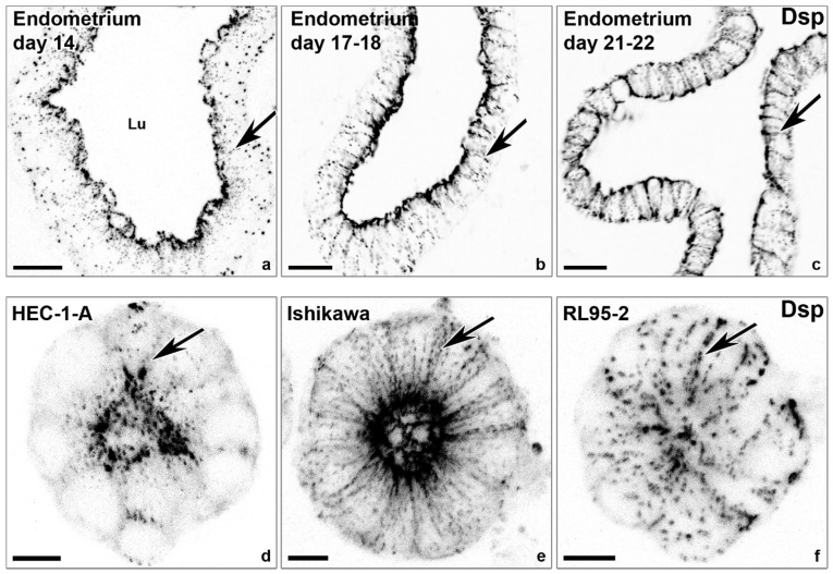
Figure 4.
The lateralization of desmosomal cell–cell adhesions coincides with the window of implantation and polarization of endometrial epithelial cell lines. The confocal fluorescence micrographs (inverse presentation; single focal planes are shown in (a–c); the projections of 8 consecutive focal planes shown in (d–f)) reveal anti-desmoplakin (Dsp) reactivity that detects punctate desmosomes in the endometrial epithelial cell layer of the human endometrium obtained at different days of the menstrual cycle (a–c) and in gland-like spheroids (d–f) derived from endometrial adenocarcinoma cell lines with high polarity (HEC-1-A), intermediate polarity (Ishikawa), and low polarity (RL95-2). Note the different distributions of desmoplakin-positive desmosomes along the basolateral plasma membrane (arrows). Lu, lumen. Scale bars: 20 μm (a–c), 10 μm (d–f). The images were modified from [27,31].

