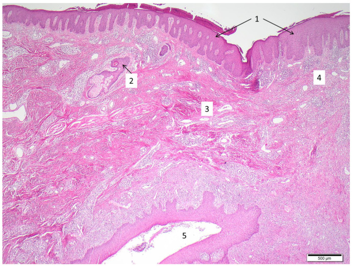
Figure 2.
Typical histological features of HS. Sample acquired by punch biopsy from gluteal region. Hyperparakeratosis and papillomatosis (1), follicular hyperkeratosis and perifolliculitis (2), fibrosis (3), abscess-like accumulation of neutrophils and spotted infiltrate of lymphocytes/plasma cells (4), epithelialized sinus tract with surrounding inflammatory reaction (5).

