Abstract
Background
The impact of primary tumour location on the prognosis of patients with peritoneal metastasis (PM) arising from colorectal cancer (CRC) after cytoreductive surgery (CRS) and hyperthermic intraperitoneal chemotherapy (HIPEC) is rarely discussed, and the evidence is still limited.
Methods
Patients with PM arising from CRC treated with CRS and HIPEC at the China National Cancer Center and Huanxing Cancer Hospital between June 2017 and June 2019 were systematically reviewed. Clinical characteristics, pathological features, perioperative parameters, and prognostic data were collected and analysed.
Results
A total of 70 patients were divided into two groups according to either colonic or rectal origin (18 patients in the rectum group and 52 patients in the colon group). Patients with PM of a colonic origin were more likely to develop grade 3–4 postoperative complications after CRS+HIPEC (38.9% vs 19.2%, P = 0.094), but this difference was not statistically significant. Patients with colon cancer had a longer median overall survival (OS) than patients with rectal cancer (27.0 vs 15.0 months, P = 0.011). In the multivariate analysis, the independent prognostic factors of reduced OS were a rectal origin (HR 2.15, 95% CI 1.15–4.93, P = 0.035) and incomplete cytoreduction (HR 1.99, 95% CI 1.06–4.17, P = 0.047).
Conclusion
CRS is a complex and potentially life-threatening procedure, and we suggest that the indications for CRS+HIPEC in patients with PM of rectal origin be more restrictive and that clinicians approach these cases with caution.
Keywords: Cytoreductive surgery, Hyperthermic intraperitoneal chemotherapy, Primary tumour location, Prognosis
Introduction
Colorectal cancer (CRC) is one of the most common malignant tumours in the world, and its morbidity and mortality rank third and fourth, respectively [1–3]. Among patients with CRC, 5–15% have synchronous peritoneal metastasis (PM), and the incidence of metachronous PM is as high as 20 to 50% [4]. PM arising from CRC is an indicator of terminal stage disease and carries a very poor prognosis. In the past, palliative surgery and systemic chemotherapy were mostly adopted, but the therapeutic effect was poor, and the median survival time was only 5 to 7 months [5]. Currently, cytoreductive surgery (CRS) combined with hyperthermic intraperitoneal chemotherapy (HIPEC) has shown good clinical efficacy in the comprehensive treatment of various malignant peritoneal diseases, including CRC, ovarian cancer, and appendiceal mucous adenocarcinoma, and it has been considered a standard therapy for prolonging the survival of patients with PM arising from CRC [6–10].
At present, most of the extant literature has demonstrated that a high peritoneal cancer index (PCI), incomplete cytoreduction, young age, lymphovascular invasion, and postoperative complications are poor prognostic factors after CRS+HIPEC [11–15]. It has been well established that different primary tumour locations have different biological behaviours and prognosis [16–19]. However, previous studies have only compared the prognostic differences between the colonic origin and rectal origin of the primary tumour; the impact of the primary tumour location on the prognosis of patients with PM arising from CRC after CRS+HIPEC is rarely discussed, and clinical evidence remains scarce [20]. It is highly desirable to optimize patient selection to include only those who are most likely to benefit from this complex and potentially life-threatening procedure. Therefore, the aim of this study was to explore the impact of primary tumour location according to colon or rectal origin on the prognosis of patients with PM arising from CRC treated with CRS+HIPEC in our institution.
Methods
Study design and patients
The study protocol was approved by the Ethics Committee of the Cancer Hospital, Chinese Academy of Medical Sciences (NCC2017-YZ-026, October 17, 2017). The data of all patients with synchronous or metachronous PM arising from CRC who underwent CRS with HIPEC at the National Cancer Center and Huanxing Cancer Hospital were retrospectively obtained from a prospectively maintained database between June 2017 and June 2019. The inclusion criteria of this study were as follows: (1) patients with PM of a colonic or rectal origin, (2) pathologically confirmed PM after operation, and (3) age between 18 and 75 years. The exclusion criteria were as follows: (1) complications with the liver, lung, or other sites of distant metastasis; (2) history of other malignant tumours; and (3) malignant tumour of appendix origin. According to the location of the primary tumour, all enrolled patients were divided into a colon group (n = 52) and a rectal group (n = 18). The colon was regarded as the caecum, ascending colon, transverse colon descending colon, and sigmoid colon, while the rectum was regarded as the intestinal canal below 15 cm from the anal margin.
Demographic and clinical variables, as well as perioperative and long-term survival outcomes, were collected and compared. All enrolled patients underwent a routine preoperative evaluation, which included laboratory examinations, abdominal contrast-enhanced computed tomography, pelvic magnetic resonance imaging, and fluorodeoxyglucose positron emission tomography, to assess their general condition. According to the Sugarbaker/Jacquet classification, peritoneal disease burden and the completeness of cytoreduction were assessed using the PCI and completeness of cytoreduction (CC) score, respectively [21, 22]. All postoperative complications were graded using the Clavien-Dindo classification according to the treatment received [23].
Surgical technique
The surgical techniques adopted at our institution have been previously described [9, 21]. Briefly, three outflow drains and one inflow drain were routinely placed in the abdomen in preparation for HIPEC. HIPEC was administered in a closed fashion, with oxaliplatin (200 mg/m2) and raltitrexed (3 mg/m2), combined with or without lobaplatin (50 mg/m2). Then, patients were treated with a mixed solution of chemotherapy agents and 3 l of saline solution infused into the abdominal and pelvic cavity for 60 min at 42–43 °C. Next, two additional HIPEC procedures were performed in the ward on the second and fourth days after surgery in both groups. Furthermore, two surgical specialists with more than 20 years of experience in gastrointestinal surgery performed the operations at the two centres, and the HIPEC technique and postoperative treatment were identical.
Statistical analysis
Data between two groups were analysed with SPSS 24.0 software (IBM, Armonk, NY, USA). Categorical data are expressed as percentages and were compared using the χ2 test or Fisher’s exact test as appropriate. Continuous data are expressed as the mean ± standard deviation and were compared using Student’s t-test and the Mann-Whitney U test for independent values with normally and nonnormally distributed values, respectively. Overall survival (OS) was defined as the time from surgery to the time of death from any cause or July 31, 2020, whichever came first. The Kaplan-Meier method and log-rank test were utilized to evaluate associations between individual factors and OS. Variables found to be significant (P value < 0.20) in the univariate analysis were incorporated into the multivariate analysis to identify independent predictors of OS. A P value < 0.05 was considered statistically significant.
Results
Demographic and clinical variables
A total of 70 patients with PM of CRC origin who underwent CRS+HIPEC were included in the present study. Of these patients, 18 (25.7%) had rectal cancer and 52 (74.3%) had colon cancer. The mean age of all patients was 54.5 years, and the majority (55.7%) of patients in the study were male. Patients were well balanced across the two groups in terms of age, sex, body mass index (BMI), preoperative comorbidity, preoperative chemotherapy, presentation of PM, preoperative CEA level, preoperative CA19-9 level, histology, tumour grade, adjuvant chemotherapy, BRAF status, and MSI (P > 0.05) (Table 1).
Table 1.
Patient characteristics
| Characteristics | Total (n = 70) | Rectum (n = 18) | Colon (n = 52) | P |
|---|---|---|---|---|
| Age at operation (years, mean ± SD) | 54.5 ± 11.6 | 52.7 ± 12.2 | 55.2 ± 11.6 | 0.451 |
| Sex (%) | 0.571 | |||
| Male | 39 (55.7) | 9 (50.0) | 30 (57.7) | |
| Female | 31 (44.3) | 9 (50.0) | 22 (42.3) | |
| Body mass index (kg/m2, mean ± SD) | 22.7 ± 3.6 | 23.8 ± 3.9 | 22.8 ± 3.5 | 0.665 |
| Comorbidity | 18 (25.7) | 4 (22.2) | 14 (26.9) | 0.936 |
| Hypertension | 10 (14.3) | 2 (11.1) | 8 (15.4) | |
| Diabetes | 6 (8.6) | 2 (11.1) | 4 (7.7) | |
| Coronary heart disease | 2 (2.9) | 1 (5.5) | 1 (1.9) | |
| Arrhythmia | 4 (5.7) | 0 (0) | 4 (7.7) | |
| Others | 6 (8.6) | 1 (5.5) | 5 (9.6) | |
| Preoperative chemotherapy (%) | 0.477 | |||
| Presence | 30 (42.9) | 9 (50.0) | 21 (40.4) | |
| Absence | 40 (57.1) | 9 (50.0) | 31 (59.6) | |
| Presentation of PM (%) | 0.118 | |||
| Synchronous | 42 (60.0) | 8 (44.4) | 34 (65.4) | |
| Metachronous | 28 (40.0) | 10 (55.6) | 18 (34.6) | |
| T stage | 0.954 | |||
| T1–T2 | 5 (11.9) | 1 (12.5) | 4 (11.8) | |
| T3–T4 | 37 (88.1) | 7 (87.5) | 30 (88.2) | |
| N stage | 0.482 | |||
| N0 | 2 (4.8) | 0 (0) | 2 (5.9) | |
| N1–N2 | 40 (95.2) | 8 (100.0) | 32 (94.1) | |
| Preoperative CEA level (ng, mean ± SD) | 31.9 ± 61.5 | 16.6 ± 27.2 | 37.2 ± 69.8 | 0.290 |
| Preoperative CA19-9 level (ng, mean ± SD) | 75.4 ± 93.3 | 69.2 ± 112.4 | 77.5 ± 88.4 | 0.780 |
| Histology (%) | 0.275 | |||
| Adenocarcinoma | 43 (61.4) | 13 (72.2) | 30 (57.7) | |
| Mucinous | 27 (38.6) | 5 (27.8) | 22 (42.3) | |
| Tumour grade | 0.370 | |||
| Moderate | 26 (37.1) | 8 (44.4) | 17 (32.7) | |
| Poor | 44 (62.9) | 10 (55.6) | 35 (67.3) | |
| Adjuvant chemotherapy | 0.924 | |||
| Presence | 55 (78.6) | 14 (77.8) | 41 (78.8) | |
| Absence | 15 (21.4) | 4 (22.2) | 11 (21.2) | |
| BRAF status | 0.809 | |||
| Mutation | 13 (18.6) | 3 (16.7) | 10 (19.2) | |
| No mutation | 57 (81.4) | 15 (83.3) | 42 (80.8) | |
| MSI | 0.961 | |||
| MSI-H | 8 (11.4) | 2 (11.1) | 6 (11.5) | |
| MSS | 62 (88.6) | 16 (88.9) | 46 (88.5) |
Note: SD standard deviation, PM standard peritoneal metastasis
Operative and perioperative data
The operative details and postoperative courses are listed in Table 2. The mean PCI of all enrolled patients was 11.1, and complete cytoreduction (CC 0–1) was achieved in most patients (68.6%). Patients in both groups had comparable mean operative times (255.5 vs 257.4 min, P = 0.922) and estimated blood loss (98.9 vs 130.2 ml, P = 0.301). Patients in the rectal group were more likely to undergo colostomy or ileostomy (66.7% vs 30.8%, P = 0.007) during the operation. Compared with patients in the colon group, patients in the rectum group were more likely to develop grade 3–4 postoperative complications (38.9% vs 19.2%, P = 0.094), but this difference was not statistically significant. Ileus (7.1%) and pelvic cavity abscesses (7.1%) were the most common postoperative complications, followed by anastomotic leakage (5.7%), wound infection (2.9%), pneumonia (5.4%), pleural effusion (1.4%), cardiac arrhythmia (1.4%), urinary retention (1.4%), and rectovaginal leakage (1.4%). Two patients (2.9%) required revision surgery due to extensive pelvic cavity abscesses and postoperative bleeding.
Table 2.
Operative and perioperative data
| Characteristic | Total (n = 70) | Rectum (n = 18) | Colon (n = 52) | P |
|---|---|---|---|---|
| Operative method | 0.538 | |||
| Laparoscopic surgery | 14 (20.0) | 5 (27.8) | 9 (17.3) | |
| Open surgery | 56 (80.0) | 13 (72.2) | 43 (82.7) | |
| HIPEC regimen | 0.900 | |||
| Lobaplatin + oxaliplatin + raltitrexed | 32 (45.7) | 8 (44.4) | 24 (46.2) | |
| Oxaliplatin + raltitrexed | 38 (54.3) | 10 (55.6) | 28 (53.8) | |
| Colostomy or ileostomy | 0.007 | |||
| Presence | 28 (40.0) | 12 (66.7) | 16 (30.8) | |
| Absence | 42 (60.0) | 6 (33.3) | 36 (69.2) | |
| PCI score (mean ± SD) | 11.1 ± 6.0 | 11.7 ± 6.9 | 10.8 ± 5.8 | 0.600 |
| Presence of ascites | 0.693 | |||
| Presence | 30 (42.9) | 7 (38.9) | 23 (44.2) | |
| Absence | 40 (57.1) | 11 (61.1) | 29 (55.8) | |
| CC score | 0.168 | |||
| CC 0–1 | 48 (68.6) | 10 (55.5) | 38 (73.1) | |
| CC 2–3 | 22 (31.4) | 8 (44.5) | 14 (26.9) | |
| Operative time, min (mean ± SD) | 256.9 ± 66.0 | 255.5 ± 83.5 | 257.4 ± 60.4 | 0.922 |
| Estimated blood loss, ml (mean ± SD) | 122.1 ± 109.2 | 98.9 ± 82.9 | 130.2 ± 117.5 | 0.301 |
| Postoperative complications (grades III, IV) | 17 (24.3) | 7 (38.9) | 10 (19.2) | 0.094 |
| Postoperative bleeding | 2 (2.9) | 1 (5.6) | 1 (1.9) | |
| Anastomotic leakage | 4 (5.7) | 2 (11.1) | 2 (3.8) | |
| Pelvic cavity abscess | 5 (7.1) | 2 (11.1) | 3 (5.8) | |
| Ileus | 5 (7.1) | 2 (11.1) | 3 (5.8) | |
| Pneumonia | 1 (1.4) | 1 (5.6) | 0 (0) | |
| Pleural effusion | 1 (1.4) | 1 (5.6) | 0 (0) | |
| Cardiac arrhythmia | 1(1.4) | 0 (0) | 1 (1.9) | |
| Wound infection | 2 (2.9) | 1 (5.6) | 1 (1.9) | |
| Urinary retention | 1 (1.4) | 0 (0) | 1 (1.9) | |
| Rectovaginal leakage | 1 (1.4) | 0 (0) | 1 (1.9) | |
| Postoperative hospital stay, days (mean ± SD) | 14.6 ± 5.3 | 15.4 ± 4.7 | 14.3 ± 5.6 | 0.380 |
| Re-operation | 2 (2.9) | 1 (5.6) | 1 (5.6) | 1.000 |
| Mortality | 0 (0) | 0 (0) | 0 (0) | – |
Note: HIPEC standard hyperthermic intraperitoneal chemotherapy, PCI standard peritoneal cancer index, CC standard complete cytoreduction
Overall survival
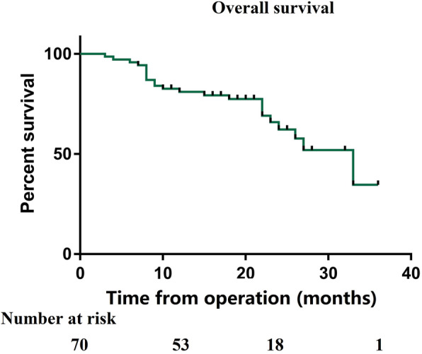
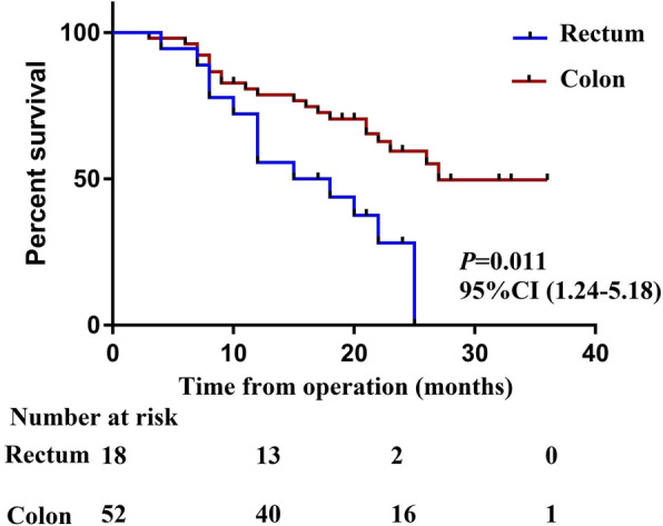
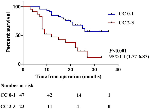
The median estimated follow-up period from CRS/HIPEC for the study population was 28 months. The median survival period for all patients was 25 months, and the estimated 1-, 2- and 3-year OS rates for the entire cohort were 72.6%, 51.4%, and 40.1%, respectively (Fig. 1). The median OS for those with colon cancer was 27 months compared with 15 months for those with rectal cancer (P = 0.011) (Fig. 2). The median OS from CRS+HIPEC in patients undergoing incomplete cytoreduction (CC 2–3) was 12 months, while the median OS was not reached in patients undergoing complete cytoreduction (CC 0–1) (Fig. 3). Variables with P < 0.20 in the univariate regression analysis, such as increasing PCI (HR 1.09, 95% CI 1.03–1.14, P = 0.002), rectal origin (HR 2.54, 95% CI 1.24–5.18, P = 0.011), incomplete cytoreduction (HR 3.49, 95% CI 1.77–6.87, P < 0.001), and HIPEC regimen (HR 0.63, 95% CI 0.31–1.28, P = 0.199), were included in the multivariate analysis. In the multivariate analysis, independent prognostic factors of reduced OS were rectal origin (HR 2.15, 95% CI 1.15–4.93, P = 0.035) and incomplete cytoreduction (HR 1.99, 95% CI 1.06–4.17, P = 0.047) (Table 3).
Fig. 1.

Overall survival of the entire cohort
Fig. 2.

The median OS for those with colon cancer vs those with rectal cancer
Fig. 3.

The median OS from CRS+HIPEC in patients undergoing incomplete cytoreduction vs complete cytoreduction
Table 3.
Univariate analysis and multivariate analysis
| Variables | Overall survival | |||
|---|---|---|---|---|
| Univariate analysis | Multivariate analysis | |||
| HR (95% CI) | P | HR (95% CI) | P | |
| Sex: male/female | 1.32 (0.66–2.64) | 0.434 | ||
| Age at operation | 1.02 (0.98–1.05) | 0.283 | ||
| Preoperative chemotherapy (no/yes) | 1.22 (0.53–2.80) | 0.635 | ||
| Synchronous/metachronous | 1.46 (0.73–2.93) | 0.288 | ||
| Site of original (rectum/colon) | 2.54 (1.24–5.18) | 0.011 | 2.15 (1.15–4.93) | 0.035 |
| Histology (mucinous/adenocarcinoma) | 1.53 (0.78–3.00) | 0.215 | ||
| Preoperative CEA level | 1.00 (0.99–1.00) | 0.279 | ||
| Preoperative CA19-9 level | 1.00 (0.99–1.00) | 0.247 | ||
| HIPEC regimen (lobaplatin/non-lobaplatin) | 0.63 (0.31–1.28) | 0.199 | 1.39 (0.65–2.94) | 0.394 |
| Presence of ascites (yes/no) | 1.33 (0.68–2.60) | 0.410 | ||
| PCI score | 1.09 (1.03–1.14) | 0.002 | 1.05 (0.98–1.12) | 0.140 |
| CC score (2–3/0–1) | 3.49 (1.77–6.87) | < 0.001 | 1.99 (1.06–4.17) | 0.047 |
| Grade 3–4 postoperative complication (no/yes) | 1.63 (0.77–3.42) | 0.201 | ||
| Leukopenia (no/yes) | 0.67 (0.28–1.63) | 0.382 | ||
| Neutropenia (no/yes) | 0.80 (0.33–1.94) | 0.626 | ||
| Thrombocytopenia (no/yes) | 0.49 (0.15–1.63) | 0.245 | ||
| BRAF status (mutation/no mutation) | 1.58 (0.79–3.13) | 0.266 | ||
| MSI (MSS/MSI-H) | 1.27 (0.61–2.73) | 0.573 | ||
| Adjuvant therapy (yes/no) | 0.76 (0.35–1.65) | 0.489 | ||
| Colostomy or ileostomy (yes/no) | 1.39 (0.71–2.74) | 0.334 | ||
Discussion
Primary tumour location is recognized as an important prognostic factor for metastatic CRC, and it is also a selection factor for the administration of different targeted medicines [16–19]. However, the impact of primary tumour location on the prognosis of CRC patients undergoing CRS+HIPEC due to PM is rarely discussed, so the available evidence remains limited [20]. Therefore, we conducted this study to elucidate the differences in different primary tumour locations among patients with PM arising from CRC and focused on the significant impacts of these differences on perioperative outcomes and long-term prognosis.
In the present study, 70 enrolled patients were divided into two groups according to the origin of the primary tumour: the colon group (52 patients) and the rectal group (18 patients). The average age of the patients included in this study was only 54.5 years old, and only 25.7% of patients had comorbidities before surgery. This may be due to the aggressive tumour behaviour observed in young patients; these tumours show high invasiveness and a predilection towards distant metastases in regions such as the peritoneum in this population. Our results revealed that patients with colon cancer-derived PM had a longer median survival after CRS+HIPEC (27.0 vs 15.0 months, P = 0.011), and primary tumour location remained an independent predictor of OS (HR 2.15, 95% CI 1.15–4.93, P = 0.035). In 2018, Tonello et al. [17] published a paper in which they analysed survival in patients with colorectal PM treated with CRS+HIPEC and reported that PM of a rectal origin was associated with worse long-term survival outcomes than PM of a colonic origin (median OS 47.8 vs 22.0 months, P = 0.008). Similarly, Da Silva et al. [16] also demonstrated that the median survival in patients with PM of a colonic origin was significantly better than that in patients with PM of a rectal origin (35.0 vs 17.0 months). The above research results are basically consistent with our findings.
Several theories have been proposed to explain this difference in terms of the prognosis of PM of a rectal origin. Anatomically, rectal tumours are located in a narrow pelvic cavity, which makes resection of the primary tumour and pelvic peritoneal metastasis difficult; therefore, achieving complete cytoreduction is a challenge, and the possibility of a residual tumour is increased [24]. Low-middle rectal cancer (under the peritoneum) increases the risk of perforating the rectal wall, which is thicker than the colon wall; the thickness of the rectal wall is the reason underlying the more biologically aggressive disease characteristics observed in this population [16]. Finally, patients with peritoneal metastases originating from rectal cancer are more likely to develop postoperative complications, and the occurrence of complications negatively affects the overall condition of the patients, as well as subsequent adjuvant treatment, and thus has an impact on prognosis. However, the above mechanisms are limited to only a theoretical level; additional studies are needed in the future to further explain the differences in the prognosis of patients with PM arising from different sites of origin at the genetic level.
Close attention has been given to the morbidity and mortality associated with the CRS+HIPEC procedure. Our institution confirmed that the grade 3–4 morbidity and mortality rates after CRS+HIPEC were 24.3% and 0%, respectively, which is basically consistent with the results reported by international centres [9, 14, 25–27]. Notably, we also found that patients with PM of a rectal origin were more likely to develop grade 3–4 postoperative complications after CRS+HIPEC than patients with PM of a colonic origin (38.9% vs 19.2%, P = 0.094), but this difference was not statistically significant. CRS is an originally complex and potentially life-threatening procedure. Due to the special anatomical location of rectal tumours, the narrow operating space further increases the difficulty of CRS.
The limitations of this study are those inherent to a single institution with a limited sample size, which may underlie some of the differences observed between the rectal group and the colon group. Second, this study was also limited by its retrospective nature, which makes it difficult to control for bias and confounders. Therefore, we recommend that clinicians exert caution when making any definitive conclusions. Multicentre prospective randomized controlled studies are required to further verify our results.
Conclusion
Patients with PM of a rectal origin were more likely to develop postoperative complications after CRS+HIPEC, which is indicative of a poor prognosis. We suggest that the indications for CRS+HIPEC in patients with PM of rectal origin should be more restrictive and cautious.
Acknowledgements
Not applicable.
Abbreviations
- PM
Peritoneal metastasis
- CRC
Colorectal cancer
- CRS
Cytoreductive surgery
- HIPEC
Hyperthermic intraperitoneal chemotherapy
- PCI
Peritoneal cancer index
- CC
Completeness of cytoreduction
- OS
Overall survival
- BMI
Body mass index
Authors’ contributions
Contributions: (I) conception and design: JX and WP; (II) administrative support: JL, XW, and ZZ; (III) provision of study materials or patients: JB, QF, and ZJ; (IV) collection and assembly of data: SZ and HC; (V) data analysis and interpretation: SZ and HC. All authors read and approved the final manuscript.
Funding
This work was supported by Capital’s Funds for Health Improvement and Research (2016-2-4022). The funding source was not involved in the preparation of the article.
Availability of data and materials
The datasets generated and/or analysed during the current study are not publicly available because the data are confidential patient data but are available from the corresponding author upon reasonable request.
Declarations
Ethics approval and consent to participate
The Ethics Committee of the National Cancer Center/Cancer Hospital, Chinese Academy of Medical Sciences and Peking Union Medical College, approved this study. Prior written informed consent was obtained from all study participants.
Consent for publication
Not applicable.
Competing interests
The authors declare that they have no competing interests.
Footnotes
Publisher’s Note
Springer Nature remains neutral with regard to jurisdictional claims in published maps and institutional affiliations.
Haipeng Chen and Sicheng Zhou contributed equally to this work.
Contributor Information
Jianping Xu, Email: 13651379626@139.com.
Wei Pei, Email: peiweifbwk@163.com.
References
- 1.Chen WQ, Zheng RS, Baade PD, Zhang S, Zeng H, Bray F, Jemal A, Yu XQ, He J. Cancer statistics in China, 2015. CA Cancer J Clin. 2016;66(2):115–132. doi: 10.3322/caac.21338. [DOI] [PubMed] [Google Scholar]
- 2.Wang W, Lu K, Wang L, Jing H, Pan W, Huang S, Xu Y, Bu D, Cheng M, Liu J, Liu J, Shen W, Zhang Y, Yao J, Zhu T. Comparison of non-schistosomal colorectal cancer and schistosomal colorectal cancer. World J Surg Oncol. 2020;18(1):149. doi: 10.1186/s12957-020-01925-5. [DOI] [PMC free article] [PubMed] [Google Scholar]
- 3.Liao CK, Yu YL, Lin YC, Hsu YJ, Chern YJ, Chiang JM, You JF. Prognostic value of the C-reactive protein to albumin ratio in colorectal cancer: an updated systematic review and meta-analysis. World J Surg Oncol. 2021;19(1):139. doi: 10.1186/s12957-021-02253-y. [DOI] [PMC free article] [PubMed] [Google Scholar]
- 4.van Gestel YR, de Hingh IH, van Herk-Sukel MP, et al. Patterns of metachronous metastases after curative treatment of colorectal cancer. Cancer Epidemiol. 2014;38(4):448–454. doi: 10.1016/j.canep.2014.04.004. [DOI] [PubMed] [Google Scholar]
- 5.Kerscher AG, Chua TC, Gasser M, Maeder U, Kunzmann V, Isbert C, Germer CT, Pelz JOW. Impact of peritoneal carcinomatosis in the disease history of colorectal cancer management: a longitudinal experience of 2406 patients over two decades. Br J Cancer. 2013;108(7):1432–1439. doi: 10.1038/bjc.2013.82. [DOI] [PMC free article] [PubMed] [Google Scholar]
- 6.Bushati M, Rovers KP, Sommariva A, Sugarbaker PH, Morris DL, Yonemura Y, Quadros CA, Somashekhar SP, Ceelen W, Dubé P, Li Y, Verwaal VJ, Glehen O, Piso P, Spiliotis J, Teo MCC, González-Moreno S, Cashin PH, Lehmann K, Deraco M, Moran B, de Hingh IHJT. The current practice of cytoreductive surgery and HIPEC for colorectal peritoneal metastases: results of a worldwide web-based survey of the Peritoneal Surface Oncology Group International (PSOGI) Eur J Surg Oncol. 2018;44(12):1942–1948. doi: 10.1016/j.ejso.2018.07.003. [DOI] [PubMed] [Google Scholar]
- 7.Cashin PH, Mahteme H, Spång N, Syk I, Frödin JE, Torkzad M, Glimelius B, Graf W. Cytoreductive surgery and intraperitoneal chemotherapy versus systemic chemotherapy for colorectal peritoneal metastases: a randomised trial. Eur J Cancer. 2016;53:155–162. doi: 10.1016/j.ejca.2015.09.017. [DOI] [PubMed] [Google Scholar]
- 8.Van Driel WJ, Koole SN, Sikorska K, et al. Hyperthermic intraperitoneal chemotherapy in ovarian cancer. N Engl J Med. 2018;378(3):230–240. doi: 10.1056/NEJMoa1708618. [DOI] [PubMed] [Google Scholar]
- 9.Klaver CEL, Wisselink DD, Punt CJA, Snaebjornsson P, Crezee J, Aalbers AGJ, Brandt A, Bremers AJA, Burger JWA, Fabry HFJ, Ferenschild F, Festen S, van Grevenstein WMU, Hemmer PHJ, de Hingh IHJT, Kok NFM, Musters GD, Schoonderwoerd L, Tuynman JB, van de Ven AWH, van Westreenen HL, Wiezer MJ, Zimmerman DDE, van Zweeden AA, Dijkgraaf MGW, Tanis PJ, Andeweg CS, Bastiaenen VP, Bemelman WA, van der Bilt JDW, Bloemen J, den Boer FC, Boerma D, ten Bokkel Huinink D, Brokelman WJA, Cense HA, Consten ECJ, Creemers GJ, Crolla RMPH, Dekker JWT, Demelinne J, van Det MJ, van Diepen KK, Diepeveen M, van Duyn EB, van den Ende ED, Evers P, van Geloven AAW, van der Harst E, Heemskerk J, Heikens JT, Hess DA, Inberg B, Jansen J, Kloppenberg FWH, Kootstra TJM, Kortekaas RTJ, Los M, Madsen EVE, van der Mijle HCJ, Mol L, Neijenhuis PA, Nienhuijs SW, van den Nieuwenhof L, Peeters KCMJ, Polle SW, Pon J, Poortman P, Radema SA, van Ramshorst B, de Reuver PR, Rovers KP, Schmitz RF, Sluiter N, Sommeijer DW, Sonneveld DJA, van Sprundel TC, Veltkamp SC, Vermaas M, Verwaal VJ, Wassenaar E, Wegdam JA, de Wilt JHW, Westerterp M, Wit F, Witkamp AJ, van Woensdregt K, van der Zaag ES, Zournas M. Adjuvant hyperthermic intraperitoneal chemotherapy in patients with locally advanced colon cancer (COLOPEC): a multicentre, open-label, randomised trial. Lancet Gastroenterol Hepatol. 2019;4(10):761–770. doi: 10.1016/S2468-1253(19)30239-0. [DOI] [PubMed] [Google Scholar]
- 10.Quenet F, Elias D, Roca L, et al. A UNICANCER phase III trial of hyperthermic intra-peritoneal chemotherapy (HIPEC) for colorectal peritoneal carcinomatosis (PC): PRODIGE 7. J Clin Oncol. 2018;36:18. doi: 10.1200/JCO.2018.36.18_suppl.LBA3503. [DOI] [Google Scholar]
- 11.Zhou S, Feng Q, Zhang J, Zhou H, Jiang Z, Liu Z, Zheng Z, Chen H, Wang Z, Liang J, Pei W, Liu Q, Zhou Z, Wang X. High-grade postoperative complications affect survival outcomes of patients with colorectal cancer peritoneal metastases treated with cytoreductive surgery and hyperthermic Intraperitoneal chemotherapy. BMC Cancer. 2021;21(1):41. doi: 10.1186/s12885-020-07756-7. [DOI] [PMC free article] [PubMed] [Google Scholar]
- 12.Massalou D, Benizri E, Chevallier A, Duranton-Tanneur V, Pedeutour F, Benchimol D, Béréder JM. Peritoneal carcinomatosis of colorectal cancer: novel clinical and molecular outcomes. Am J Surg. 2017;213(2):377–387. doi: 10.1016/j.amjsurg.2016.03.008. [DOI] [PubMed] [Google Scholar]
- 13.Sipok A, Sardi A, Nieroda C, et al. Comparison of survival in patients with isolated peritoneal carcinomatosis from colorectal cancer treated with cytoreduction and melphalan or mitomycin-C as hyperthermic intraperitoneal chemotherapy agent. Int J Surg Oncol. 2018;2018:1920276. doi: 10.1155/2018/1920276. [DOI] [PMC free article] [PubMed] [Google Scholar]
- 14.Solaini L, D'Acapito F, Passardi A, et al. Cytoreduction plus hyperthermic intraperitoneal chemotherapy for peritoneal carcinomatosis in colorectal cancer patients: a single-center cohort study. World J Surg Oncol. 2019;17(1):58. doi: 10.1186/s12957-019-1602-z. [DOI] [PMC free article] [PubMed] [Google Scholar]
- 15.Creasy JM, Sadot E, Koerkamp BG, Chou JF, Gonen M, Kemeny NE, Saltz LB, Balachandran VP, Peter Kingham T, DeMatteo RP, Allen PJ, Jarnagin WR, D’Angelica MI. The impact of primary tumor location on long-term survival in patients undergoing hepatic resection for metastatic colon cancer. Ann Surg Oncol. 2018;25(2):431–438. doi: 10.1245/s10434-017-6264-x. [DOI] [PMC free article] [PubMed] [Google Scholar]
- 16.da Silva RG, Sugarbaker PH. Analysis of prognostic factors in seventy patients having a complete cytoreduction plus perioperative intraperitoneal chemotherapy for carcinomatosis from colorectal cancer. J Am Coll Surg. 2006;203(6):878–886. doi: 10.1016/j.jamcollsurg.2006.08.024. [DOI] [PubMed] [Google Scholar]
- 17.Tonello M, Ortega-Perez G, Alonso-Casado O, Torres-Mesa P, Guiñez G, Gonzalez-Moreno S. Peritoneal carcinomatosis arising from rectal or colonic adenocarcinoma treated with cytoreductive surgery (CRS) hyperthermic intraperitoneal chemotherapy (HIPEC): two different diseases. Clin Transl Oncol. 2018;20(10):1268–1273. doi: 10.1007/s12094-018-1857-9. [DOI] [PubMed] [Google Scholar]
- 18.Verwaal VJ, van Ruth S, de Bree E, van Slooten GW, van Tinteren H, Boot H, Zoetmulder FAN. Randomized trial of cytoreduction and hyperthermic intraperitoneal chemotherapy versus systemic chemotherapy and palliative surgery in patients with peritoneal carcinomatosis of colorectal cancer. J Clin Oncol. 2003;21(20):3737–3743. doi: 10.1200/JCO.2003.04.187. [DOI] [PubMed] [Google Scholar]
- 19.Zhang RX, Ma WJ, Gu YT, Zhang TQ, Huang ZM, Lu ZH, Gu YK. Primary tumor location as a predictor of the benefit of palliative resection for colorectal cancer with unresectable metastasis. World J Surg Oncol. 2017;15(1):138. doi: 10.1186/s12957-017-1198-0. [DOI] [PMC free article] [PubMed] [Google Scholar]
- 20.Kelly KJ, Alsayadnasser M, Vaida F, Veerapong J, Baumgartner JM, Patel S, Ahmad S, Barone R, Lowy AM. Does primary tumor side matter in patients with metastatic colon cancer treated with cytoreductive surgery and hyperthermic intraperitoneal chemotherapy? Ann Surg Oncol. 2019;26(5):1421–1427. doi: 10.1245/s10434-019-07255-5. [DOI] [PubMed] [Google Scholar]
- 21.Jacquet P, Sugarbaker PH. Clinical research methodologies in diagnosis and staging of patients with peritoneal carcinomatosis. Cancer Treat Res. 1996;82:359–374. doi: 10.1007/978-1-4613-1247-5_23. [DOI] [PubMed] [Google Scholar]
- 22.Koh JL, Yan TD, Glenn D, Morris DL. Evaluation of preoperative computed tomography in estimating peritoneal cancer index in colorectal peritoneal carcinomatosis. Ann Surg Oncol. 2009;16(2):327–333. doi: 10.1245/s10434-008-0234-2. [DOI] [PubMed] [Google Scholar]
- 23.Dindo D, Demartines N, Clavien PA. Classification of surgical complications: a new proposal with evaluation in a cohort of 6336 patients and results of a survey. Ann Surg. 2004;240(2):205–213. doi: 10.1097/01.sla.0000133083.54934.ae. [DOI] [PMC free article] [PubMed] [Google Scholar]
- 24.Sugarbaker PH. Update on the prevention of local recurrence and peritoneal metastases in patients with colorectal cancer. World J Gastroenterol. 2014;20(28):9286–9291. doi: 10.3748/wjg.v20.i28.9286. [DOI] [PMC free article] [PubMed] [Google Scholar]
- 25.Woeste MR, Philips P, Egger ME, et al. Optimal perfusion chemotherapy: a prospective comparison of mitomycin C and oxaliplatin for hyperthermic intraperitoneal chemotherapy in metastatic colon cancer. J Surg Oncol. 2020; [published online ahead of print, 2020 Apr 1]. [DOI] [PubMed]
- 26.Beal EW, Suarez-Kelly LP, Kimbrough CW, Johnston FM, Greer J, Abbott DE, Pokrzywa C, Raoof M, Lee B, Grotz TE, Leiting JL, Fournier K, Lee AJ, Dineen SP, Powers B, Veerapong J, Baumgartner JM, Clarke C, Mogal H, Russell MC, Zaidi MY, Patel SH, Dhar V, Lambert L, Hendrix RJ, Hays J, Abdel-Misih S, Cloyd JM. Impact of neoadjuvant chemotherapy on the outcomes of cytoreductive surgery and hyperthermic intraperitoneal chemotherapy for colorectal peritoneal metastases: a multi-institutional retrospective review. J Clin Med. 2020;9(3):748. doi: 10.3390/jcm9030748. [DOI] [PMC free article] [PubMed] [Google Scholar]
- 27.Wong JSM, Tan GHC, Chia CS, Ong J, Ng WY, Teo MCC. The importance of synchronicity in the management of colorectal peritoneal metastases with cytoreductive surgery and hyperthermic intraperitoneal chemotherapy. World J Surg Oncol. 2020;18(1):10. doi: 10.1186/s12957-020-1784-4. [DOI] [PMC free article] [PubMed] [Google Scholar]
Associated Data
This section collects any data citations, data availability statements, or supplementary materials included in this article.
Data Availability Statement
The datasets generated and/or analysed during the current study are not publicly available because the data are confidential patient data but are available from the corresponding author upon reasonable request.


