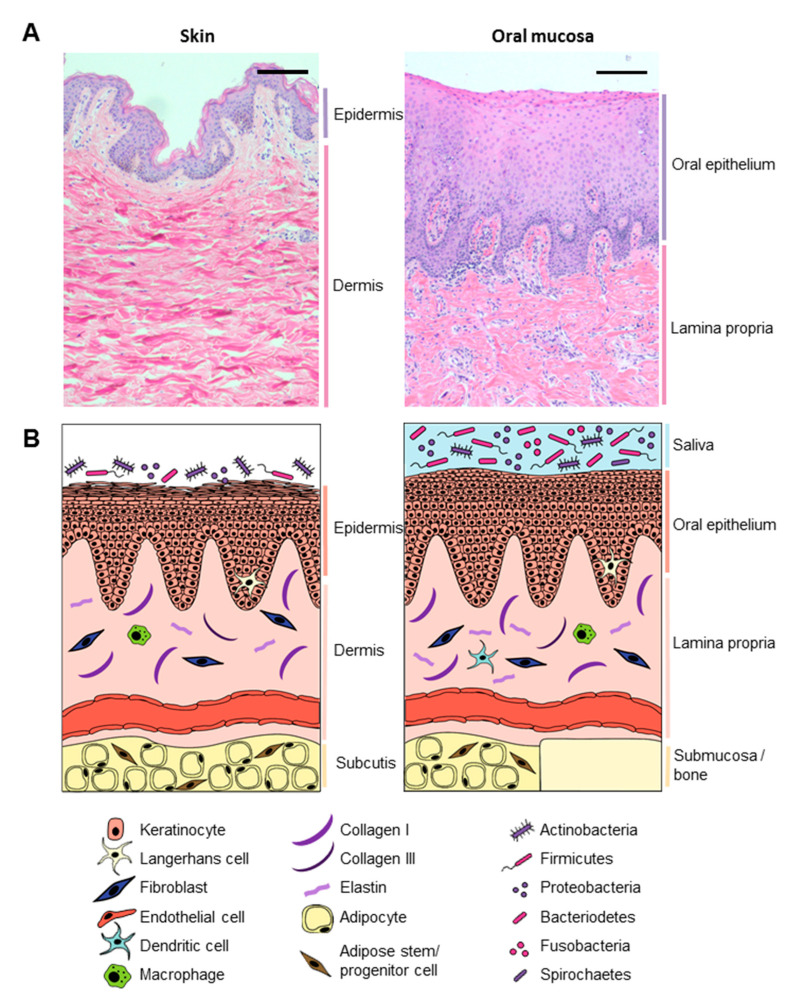
Figure 2.
Comparing healthy skin and oral mucosa. (A) Histological comparison between healthy skin (left) and gingiva (right) tissue. Hematoxylin and eosin staining of 5 µm paraffin embedded tissue sections. Scale bar: 100 µm; (B) graphical illustration comparing skin (left) and oral mucosa (right).

