Figure 7.
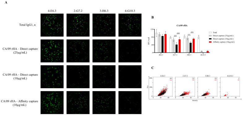
Detection of CA/09 rHA-specific FluoroSpots is influenced by protein-coating efficiency. Murine B-cell hybridomas (~100 cells/well) were evaluated for total or antigen-specific FluoroSpot formation. (A) Representative well images of murine B-cell hybridomas secreting monoclonal antibody (mAb) (IgG1, κ) with specificity for the recombinant hemagglutinin protein representing the A/California/04/2009 (CA/09) H1N1 vaccine strain. Contrast enhancements were uniformly performed on all images to aid their visualization in publication. (B) Total or CA/09 rHA-specific SFU/well (mean ±SD) for each B-cell hybridoma line. Significant differences in SFU/well were determined using an analysis of variation (ANOVA) with Sidak’s post hoc test. *** p < 0.001. (C) CA/09 rHA-specific FluoroSpots were merged into FCS files and visualized as bivariate plots. FluoroSpots originating from assay wells in which CA/09 rHA was directly captured on the membrane (black dots) or through affinity capture (red) are shown as overlays. The combined number of FluoroSpots detected in replicate wells for each of the respective donors is indicated in the inset using the same red/black color code.

