Figure 1.
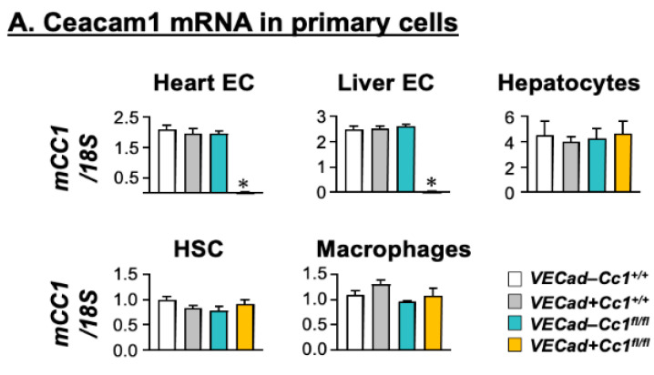
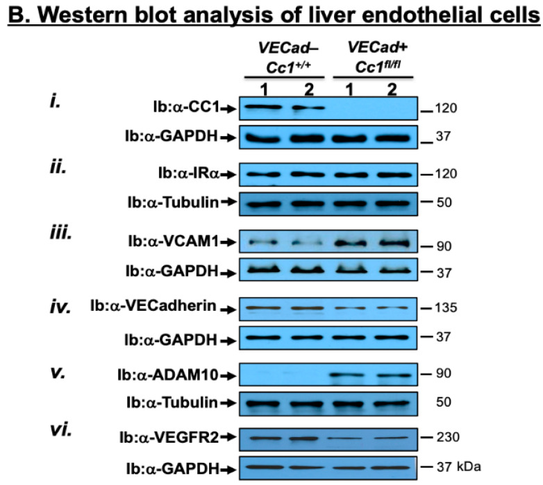
Assessing CEACAM1 expression in endothelial cells. (A) Primary cells were isolated from male mice at 2 months of age (n = 5/genotype), except for hepatic stellate cells that were derived from male mice at 8 months of age. Ceacam1 mRNA levels were analyzed by qRT-PCR in triplicate and normalized to 18S. Values are expressed as mean ± SEM. * p < 0.05 vs. all control groups; Negl, negligible. (B). Liver endothelial cells (LEC) combined from several wild-type (VECad-Cc1+/+) and null (VECad+Cc1fl/fl) mice were analyzed by immunoblotting (Ib) with specific antibodies (α-) to detect specific proteins and normalized against levels of loaded proteins by immunoblotting parallel gels or lower half-gels with α-GAPDH or α-Tubulin. The apparent molecular mass (kDa) is indicated at the right-hand side of each gel. Gels represent two separate/repeated experiments.


