Abstract
Background
This study outlines the development, implementation, and impact of a laboratory-developed, extraction-free real-time PCR assay as the primary diagnostic test for severe acute respiratory syndrome coronavirus 2 (SARS-CoV-2) in a pediatric hospital.
Methods
Clinical specimens from both upper and lower respiratory tract sources were validated, including nasopharyngeal aspirates, nasopharyngeal swabs, anterior nares swabs, and tracheal aspirates (n = 333 clinical samples). Testing volumes and laboratory turnaround times were then compared before and after implementation to investigate effects of the workflow changes.
Results
Compared to magnetic-bead extraction platforms, extraction-free real-time PCR demonstrated ≥95% positive agreement and ≥97% negative agreement across all tested sources. Implementation of this workflow reduced laboratory turnaround time from an average of 8.8 (+/−5.5) h to 3.6 (+/−1.3) h despite increasing testing volumes (from 1515 to 4884 tests per week over the reported period of testing).
Conclusions
The extraction-free workflow reduced extraction reagent cost for SARS-CoV-2 testing by 97%, shortened sample handling time, and significantly alleviated supply chain scarcities due to the elimination of specialized extraction reagents for routine testing. Overall, this assay is a viable option for laboratories to increase efficiency and navigate reagent shortages for SARS-CoV-2 diagnostic testing.
Background
Impact statement
The extraction-free severe acute respiratory syndrome coronavirus 2 (SARS-CoV-2) real-time PCR assay reported here was validated with samples from pediatric and adult patients and used to expand access to testing at the Children’s Hospital of Philadelphia for both groups. This report outlines the analytic and operational feasibility of a laboratory-developed extraction-free SARS-CoV-2 assay that does not require additional specialized equipment. A significant reduction in both turnaround time and reagent costs was observed with implementation of the assay, highlighting the potential of this approach for clinical labs seeking to increase their capacity for testing while managing resource constraints imposed by the COVID-19 pandemic.
Diagnostic testing for severe acute respiratory syndrome coronavirus 2 (SARS-CoV-2), the etiologic agent of coronavirus infectious disease 2019 (COVID-19), has become an unprecedented challenge for clinical laboratories. Because of the pathogen’s novelty and its rapid spread through the global population (1), public health and clinical laboratories have dedicated extensive resources to developing sustainable testing infrastructure. This process has been plagued by reagent shortages, personnel burnout, and frequent updates to regulatory requirements (2, 3). As a result, laboratories struggled to reach the necessary capacity, and turnaround times have lengthened considerably as demand for testing increased.
Real-time PCR testing remains the gold standard for the detection and diagnosis of viral infections. The simplest form of this technology includes separate extraction of nucleic acid material followed by amplification and quantification of nucleic acid on a thermocycler with primers/probes specific for the pathogen of interest. Lab-developed real-time PCR assays were the first implemented to detect SARS-CoV-2 early in the pandemic response (4, 5). However, as the burden of COVID-19 testing has increased, many clinical laboratories in the United States have responded by bringing on new testing platforms, and increasingly, these include commercial all-in-one options following emergency use authorization by the Food and Drug Administration. Commercial SARS-CoV-2 testing platforms are less technically demanding than manual real-time PCR assays and often capable of higher throughput and rapid turnaround times; however, they are more expensive. In addition, reagent shortages associated with commercial platforms have limited the capacity of high throughput systems, forcing laboratories to use multiple test platforms and to continually adjust workflows based on reagent availability. To avoid these hurdles and remain independent of manufacturer-specific reagents, the Infectious Disease Diagnostic Laboratory (IDDL) at the Children’s Hospital of Philadelphia maintained a laboratory-developed test consisting of manual real-time PCR and adapted this assay to include an extraction-free protocol.
Extraction-free real-time PCR assays eliminate the costly and time-intensive step of isolating nucleic acid from patient specimens. Variations of these assays have been published for the detection of SARS-CoV-2, testing varying temperatures for heat-inactivation (6–9) and buffers for stability (10). In the IDDL, we developed a modified version of a heat-inactivation protocol based on methods previously published by the Centers of Disease Control and Prevention (5), which has shown close correlation with traditional extraction procedures (7, 8, 11–13) and achieves full viral inactivation (14). In the following study, we describe the strategy undertaken by the Children’s Hospital of Philadelphia to implement an extraction-free diagnostic testing workflow. Through our validation work, the extraction-free workflow demonstrated equivalent performance to automated extraction (MagNA Pure) for a range of specimen types. Following its implementation, we observed a significant reduction in turnaround times, enabling the lab to better serve both the diagnostic needs of the hospital and the local pediatric community. Overall, the extraction-free workflow minimized cost, increased reagent flexibility, and decreased hands-on technologist handling time for SARS-CoV-2 diagnostic testing.
Methods
Heat-Inactivation as an Alternative to Magnetic Bead Extraction
An extraction-free method was used for heat inactivation of the SARS-CoV-2. Clinical samples (75 µL) were added to a 96-well plate and then firmly sealed with aluminum foil (ThermoFisher AB0626). Samples were vortexed for 1 min, then heated in a standard benchtop thermocycler (Applied Biosystems) to 95 °C for 5 min, and then cooled to 4 °C. This protocol is a modification of the heat treatment outlined in the CDC’s Emergency Use Authorization: Appendix A: Heat Treatment Alternative to Extraction (5). After cooling, samples were kept on 0 to 7 °C cold blocks and vortexed, centrifuged, and then mixed thoroughly by pipetting with a multichannel micropipette prior to plating for real-time PCR.
This heat-inactivation protocol was compared to testing using traditional nucleic acid extraction on an automated magnetic bead platform. For this method, 200 µL of clinical samples were aliquoted and then extracted using the MagNA Pure LC 2.0 System (Roche, Indianapolis, IN, USA) with the associated MagNA Pure LC Total Nucleic Acid Isolation protocol and eluted in 66 µL total volume.
Laboratory-Developed Real-Time PCR
Real-time PCR for SARS-CoV-2 was performed on a clean bench, physically separated from the extraction/inactivation instrumentation. First, 5 µL of extracted or heat-inactivated product was added to 15 µL of Mastermix consisting of Quantabioplex (QuantaBio cat 95168), COVID N2 primer/probe sets for an amplicon length of approximately 127 base pairs (forward 5-TTACAAACATTGGCCGCAAA-3′, reverse 5-GCGCGACATTCCGAAGAA-3′, probe: 5-FAM-ACAATTTGCCCCCAGCGCTTCAG-BHQ1-3; FAM = 6-carboxyfluorescein, BHQ-1 = Black Hole Quencher 1) at 450 nM/reaction, and ribonuclease P primer (forward 5-AGATTTGGACCTGCGAGCG-3′, reverse 5-GAGCGGCTGTCTCCACAAGT-3′, probe: 5-Cy5-TTCTGACCT-TAO-GAAGGCTCTGCGCG-3′lAbRQSp; Cy5 = cyanine 5, 3lAbRQSp = Iowa Black Quencher) as an internal control. Primer-probe BLAST analysis was also performed using the US National Library of Medicine Standard Nucleotide BLAST database. There were no concerning or significant findings. The N2 primer-probe sets showed 100% homology with SARS-CoV-2. Amplification and detection were performed on a QuantStudio Dx Real-Time PCR Instrument (ThermoFisher) using QuantStudio Dx software. PCR conditions were set to 50 °C for 10 min and then held at 95 °C for 5 min. Samples then went through 40 cycles of denaturation at 95 °C for 3 s and extension/annealing at 60 °C for 30 s.
Analytical Sensitivity
Validation was performed using remnant clinical samples collected from upper and lower respiratory tract sources (nasopharyngeal [NP] aspirates, NP swabs, anterior nares swabs, tracheal aspirates) that were frozen at −80°C. Samples were thawed at room temperature prior to testing.
The analytical sensitivity of the assay was determined by diluting out a strong positive sample from both the upper respiratory tract (NP aspirate) and the lower respiratory tract (tracheal aspirate). These sources were used as representative because they are more technically challenging than swab samples. These samples were diluted from their original concentration (cycle threshold [Ct] = 18.9) into pooled remnant aspirate from negative samples. An 8-member dilution series was extracted on the MagNA Pure or the extraction-free platform and then quantified 10 times over 5 days. To determine the limit of detection, probit analysis was used to extrapolate the concentration at which 95% of samples would be detected. Dilutions of ATCC VR-32765D Quantitative RNA (lot #70033491 8 × 10e5 genome equivalents/µL) were employed to make the standard curve for quantitation of the viral copies. Precision was determined by testing the same sample from each source over 5 days on both the MagNA Pure and extraction-free platforms and comparing the Ct values. Each sample was tested 10 times, and descriptive statistics were generated to determine the intraassay precision.
Clinical Accuracy Analysis
At least 40 positive and 40 negative remnant samples with a range of Ct values on initial clinical testing were collected for each sample source, with the exception of nasal aspirates. Twenty of the positive nasal aspirate samples were contrived by spiking sample from positive patients into negative nasal aspirate matrix to replicate a clinical sample with similar properties. Samples were then retested and recharacterized as positive or negative for this analysis based on their result when thawed and extracted on the MagNA Pure instrument. All samples were thawed and tested side by side using heat inactivation and extraction on the same day. Two samples (1 NP swab sample and 1 tracheal aspirate) were invalid based on lack of Ribonuclease P amplification and were therefore excluded from the analysis. One tracheal aspirate sample was classified as negative based on the MagNA Pure reaction but amplified using the extraction-free platform. This sample was included in the footnote of Table 1.
Table 1.
Clinical accuracy comparison analysis for samples extracted on the MagNA Pure compared to extraction-free workflow.
|
| ||||||
|---|---|---|---|---|---|---|
| Extraction-free |
||||||
| MagNA Pure extraction | Positive | Negative | Total | Positive % agreement (total) | Positive % agreement (Ct < 35) | Negative % agreement (total) |
| NP swabs | ||||||
| Total positive | 38 | 2 | 40 | 95.0% | ||
| Positive (Ct < 35) | 35 | 1 | 36 | 97.2% | ||
| Total negative | 0 | 43 | 43 | 100.0% | ||
| Anterior nares swabs | ||||||
| Total positive | 38 | 2 | 40 | 95.0% | ||
| Positive (Ct < 35) | 36 | 1 | 37 | 97.3% | ||
| Total Negative | 0 | 46 | 46 | 100.0% | ||
| NP aspirates | ||||||
| Total positive | 39 | 1 | 40 | 97.5% | ||
| Positive (Ct < 35) | 38 | 0 | 38 | 100.0% | ||
| Total negative | 0 | 44 | 44 | 100.0% | ||
| Tracheal aspiratesa | ||||||
| Total positive | 39 | 1 | 40 | 97.5% | ||
| Positive (Ct < 35) | 33 | 0 | 33 | 100.0% | ||
| Total negative | 1 | 40 | 41 | 97.6% | ||
Sources tested include NP swabs, anterior nares swabs, NP aspirates, and tracheal aspirates. PPA shown for total positive samples and for subset of samples with Ct < 35 to exclude the weakest positive samples. Negative percentage agreement calculated for total negative samples.
One sample was not detected using the MagNA Pure but was detected at Ct = 38.11 on the extraction-free platform.
A comparison of clinical accuracy was determined by comparing at least 40 known positive and negative samples across the following testing sources: NP aspirates, NP swabs, anterior nares swabs, and tracheal aspirates. Negative percentage agreement was calculated for a comparison between magnetic-bead extraction and extraction-free platforms. Similarly, positive percentage agreement (PPA) was calculated for all samples across these platforms as well as for all samples excluding the weakest positives defined by having Ct values <35 on the magnetic bead extraction platforms.
Direct comparison of extraction methods was similarly determined by comparing the Ct values for positive SARS-CoV-2 specimens extracted using a magnetic-bead platform compared to those tested using the extraction-free workflow.
Implementation of Extraction-Free Workflow
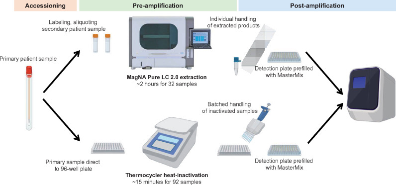
The extraction-free test was put into clinical use as the primary molecular SARS-CoV-2 assay for the laboratory following validation, allowing several ancillary workflow changes in addition to the elimination of the nucleic acid extraction step (Fig. 1). Initially, testing included labeling secondary tubes after sample receipt, aliquoting the samples into the secondary tubes, and passing the secondary tubes to another bench for extraction. Following extraction, product was plated to a 96-well plate for real-time PCR. With the implementation of the test modification, samples were plated directly to a 96-well plate after receipt in the laboratory. Plates were then covered with pierceable foil films to minimize contamination due to splashing during subsequent plate handling steps. After heat inactivation, samples were then transferred to a second 96-well plate for real-time PCR using a multichannel pipette to pierce the foil. In the event of an invalid sample (ribonuclease P amplification not detected), samples were reflexed to the magnetic bead extraction platform and repeated.
Fig. 1.
Schematic of SARS-CoV2 testing workflow in the IDDL. Samples were extracted using either magnetic bead extraction on the MagNA Pure LC 2.0 extraction platform or heat inactivated using extraction-free platform. Graphics were created with Biorender.com.
Laboratory Workflow Metric Analysis
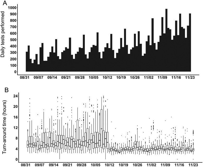
Analysis of laboratory workflow metrics was accomplished using data compiled by the Children’s Hospital of Philadelphia Department of Pathology and Laboratory Medicine Division of Pathology Informatics, using R and Rstudio for analysis and visualization (15, 16). These data include a comparison of testing volumes and average turnaround time for all tests run on the laboratory developed test 6 weeks prior to and 6 weeks post implementation of the extraction-free workflow (implemented on October 12, 2020). During this time period, there were 4 outliers with turnaround times >24 h due to atypical circumstances which were excluded from the data visualized in Fig. 3 but are included in Supplemental Table 3.
Fig. 3.
SARS-CoV-2 testing performance analysis in the IDDL. (A) Daily volume of testing performed by the laboratory developed real-time PCR assay .(B) Laboratory turnaround time in hours from receipt of samples to resulting. Data were compiled from 6 weeks prior to and post implementation of the extraction-free workflow in the laboratory. Boxplot data are shown with minimum, maximum, sample median, and quartiles plotted, and outliers are shown as dots.
Statistical Analysis
Descriptive statistics (average, SD, standard error of the mean) were generated in Excel for sensitivity and intraassay precision. For comparison of methods, a Student’s t-test and linear regression analysis were used to determine significant differences and confidence intervals between extraction platforms. These analyses were performed in GraphPad Prism version 9.
Ethics
This project was undertaken as a quality improvement initiative and as such does not constitute human subjects research, as documented with the Children's Hospital of Philadelphia Institutional Review Board.
Results
Analytical Sensitivity of Extraction-Free Protocol for SARS-CoV-2 Detection
To validate the extraction-free platform for clinical use, we first determined the analytical sensitivity using patient samples from both upper and lower respiratory sources. We generated an 8-member dilution panel from a strong positive nasal aspirate and tracheal aspirate sample (Supplemental Table 1). We then extrapolated the limit at which 95% of samples could be reliably detected. For the extraction-free workflow, the nasal aspirate sample limit of detection was 2.6E3 copies/mL, and the tracheal aspirate sample limit of detection was 3.0E3 copies/mL. For comparison, the same dilution panel run after MagNA Pure extraction found the limit of detection at 3.9E3 copies/mL for the nasal aspirate and at 4.0E3 copies/mL for tracheal aspirates. Next, the precision of the assay was calculated by comparing 10 replicates of the same sample run over 5 days (Supplemental Table 2). From these results, the Ct SD of NP aspirates from the extraction-free workflow was 0.34, compared to 0.37 when extracted with the MagNA Pure. Tracheal aspirates, which are known to be a technically challenging respiratory source, demonstrated an SD of 0.76 with the extraction-free workflow compared to 0.78 when extracted with the MagNA Pure. These results show that the extraction-free platform had comparable sensitivity and intraassay precision to extraction with the MagNA Pure.
Clinical Accuracy of SARS-CoV-2 Detection Using Extraction-Free Platform for Upper and Lower Respiratory Specimens
Samples collected from nasal aspirates and tracheal aspirates demonstrated 97.5% PPA (39/40) with the extraction-free workflow compared to the MagNA Pure extraction (Table 1). NP swabs and anterior nares swabs demonstrated a slightly lower PPA of 95% (38/40). Analysis of a subset of strong positive samples, defined as having a Ct value <35, demonstrated 100% PPA for nasal aspirates and tracheal aspirates and 97.3% PPA for NP swabs and anterior nares swabs. All samples that were negative by the MagNA Pure extraction replicated on the extraction-free platform, demonstrating ≥ 97% negative percentage agreement. Together, these data support the extraction-free protocol as a robust testing strategy for identifying the presence or absence of SARS-CoV-2 RNA in clinical samples.
To directly compare the extraction-free protocol to the traditional extraction platform, we analyzed the Ct values of known positive samples. In this analysis, samples tested from anterior nares swabs were the only source that demonstrated a significant Ct change of 0.92, indicating a slight loss in sensitivity when heat inactivating this sample type. In contrast, samples from NP swabs, nasal aspirates, and tracheal aspirates tested with the extraction-free protocol were not statistically different from when tested with the MagNA Pure extraction platform (Fig. 2). To determine if this relationship held across the entire range of data, we performed a linear regression analysis (Supplemental Fig. 1) and found that the extraction-free protocol and the MagNA Pure-generated Ct values were not statistically different from a perfect correlation. This analysis further highlighted the consistency in samples tested for NP swabs, anterior nares swabs, and nasal aspirates. In contrast, analysis of tracheal aspirates detected 4 outliers with high variability, which was unbiased toward either platform. This variability was not surprising as these are technically difficult samples with high viscosity and are a challenging source for respiratory viral testing.
Fig. 2.
Comparison of methods for samples extracted on the MagNA Pure compared to extraction-free protocols. Sources tested include (A) anterior nares swabs, (B) nasopharyngeal swabs, (C) nasopharyngeal aspirates, and (D) tracheal aspirates. Descriptive statistics include mean and SD between samples run on both platforms. A Student’s t-test was used to determine significant differences between protocols.
Workflow Impact
The extraction-free workflow conferred advantages in reduced cost, increased reagent flexibility, and shortened sample handling time. The technique requires few reagents or materials and only a vortex and thermocycler for instrumentation, making it significantly more cost-effective, reducing total extraction reagent cost for each test by 97%. It also lessens dependence on manufacturer-specific reagents for automated extraction platforms. The IDDL previously required 2 types of commercial extraction platforms from different manufacturers (1 not directly analyzed in this study) to meet volume demands in the face of ongoing reagent shortages. Last, by handling samples in a 96-well plate from the initial aliquot through the heat-inactivation step, technicians plating the real-time PCR assay could use multichannel pipettes, which was more labor-intensive when putting together plates from individual tubes of extracted product. This change to the protocol, while not technically advanced, significantly reduced the time for technicians handling high volumes of postamplification samples.
We were interested in understanding how these workflow changes affected the efficiency of COVID-19 testing. To measure this, we quantified the volume and turnaround time of testing 6 weeks before and 6 weeks following the extraction-free protocol implementation (Fig. 3). Over these 12 weeks, volumes increased continuously as the demand for testing escalated, from a weekly volume of 1515 to 4884. Despite this increase, a dramatic drop-off in turnaround time was evident starting on October 12, 2020, when the extraction-free workflow was implemented. Specifically, within-laboratory turnaround time during the week of October 5–11 averaged 8.8 (+/−5.5) h and dropped to 3.6 (+/−1.3) h during the week of October 12–18. There were no other major workflow or staffing changes during these 12 weeks that would confound this analysis of turnaround time. Indeed, 2 technologists left the laboratory during these 12 weeks, underscoring the time-saving benefits of this extraction-free platform. Beyond the increased processing speed, variability in turnaround time was also greatly reduced and remained consistently decreased in the weeks following implementation. Overall, these data demonstrate the extraction-free workflow dramatically increased laboratory efficiency in SARS-CoV-2 testing.
Conclusion
Due to the continuously increasing demand for SARS-CoV-2 testing, clinical laboratories have employed a variety of strategies to improve testing capacity and efficiency. The IDDL is experienced with laboratory-developed molecular assays using real-time PCR technology, which provides flexibility to adapt to pathogens of clinical interest in a pediatric population. However, these assays are extremely labor intensive and not well-suited for the demands of SARS-CoV-2 testing volumes. Extraction-free real-time PCR assays offer a viable solution to these challenges by (a) eliminating a costly and time-intensive extraction protocol, (b) reducing dependence on manufacturer-specific reagents and supplies, and (c) streamlining the workflow to decrease technologist effort.
Our results demonstrate that heat-inactivation is a viable alternative to traditional extraction methods for all sources tested, with >95% PPA compared to conventional extraction protocols and no more than a 1 Ct reduction in sensitivity. This is consistent with prior reports of this technology (7, 11, 12), and adds newly assessed respiratory sample types (NP and tracheal aspirates) for statistical power. As a result of these changes, our turnaround time for SARS-CoV-2 testing was more than halved. In practice, these improvements allowed us to meet the growing demand for rapid testing, particularly for screening patients before aerosol-generating procedures, while also supporting efforts to expand access to pediatric testing within our community, which increased our testing volume.
In addition to improving efficiency, this project also decreased dependence on manufacturer-specific reagents. Reagent flexibility is especially challenging under pandemic conditions, as consistent supply shortages have required laboratories to pivot between platforms and suppliers to stock reagents necessary for in-house testing. Because this workflow requires generalized real-time PCR reagents and simple instrumentation, reliance on specific manufacturers to provide reagent kits, collection tubes, and maintenance was decreased. Last, through simple modifications to sample handling workflows, a large part of the burden of aliquoting and pipetting was eliminated, reducing technologist hands-on time and improving efficiency within the laboratory. Our findings suggest that heat inactivation as an alternative to automated extractors may have utility for other laboratories challenged by supply chain shortages and can provide flexibility under current pandemic conditions where the supply chain for manufacturer-specific reagents is unpredictable.
Supplemental Material
Supplemental material is available at The Journal of Applied Laboratory Medicine online.
Supplementary Material
Nonstandard Abbreviations: SARS-CoV-2, severe acute respiratory syndrome coronavirus 2; COVID-19, coronavirus infectious disease 2019; IDDL, Infectious Disease Diagnostic Laboratory; NP, nasopharyngeal; Ct, cycle threshold; PPA, positive percentage agreement.
Author Contributions: All authors confirmed they have contributed to the intellectual content of this paper and have met the following 4 requirements: (a) significant contributions to the conception and design, acquisition of data, or analysis and interpretation of data; (b) drafting or revising the article for intellectual content; (c) final approval of the published article; and (d) agreement to be accountable for all aspects of the article thus ensuring that questions related to the accuracy or integrity of any part of the article are appropriately investigated and resolved.
R.E. Dumm, statistical analysis; M. Elkan, provision of study material or patients.
Authors’ Disclosures or Potential Conflicts of Interest: Upon manuscript submission, all authors completed the author disclosure form. Disclosures and/or potential conflicts of interest:Employment or Leadership: M. Elkan, Children's Hospital of Philadelphia. Consultant or Advisory Role: None declared. Stock Ownership: None declared. Honoraria: None declared. Research Funding: None declared. Expert Testimony: None declared. Patents: None declared.
Role of Sponsor: No sponsor was declared.
Acknowledgments: We would like to acknowledge the incredible dedication of the Infectious Disease Diagnostic Laboratory staff throughout this pandemic. Their commitment to patient care and perseverance make this testing possible. We would like to acknowledge Kenneth Smith, Robert Doms and Stephen Master for critical reading of this manuscript. And we would like to acknowledge Surabhi Mulchandani, Suzanne Mount, Brandy Neide, Kevin Weller and Kara Thompson for their steady management throughout the implementation of this workflow.
REFERENCES
- 1.Zhu N, Zhang D, Wang W, Li X, Yang B, Song J, et al. ; China Novel Coronavirus Investigating and Research Team. A novel coronavirus from patients with pneumonia in China, 2019. N Engl J Med 2020;382:727–33. [DOI] [PMC free article] [PubMed] [Google Scholar]
- 2.Binnicker MJ.Challenges and controversies to testing for COVID-19. J Clin Microbiol 2020;58(11):e01695–20. doi: 10.1128/JCM.01695-20. [DOI] [PMC free article] [PubMed] [Google Scholar]
- 3.Tang YW, Schmitz JE, Persing DH, Stratton CW.. Laboratory diagnosis of COVID-19: current issues and challenges. J Clin Microbiol 2020;58(6):e00512-20. doi: 10.1128/JCM.00512-20. [DOI] [PMC free article] [PubMed] [Google Scholar]
- 4.Corman VM, Landt O, Kaiser M, Molenkamp R, Meijer A, Chu DK, et al. Detection of 2019 novel coronavirus (2019-nCoV) by real-time RT-PCR. Euro Surveill 2020;25(3):2000045. doi: 10.2807/1560-7917.ES.2020.25.3.2000045. [DOI] [PMC free article] [PubMed] [Google Scholar]
- 5.Centers for Disease Control and Prevention, Division of Viral Diseases. CDC 2019-novel coronavirus (2019-nCoV) real-time RT-PCR diagnostic panel. December 1, 2020. https://www.fda.gov/media/134922/download (Accessed January 15, 2021).
- 6.Avetyan D, Chavushyan A, Ghazaryan H, Melkonyan A, Stepanyan A, Zakharyan R, et al. SARS-CoV-2 detection by extraction-free qRT-PCR for massive and rapid COVID-19 diagnosis during a pandemic. [Epub ahead of print] J Virol Methods June 4, 2021, as doi: 10.1016/j.jviromet.2021.114199. [DOI] [PMC free article] [PubMed]
- 7.Bruce EA, Huang ML, Perchetti GA, Tighe S, Laaguiby P, Hoffman JJ, et al. Direct RT-qPCR detection of SARS-CoV-2 RNA from patient nasopharyngeal swabs without an RNA extraction step. PLoS Biol 2020;18:e3000896. [DOI] [PMC free article] [PubMed] [Google Scholar]
- 8.Grant PR, Turner MA, Shin GY, Nastouli E, Levett LJ. Extraction-free COVID-19 (SARS-CoV-2) diagnosis by RT-PCR to increase capacity for national testing programmes during a pandemic. Preprint at https://www.biorxiv.org/content/10.1101/2020.04.06.028316v2 (Accessed April 15, 2020).
- 9.Lista MJ, Page R, Sertkaya H, Matos P, Ortiz-Zapater E, Maguire TJA, Poulton K, et al. Resilient SARS-CoV-2 diagnostics workflows including viral heat inactivation. Preprint at https://www.medrxiv.org/content/10.1101/2020.04.22.20074351v4 (Accessed April 15, 2021). [DOI] [PMC free article] [PubMed]
- 10.Alcoba-Florez J, Gonzalez-Montelongo R, Inigo-Campos A, de Artola DG, Gil-Campesino H, Ciuffreda L, et al. ; The Microbiology Technical Support T. Fast SARS-CoV-2 detection by RT-qPCR in preheated nasopharyngeal swab samples. Int J Infect Dis 2020;97:66–8. [DOI] [PMC free article] [PubMed] [Google Scholar]
- 11.Fomsgaard AS, Rosenstierne MW.. An alternative workflow for molecular detection of SARS-CoV-2—escape from the NA extraction kit-shortage, Copenhagen, Denmark, March 2020. Euro Surveill 2020;25(14):2000398. doi: 10.2807/1560-7917.ES.2020.25.14.2000398. [DOI] [PMC free article] [PubMed] [Google Scholar]
- 12.Lubke N, Senff T, Scherger S, Hauka S, Andree M, Adams O, et al. Extraction-free SARS-CoV-2 detection by rapid RT-qPCR universal for all primary respiratory materials. J Clin Virol 2020;130:104579. [DOI] [PMC free article] [PubMed] [Google Scholar]
- 13.Merindol N, Pepin G, Marchand C, Rheault M, Peterson C, Poirier A, et al. SARS-CoV-2 detection by direct rRT-PCR without RNA extraction. J Clin Virol 2020;128:104423. [DOI] [PMC free article] [PubMed] [Google Scholar]
- 14.Smyrlaki I, Ekman M, Lentini A, Rufino de Sousa N, Papanicolaou N, Vondracek M, et al. Massive and rapid COVID-19 testing is feasible by extraction-free SARS-CoV-2 RT-PCR. Nat Commun 2020;11:4812. [DOI] [PMC free article] [PubMed] [Google Scholar]
- 15.Team R. RStudio: Integrated development for R. http://www.rstudio.com/ (Accessed December 1, 2020).
- 16.Team RC. R: A language and environment for statistical computing. http://www.R-project.org/ (Accessed December 1, 2020).
Associated Data
This section collects any data citations, data availability statements, or supplementary materials included in this article.


