Abstract
Oropouche virus (OROV), a member of the Orthobunyavirus genus, is an arthropod-borne virus (arbovirus) and is the etiologic agent of human and animal disease. The primary vector of OROV is presumed to be the biting midge, Culicoides paraenesis, though Culex quinquefasciatus, Cq. venezuelensis, and Aedes serratus mosquitoes are considered secondary vectors. The objective of this systematic review is to characterize locations where OROV and/or its primary vector have been detected. Synthesis of known data through review of published literature regarding OROV and vectors was carried out through two independent searches: one search targeted to OROV, and another targeted towards the primary vector. A total of 911 records were returned, but only 90 (9.9%) articles satisfied all inclusion criteria. When locations were characterized, some common features were noted more frequently than others, though no one characteristic was significantly associated with presence of OROV using a logistic classification model. In a separate correlation analysis, vector presence was significantly correlated only with the presence of restingas. The lack of significant relationships is likely due to the paucity of data regarding OROV and its eco-epidemiology and highlights the importance of continued focus on characterizing this and other neglected tropical diseases.
Keywords: Oropouche, Culicoides paraenesis, arbovirus, febrile illness
1. Introduction
The Orthobunyavirus genus, which consists of 18 serogroups, has been isolated from numerous countries in South America, most commonly Brazil [1,2,3,4,5,6,7,8,9]. The largest serogroup in the Orthobunyavirus genus, the Simbu sergroup, includes viruses associated with both human and animal diseases and is comprised of 25 viruses, including Oropouche virus (OROV) [10,11]. OROV is an enveloped, negative-sense, single-stranded RNA virus, and the genome consists of three separate segments: small (S), medium (M), and large (L). These S, M, and L segments encode for the nucleocapsid protein, surface glycoproteins (Gn and Gc), and the RNA-dependent RNA polymerase, respectively [12]. Additionally, the OROV N gene encodes for non-structural proteins NSs and NSm [13,14].
OROV has been associated with a human febrile illness with generalized symptoms such as fever, headache, and myalgia [7,15,16,17]. The first known case of Oropouche fever was described in Trinidad in 1955. The patient was a young male, whose occupation was to burn coal in the Melajo Forest, where he often slept [4]. Neutralizing antibodies were detected in blood specimens from forest workers in various forested sites in Trinidad. Neutralizing antibodies were also found in serum samples of 7/52 monkeys throughout the area [4]. Since then, there have been outbreaks of Oropouche Fever in Brazil, Ecuador, and Peru [8,17,18]. In the past two decades, there has been progress in developing diagnostic tools for OROV and understanding of the pathogenesis of this virus [19,20,21,22].
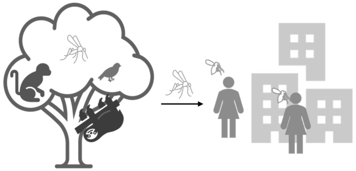
OROV is maintained in a zoonotic cycle (also called the sylvatic cycle) and an enzootic cycle. In the zoonotic cycle, the virus is transmitted between non-human vertebrate animals and associated vectors; while in the enzootic cycle, the virus cycles between human-biting vectors and humans [23,24,25]. When these cycles overlap, a spillover event occurs, spurred either when a human encroaches on the zoonotic cycle or when vector or animals from the sylvatic cycle overlap into the enzootic setting. This occurs through processes such as agricultural expansion or urbanization, or sometimes through a bridge vector that may vacillate between the two settings [26,27]. The primary enzootic vector for OROV has been identified as the biting midge Culicoides paraenesis [23,28,29,30]. In addition to the primary vector, Culex quinquefasciatus, Cq. venezuelensis, and Aedes serratus are mosquitoes that have been found to transmit OROV but are considered secondary vectors in the enzootic cycle [23,31,32]. Several species of mosquitoes, specifically Coquillettidia venezuelensis and Cx. quinquefasciatus, have been implicated in maintaining the sylvatic cycle of OROV in non-human mammalian hosts such as non-human primates and sloths, as well as avian species [23,32] (Figure 1).
Figure 1.
OROV transmission cycles as currently understood. In the sylvatic cycle, an arthropod vector takes a bloodmeal from a viremic vertebrate animal host to maintain the zoonotic cycle. Mosquitoes have been suggested as possible enzootic vectors and/or bridge vectors. Conversely, Culicoides paraenesis has been identified as the primary enzootic vector whereby the virus is maintained between humans and midges.
Before the emergence of Zika and chikungunya in the western hemisphere, OROV was recognized as being the second most common arboviral cause of human febrile illness in Brazil, second to dengue virus [33]. Though this virus is recognized as the etiologic agent of human disease and is known to infect animals, there is a scarcity in screening and detection outside of known areas associated with detected outbreaks [34,35,36]. Nevertheless, there is increased call for expanded screening of OROV across South America due to the significant number of cases and symptom similarity to other arboviruses to accurately attribute disease with specific pathogens [15,35]. However, efficient surveillance of under-detected pathogens requires sufficient data to target efforts either in specific locations or at specific times. The objective of this systematic review (SR) is to characterize the locations where OROV surveillance efforts have either confirmed OROV presence or not, as well as those locations where surveillance studies have detected the OROV vector independent of virus detection.
2. Material and Methods
Searches were conducted in August 2020. To assemble the published literature regarding OROV, an online search in the PubMed database was performed using the term “Oropouche”, which returned 143 records. We recognized that perhaps not all relevant publications were indexed in PubMed, so an additional search was performed using Google Scholar using the terms “Oropouche Virus” AND “South America”, which returned 598 records. Because many entomological studies are not indexed in PubMed, we used Google Scholar as the primary source of candidate studies for the search of the primary vector. Thus, to characterize the ecological sites where the primary vector has been studied in South America, a second literature search was performed using Google Scholar with the search terms: “Culicoides paraensis” AND “South America”, which returned 173 records.
2.1. Study Selection
Inclusion criteria for the OROV studies were defined as the following: molecular and/or serologic detection, and epidemiological data. Exclusion criteria were defined as the following: reviews, studies without primary data (modeling, assay design, phylogenetics, immunology, physiology, insecticide investigations), duplicate study populations, Oropouche location (not virus), thesis/dissertation, book/book chapter, inaccessible/untranslatable, vector competence studies. The number of records was recorded, and they were included or excluded based on the satisfaction of the defined criteria. Titles were identified and, if obvious, used to exclude studies; abstracts were then read of the remaining studies to determine if exclusion criteria were met; finally, whole papers were read to determine if the studies satisfied inclusion or exclusion criteria.
Inclusion criteria for the arthropod vector of OROV were defined as the following: epidemiological studies and entomological studies including surveillance, field observations, and abundance. Exclusion criteria were defined as the following: reviews, laboratory-based vector competence studies, investigations without primary data, duplicate study populations, thesis/dissertation, book/book chapter, and inaccessible or untranslatable articles. The total number of articles was recorded, and they were either included or excluded based on the satisfaction of the defined criteria. Titles were first examined, followed by abstracts, and then whole paper analysis.
2.2. Data Extraction and Synthesis
The records that satisfied inclusion criteria were examined for data extraction and synthesis over all studies. Documented study features included: study date and location, detection of nucleic acid and/or antibodies, laboratory assay performed or vector competence studies, hosts tested and confirmed positive, and ecological and environmental settings. Similarly, the data extracted from C. paraensis literature included: surveillance date and location, vector species collected, number of vectors collected, collection methods, and ecological settings. Articles that were returned from the Google Scholar database with the search terms “Culicoides paraensis” and “South America” that concentrated on any secondary vectors (Ae. serratus, Cq. venezuelensis, Cx. quinquefasciatus) for OROV were also included. Despite the search criteria targeting the primary vector, several records (n = 14) were returned that focused on secondary vectors. These papers were included in the review.
Variables were identified by descriptive mention in the studies and a master list of variables made. Papers were then reread and tallied for mention of each variable in the list. We considered the presence and absence of the following as indicators of different eco-environmental settings: crops, livestock, military facilities, mining, swamps, coasts, streams, springs, lakes, rivers, dams, restingas (coastal broadleaf forest), floodplains, mangroves, primary forest, secondary forest, cerrado (tropical savanna), and pantanal (tropical wetland). Maps were constructed using QGIS version 3.14.15-Pi [37] and shape files were downloaded from http://tapiquen-sig.jimdofree.com (Carlos Efraín Porto Tapiquén. Geografía, SIG y Cartografía Digital. Valencia, Spain, 2020, accessed on 30 June 2021).
2.3. Classification Models
The data were separated into two separate sets: acute (e.g., PCR detected) or convalescent (e.g., antibody) cases. To determine whether any of the eco-environmental variables affected the odds of an acute or past OROV case detection, we built a suite of logistic classification models, employing variable selection methods, and each dataset was independently analyzed. Proportion of people tested that were positive for OROV acute infection, convalescent/past infection was used as the dependent variable and the binary (noted/not mentioned) ecological variables were used as the independent variables. Case studies and case series with total number of patients tested being fewer than 10 were excluded from the statistical analyses from the OROV search dataset. Likewise, to determine associations between eco-environmental variables and the presence of C. paraensis, we calculated the proportion of C. paraensis found among all insects reported, which was used as the dependent variable and the binary ecological variables were again used as the independent variables. No vector studies were excluded in that corresponding statistical analyses.
The following model variations were considered:
-
1.
All eco-environmental variables were run as independent variables.
-
2.Reduced models whereby several variables were combined as below and run as a uni-, bi-, and tri-variate models for acute and past infections:
-
a.“livestock”, “military”, “crops”, and “mining” were combined into a single “anthropological” variable.
-
b.“river”, “dam”, and “pantanal” were combined into a single “water” variable.
-
c.“primary” or “secondary” forests were combined into a single “forest” variable.
-
a.
-
3.Reduced models whereby the several variables were combined as below and run as a uni-, bi-, and tri-variate models for the presence of the vector:
-
a.“livestock”, “military”, and “crops”, were combined into a single “anthropological” variable.
-
b.“river”, “dam”, “pantanal”, “swamp”, “streams”, “springs”, and “lakes” were combined into a single “water” variable.
-
c.“primary”, “secondary”, “mangrove”, and “restinga” forests were combined into a single “forest” variable.
-
a.
The logistf function was used (package logistf) to fit the models using R Studio (version 1.3.1093) and R version 3.6.3. Significance was assessed at the α = 0.05 confidence level.
2.4. Correlation Analyses
For eco-environmental variables, we assigned the value of 1 for present and 0 for absent and calculated the Spearman’s rank correlation coefficient of eco-environmental variables with (1) the proportion of people tested that were positive for OROV acute infection or convalescent/past infection in the PCR and antibody data sets, and (2) the proportion of C. paraensis found in the vector data sets. We tested for the significance of each correlation with a two-tailed t-test at the α = 0.05 confidence level against the null hypothesis that a correlation was equal to zero.
3. Results
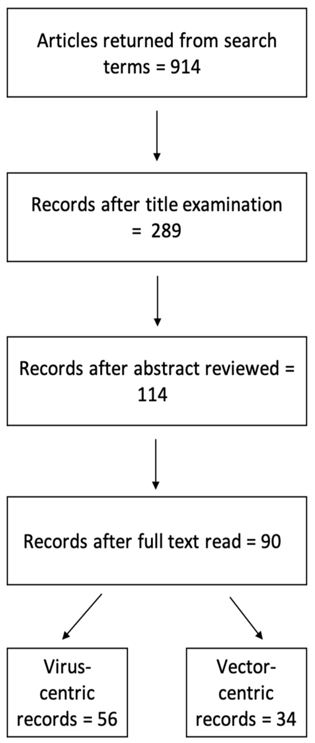
A literature search in PubMed with the term “Oropouche” returned 143 records. A separate literature search in Google Scholar with the terms “Oropouche virus” AND “South America” returned 598 records. After accounting for duplicate records from the dual database search, a total of 90 articles (only 9.9% of all returned records) satisfied all inclusion criteria. The Google Scholar search regarding the primary arthropod vector for OROV which was carried out using the terms “Culicoides paraensis” and “South America” returned 173 records. Of the 90 articles included, 62.2% involved the investigation of OROV while 37.8% of the studies were focused on entomological surveillance (Figure 2).
Figure 2.
Flow-chart of record processing for inclusion in systematic review.
3.1. OROV Surveillance
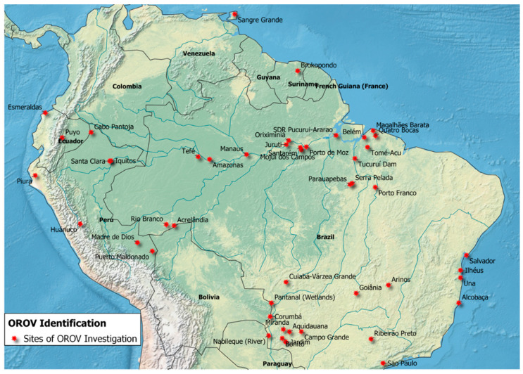
Across all 56 articles included for OROV surveillance, there were eight separate countries where these investigations took place (Figure 3). There were 37 studies performed in Brazil (63%) [2,5,7,16,17,24,32,38,39,40,41,42,43,44,45,46,47,48,49,50,51,52,53,54,55,56,57,58,59,60,61,62,63,64,65,66,67], followed by 11 studies in Peru (18.6%) [6,18,34,68,69,70,71,72,73,74,75], with the remaining (18.6%) taking place in Ecuador [3,8,15,70,76], Paraguay [70,77], Suriname [78], Trinidad [4], Bolivia [70], and Costa Rica [79].
Figure 3.
Locations identified where OROV has been detected in South America based on included records.
3.2. Arthropod Vector Surveillance
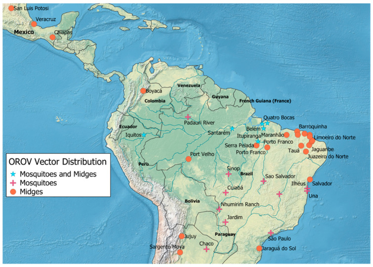
There were 34 articles that met all inclusion criteria for the C. paraensis search. There was a total of seven separate countries in which these surveillance efforts took place in literature included in this review (Figure 4). There were 21 (60%) articles that were based in Brazil [80,81,82,83,84,85,86,87,88,89,90,91,92,93,94,95,96,97,98,99,100], 6 (17.1%) studies in Peru [101,102,103,104,105,106], 4 (11.4%) in Argentina [107,108,109,110], with the remaining (11.4%) taking place in Colombia [30], Paraguay [109], Mexico [111], and Suriname [112].
Figure 4.
Locations where OROV vectors have been observed in Mexico and South America according to records included in this review. Map generated in ArcGIS.
3.3. OROV Detection
OROV detection studies involved both serological and molecular assays across a range of host species. OROV screening was performed on samples from humans, non-human primates, arthropods, mammals, avian species, and reptiles. Of the 56 articles that were focused on the detection of OROV, there were 46 serologic [2,3,5,6,7,8,16,17,18,32,34,38,39,40,41,42,43,45,46,48,49,50,52,53,56,57,58,59,60,62,63,64,65,66,67,68,69,70,71,72,73,74,75,77,78,79], 7 disease etiology investigations where OROV was confirmed [4,15,24,44,47,51,76], and 3 diagnostic development studies [54,55,61].
Several methods were used for the detection of both nucleic acid and antibodies: complement fixation (CF), hemagglutinatin inhibition (HI), neutralization tests, enzyme-linked immunosorbent assay (ELISA), plaque reduction neutralization test (PRNT), indirect immunofluorescence assay (IFA), virus isolation assays, real-time reverse transcription polymerase chain reaction (RT-PCR) and quantitative real-time RT-PCR (qRT-PCR), and Illumina-based sequencing. Of these methods, the most commonly used for antibody detection was ELISA, whereas the most commonly used method for the presence of OROV nucleic acid was RT-PCR.
From the 56 articles included in OROV investigations, 9 (16%) of the total 56 studies found no presence of OROV [43,48,59,62,65,67,75,77,79], whereas 47 (84%) of the studies that screened for OROV found either antibodies to OROV (18 studies) [2,5,18,38,39,40,41,45,46,50,52,58,63,64,68,69,71,78], OROV nucleic acid (16 studies) [3,6,8,15,34,47,49,53,54,55,60,61,66,72,73,76], or both (13 studies) [4,7,16,17,24,32,42,44,51,56,57,65,70]. Humans were tested for OROV antibodies (11 studies) [5,18,38,39,40,58,63,64,68,69,78], OROV nucleic acid (19 of studies) [3,6,8,34,43,47,48,49,53,54,55,59,61,62,66,72,73,76,77], or both (17 studies) [4,7,15,16,17,24,32,41,42,44,51,52,56,57,70,71,74].
Non-human mammals that were tested for OROV include: non-human primates, sloths, avian species, rodents, sheep, reptiles, and horses. There were seven studies that tested non-human primates, five studies tested for antibodies [45,46,50,65,75], one for OROV nucleic acid [60], and one for both [67]. There were three studies that tested samples obtained from sloths where two of these studies tested for antibodies [65,79] and one study tested for OROV nucleic acid [32]. Three studies tested avian samples for antibodies [16,17,58], two studies tested rodents for antibodies [17,58], and one study that tested sheep, caiman, and equine samples for antibodies [2].
4. Ecological Factors Associated with OROV Presence
4.1. Vector Presence and Diversity
In the 56 studies returned by the search for the virus, several contained information about primary or secondary vectors of OROV. In 8/56 studies (14.3%), surveillance for known OROV vectors was also conducted and presence confirmed of at least one competent species [4,16,17,32,45,49,56,57]. In four of these three studies—three in Para, Brazil [17,49,57] and one in Maranhão, Brazil [56]—in which OROV was found, the presence of C. paraensis was documented. Further, 7/56 studies observed mosquito vectors: Ae. serratus in four studies located in Trinidad [4] and Brazil (two in Para State [16,32] and one in Mato Grosso do Sul State [45]), and Cx. quinquefasciatus in three studies in Brazil (Para State [17,57] and Mato Grosso State [49]). Of the 34 entomology articles, C. paraensis was found in 18 studies (52.9%) [30,80,81,90,91,93,94,95,96,97,98,103,104,108,109,110,111,112], and each of the three mosquito vectors were present in seven studies (2.9%), some of which overlapped [82,83,84,85,86,87,88,89,93,101,102,107].
4.2. Anthropogenic Land Use
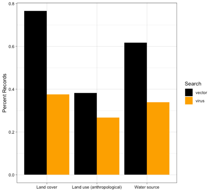
For the OROV search, 15/54 (27.8%) articles provided information regarding anthropogenic land use (Figure 5). The uses of land in these locations were crop cultivation, livestock rearing, military outposts, and mining, present in eleven [4,7,16,17,56,57,63,65,68,69,79], six [2,7,17,56,63,79], three [8,18,75], and one [57] studies, respectively. The two most common land uses, crop cultivation and livestock rearing, were both present in five of studies [7,17,56,63,79].
Figure 5.
The proportion of records that had observations about land cover, anthropogenic land use, and/or water sources contained within the text according to which search they were returned from. Black represents the proportion of records in the vector data (N = 34) and gold represents the proportion of records in the virus data (N = 56).
There were 13/34 studies from the vector search that described land use (Figure 5). The most common use of land was crop cultivation (nine studies)) [30,80,85,88,89,104,108,110,112], followed by livestock (seven studies) [80,91,97,99,102,104,108]. In studies, crop cultivation and livestock were noted [80,104,108].
4.3. Water Source
In 19 of the 56 articles focusing on OROV surveillance, a nearby water source was noted (Figure 5). In 14 locations there was at least one river present [17,18,38,45,56,58,59,63,67,68,69,71,78,79]. Other sources of water were noted but to a much lesser extent with three locations in the Pantanal (the largest tropical wetland in the world) [2,46,49], two locations in a coastal area [51,55], and two locations near a dam [64,78]. We found that water sources were mentioned in 21 of the 34 articles regarding C. paraenesis surveillance (Figure 5). The presence of at least one river was described in 11/21 articles [80,81,82,85,93,94,102,106,109,110,112]. Streams were the second most frequently mentioned in five articles [30,89,90,108,110]. There were four studies that noted marshes/swamps [89,99,107,112], whereas coastal locations [91,94], lakes [83,86,89], floodplains [83,95], and springs [86,89] were all water sources mentioned twice each. There was one location where surveillance took place at the site of a dam [82] and one study that took place in the Pantanal [83].
4.4. Land Cover
There were 21 (38.9%) articles that made observations regarding land cover from the 56 studies that focused on OROV surveillance (Figure 5). Locations that have primary or undisturbed forest were most common, mentioned in 16 studies [4,6,8,15,16,17,18,24,59,65,66,67,68,70,72,75]. Secondary forests, or forest locations that had been previously used for anthropogenic purposes were found in two studies [4,69]. A cerrado land cover, which consists of bushy, small bent and twisted trunk trees, was described in four locations [45,46,49,66], while two locations described mountainous terrain [6,57].
Of the 34 studies from the C. paraensis search, 26 (76.5%) made note of the land cover (Figure 5). Primary and secondary forest locations were noted in sexteen [30,81,83,85,89,90,95,98,99,102,104,107,108,109,110,112] and eleven studies [80,82,84,86,88,92,99,101,102,104,106], respectively. There were three surveillance studies that took place in locations described as cerrado [83,84,112], while both mangrove [91,112] and restinga land cover [98,100] types were mentioned. Restinga refers to coastal forests with the soil mostly consisting of sand and salt. There were several studies that had overlapping types of land cover which included cerrado, primary forest, and secondary forest locations [83,84,98,99,102,104,112].
4.5. Odds of Detecting Acute and Past Infections
Logistic regression run on the full model run with all 12 variables did not reveal any significant predictors for odds of either pcr prevalence (acute infections) or antibody prevalence (past infections). Due to the small dataset and the large number of variables, we next employed a data reduction method and ran the model with uni-, bi-, and trivariate combinations of the variables related to anthropological land-use, the presence of water, and/or forests. In none of the models were there any variables significant for either the acute or past infections. Correlation analysis revealed a single significant correlation between acute infections and the presence of restingas (Spearman’s rho ρ = −0.41, p-value = 0.0398). All other correlations were statistically insignificant at the α = 0.05 confidence level.
4.6. Odds of Vector Presence
Similarly, the full model of 17 eco-environmental variables for determining differences in odds of detection of the vector species did not reveal any statistically significant predictors for the proportion of C. paraensis found. None of the uni-, bi-, or trivariate models run using the data reduction methods returned significant results. This suggests that, in South America, there is still not enough data available to determine whether the detection of C. paraensis is likely to be associated with specific eco-environmental variables. Correlation analyses, likewise, found no significant correlations between the proportion of C. paraensis found and the eco-environmental variables.
5. Discussion
Our primary objective was to assemble the existing data regarding the environmental and ecological factors associated with known detection of OROV in South America, and to identify factors associated with the presence of the primary enzootic vector. Our review of the published data demonstrates that there are similarities among the factors observed in OROV and vector detection. First, the most prominent anthropogenic land use observed was crop cultivation and livestock for both OROV and vector presence. Secondly, rivers were the most common source of water in locations where OROV and arthropod vectors were documented. Lastly, both OROV and vectors were more frequently found in primary forest locations. However, our analyses of these data show that there is no significant signal among these variables and OROV and/or vector presence/absence. This is likely due to a lack of power in the analyses given the relatively small number of records and overall data. Again, this points to the need for further studies to characterize the ecological and environmental drivers of OROV detection and its associated vector(s). Further, more data are needed to determine whether the qualitative patterns detected in the characteristics observed is a result of sampling bias or whether these characteristics truly could be associated with OROV detection.
Because OROV is an arbovirus, the presence of a vector is highly correlated with the likelihood of virus detection [28,29]. Arbovirus transmission potential is measured largely by the vectorial capacity equation, which investigates key variables including such factors as vector density relative to humans, and extrinsic and intrinsic factors that govern the virus–vector dynamics [113]. Often, the processes of transmission within the vector—vector competence and the EIP—are affected by environmental factors, most notably temperature, which can vary in response to anthropogenic land use, land cover, and proximity to water [114,115,116,117]. Therefore, these environmental and ecological characteristics are crucial factors that should be considered in OROV transmission and when planning efforts aimed at detecting this potentially emergent arbovirus.
For example, because a good portion of the C. paraenesis lifecycle includes semi-aquatic stages, these arthropods are often found in moist, decaying vegetation such as that from recently harvested farms [117]. C. paraensis often lays eggs in moist areas, where developmental stages progress through the lifecycle by feeding on decomposing organic matter, algae, and small vertebrate prey [117]. The necessity of water to the lifecycle of C. paraensis (as well as the secondary mosquito vectors) increases the likelihood of finding these arthropods in close proximity to water sources [117]. In addition, water is an attractant to susceptible vertebrate hosts in both uninhabited and inhabited locations, which leads to host–vector interactions necessary for arboviral transmission in the context of potential reservoir hosts, such as avians, non-human primates, and rodents [116]. While C. paraensis is known to feed on humans, their opportunistic feeding patterns lead them to feed on other vertebrate hosts in lieu of human presence [117]. In addition, forested environments suitable for reservoir hosts enables arthropod vectors to both acquire and transmit OROV [116,117].
The increasing demand for agricultural and industrial products has resulted in worldwide globalization [118]. The proceeding economic expansion into South America centers around crop production, especially soy, while some areas also take part in the livestock trade [118,119]. In addition, mining for bauxite, nickel, and gold have had a positive impact on economic development in this region [120]. However, these economic gains have had unintended consequences whereby previously uninhabited forests are now in closer proximity to human populations and/or human enterprises. This, in turn, increases the chances of zoonotic spillover events by increasing contact between vector and reservoir vertebrate host populations [26].
There are numerous locations across regions of South America where investigations into the presence of OROV have successfully identified circulation of the virus; however, it is noteworthy that there are geographic gaps in OROV surveillance efforts. These data demonstrate that there was no evidence of surveillance for OROV noted in Colombia, Venezuela, and Guyana, even though all three of these countries border Brazil. Colombia, particularly, borders three countries where OROV has been identified (Figure 3). Further, of the included records from the Google Scholar “Culicoides paraensis” and “South America” search, entomological surveillance efforts for the primary vector of OROV in South America are predominantly in Brazil, especially on the easternmost portion of the country (Figure 4).
There are obvious gaps in the literature concerning OROV, a neglected tropical, zoonotic disease of emergent public health importance. The most surveillance has been conducted in Brazil (Figure 3). While one C. paraensis surveillance study in Colombia verified the presence of the vector, entomological surveillance specific to C. paraensis was also found to be primarily concentrated in Brazil, with sporadic surveillance locations in Peru and Argentina [90,103,110]. Additionally, our review provided a preliminary characterization of ecological and environmental factors that may be associated with increased chances of OROV detection and perhaps circulation. However, continued research is needed to fully elucidate OROV transmission dynamics, arthropod vector competence, and OROV distribution to prepare for future emergence or expansion of this public health threat.
6. Conclusions
There were notable common ecological aspects associated with both the locations involved in OROV detection and the presence of the vector in the 90 studies included in this systematic review. Ecological characteristics included in both OROV and arthropod vector surveillance studies had crop cultivation as the most common anthropogenic use of land, rivers as the most common water source, and primary forest as the most prominent type of land cover. The observations found within this systematic review illustrate the importance of continued research into the ecological factors associated with the distribution of under-characterized arboviruses. This review reveals the need for continued surveillance of OROV and its arthropod vector, and the description of factors associated with detection may help to prioritize OROV surveillance initiatives.
Acknowledgments
We want to thank Erik Turner for his technical assistance, as well as whoever decided a sloth icon should be included in slide show software. In addition, we would like to thank Patrick O’Dell for his comments and insights.
Author Contributions
Conceptualization—R.C.C. and C.E.S.W.; Data curation—C.E.S.W.; Formal analysis—R.C.C., C.E.S.W. and M.A.R.; Project administration—R.C.C.; Original draft preparation—C.E.S.W. and R.C.C.; Review and editing—C.E.S.W., R.C.C. and M.A.R. All authors have read and agreed to the published version of the manuscript.
Funding
This research was funded by NIH/NIGMS R01 GM122077.
Institutional Review Board Statement
Not applicable.
Informed Consent Statement
Not applicable.
Data Availability Statement
Shape files were downloaded from a publically available resource (http://tapiquen-sig.jimdofree.com) and otherwise all data is available within the manuscript.
Conflicts of Interest
The authors declare no conflict of interest.
Footnotes
Publisher’s Note: MDPI stays neutral with regard to jurisdictional claims in published maps and institutional affiliations.
References
- 1.Hughes H.R., Adkins S., Alkhovskiy S., Beer M., Blair C., Calisher C.H., Drebot M., Lambert A.J., de Souza W.M., Marklewitz M., et al. ICTV Virus Taxonomy Profile: Peribunyaviridae. J. Gen. Virol. 2020;101:1–2. doi: 10.1099/jgv.0.001365. [DOI] [PMC free article] [PubMed] [Google Scholar]
- 2.Pauvolid-Correa A., Campos Z., Soares R., Nogueira R.M.R., Komar N. Neutralizing antibodies for orthobunyaviruses in Pantanal, Brazil. PLoS Negl. Trop. Dis. 2017;11:e0006014. doi: 10.1371/journal.pntd.0006014. [DOI] [PMC free article] [PubMed] [Google Scholar]
- 3.Wise E.L., Marquez S., Mellors J., Paz V., Atkinson B., Gutierrez B., Zapata S., Coloma J., Pybus O.G., Jackson S.K., et al. Oropouche virus cases identified in Ecuador using an optimised qRT-PCR informed by metagenomic sequencing. PLoS Negl. Trop. Dis. 2020;14:e0007897. doi: 10.1371/journal.pntd.0007897. [DOI] [PMC free article] [PubMed] [Google Scholar]
- 4.Anderson C.R., Spence L., Downs W.G., Aitken T.H. Oropouche virus: A new human disease agent from Trinidad, West Indies. Am. J. Trop. Med. Hyg. 1961;10:574–578. doi: 10.4269/ajtmh.1961.10.574. [DOI] [PubMed] [Google Scholar]
- 5.Mouraao M.P., Bastos M.S., Gimaqu J.B., Mota B.R., Souza G.S., Grimmer G.H., Galusso E.S., Arruda E., Figueiredo L.T. Oropouche fever outbreak, Manaus, Brazil, 2007–2008. Emerg. Infect. Dis. 2009;15:2063–2064. doi: 10.3201/eid1512.090917. [DOI] [PMC free article] [PubMed] [Google Scholar]
- 6.Silva-Caso W., Aguilar-Luis M.A., Palomares-Reyes C., Mazulis F., Weilg C., Del Valle L.J., Espejo-Evaristo J., Soto-Febres F., Martins-Luna J., Del Valle-Mendoza J. First outbreak of Oropouche Fever reported in a non-endemic western region of the Peruvian Amazon: Molecular diagnosis and clinical characteristics. Int. J. Infect. Dis. 2019;83:139–144. doi: 10.1016/j.ijid.2019.04.011. [DOI] [PubMed] [Google Scholar]
- 7.Vasconcelos H.B., Azevedo R.S., Casseb S.M., Nunes-Neto J.P., Chiang J.O., Cantuaria P.C., Segura M.N., Martins L.C., Monteiro H.A., Rodrigues S.G., et al. Oropouche fever epidemic in Northern Brazil: Epidemiology and molecular characterization of isolates. J. Clin. Virol. 2009;44:129–133. doi: 10.1016/j.jcv.2008.11.006. [DOI] [PubMed] [Google Scholar]
- 8.Izurieta R.O., Macaluso M., Watts D.M., Tesh R.B., Guerra B., Cruz L.M., Galwankar S., Vermund S.H. Assessing yellow Fever risk in the ecuadorian Amazon. J. Glob. Infect. Dis. 2009;1:7–13. doi: 10.4103/0974-777X.49188. [DOI] [PMC free article] [PubMed] [Google Scholar]
- 9.Groot H. Estudios sobre virus transmitidos por arthropodos en Colombia. Rev. Acad. Colomb. Cienc. Exactas Físicas Nat. 1964;12:46. [Google Scholar]
- 10.Rogers M.B., Gulino K.M., Tesh R.B., Cui L., Fitch A., Unnasch T.R., Popov V.L., Travassos da Rosa A.P.A., Guzman H., Carrera J.P., et al. Characterization of five unclassified orthobunyaviruses (Bunyaviridae) from Africa and the Americas. J. Gen. Virol. 2017;98:2258–2266. doi: 10.1099/jgv.0.000899. [DOI] [PMC free article] [PubMed] [Google Scholar]
- 11.Saeed M.F., Wang H., Nunes M., Vasconcelos P.F., Weaver S.C., Shope R.E., Watts D.M., Tesh R.B., Barrett A.D. Nucleotide sequences and phylogeny of the nucleocapsid gene of Oropouche virus. J. Gen. Virol. 2000;81:743–748. doi: 10.1099/0022-1317-81-3-743. [DOI] [PubMed] [Google Scholar]
- 12.Elliott R.M. Orthobunyaviruses: Recent genetic and structural insights. Nat. Rev. Microbiol. 2014;12:673–685. doi: 10.1038/nrmicro3332. [DOI] [PubMed] [Google Scholar]
- 13.Dong H., Li P., Elliott R.M., Dong C. Structure of Schmallenberg orthobunyavirus nucleoprotein suggests a novel mechanism of genome encapsidation. J. Virol. 2013;87:5593–5601. doi: 10.1128/JVI.00223-13. [DOI] [PMC free article] [PubMed] [Google Scholar]
- 14.Dutuze M.F., Nzayirambaho M., Mores C.N., Christofferson R.C. A Review of Bunyamwera, Batai, and Ngari Viruses: Understudied Orthobunyaviruses With Potential One Health Implications. Front. Vet. Sci. 2018;5:69. doi: 10.3389/fvets.2018.00069. [DOI] [PMC free article] [PubMed] [Google Scholar]
- 15.Manock S.R., Jacobsen K.H., de Bravo N.B., Russell K.L., Negrete M., Olson J.G., Sanchez J.L., Blair P.J., Smalligan R.D., Quist B.K., et al. Etiology of acute undifferentiated febrile illness in the Amazon basin of Ecuador. Am. J. Trop. Med. Hyg. 2009;81:146–151. doi: 10.4269/ajtmh.2009.81.146. [DOI] [PubMed] [Google Scholar]
- 16.LeDuc J.W., Hoch A.L., Pinheiro F.P., da Rosa A.P. Epidemic Oropouche virus disease in northern Brazil. Bull. Pan Am. Health Organ. 1981;15:97–103. [PubMed] [Google Scholar]
- 17.Pinheiro F.P., Travassos da Rosa A.P., Travassos da Rosa J.F., Bensabath G. An outbreak of Oropouche virus diease in the vicinity of santarem, para, barzil. Tropenmed. Parasitol. 1976;27:213–223. [PubMed] [Google Scholar]
- 18.Watts D.M., Lavera V., Callahan J., Rossi C., Oberste M.S., Roehrig J.T., Cropp C.B., Karabatsos N., Smith J.F., Gubler D.J., et al. Venezuelan equine encephalitis and Oropouche virus infections among Peruvian army troops in the Amazon region of Peru. Am. J. Trop. Med. Hyg. 1997;56:661–667. doi: 10.4269/ajtmh.1997.56.661. [DOI] [PubMed] [Google Scholar]
- 19.Tilston-Lunel N.L., Acrani G.O., Randall R.E., Elliott R.M. Generation of Recombinant Oropouche Viruses Lacking the Nonstructural Protein NSm or NSs. J. Virol. 2015;90:2616–2627. doi: 10.1128/JVI.02849-15. [DOI] [PMC free article] [PubMed] [Google Scholar]
- 20.Gutierrez B., Wise E.L., Pullan S.T., Logue C.H., Bowden T.A., Escalera-Zamudio M., Trueba G., Nunes M.R.T., Faria N.R., Pybus O.G. Evolutionary Dynamics of Oropouche Virus in South America. J. Virol. 2020;94:5. doi: 10.1128/JVI.01127-19. [DOI] [PMC free article] [PubMed] [Google Scholar]
- 21.Naveca F.G., Nascimento V.A.D., Souza V.C., Nunes B.T.D., Rodrigues D.S.G., Vasconcelos P. Multiplexed reverse transcription real-time polymerase chain reaction for simultaneous detection of Mayaro, Oropouche, and Oropouche-like viruses. Mem Inst. Oswaldo Cruz. 2017;112:510–513. doi: 10.1590/0074-02760160062. [DOI] [PMC free article] [PubMed] [Google Scholar]
- 22.Proenca-Modena J.L., Sesti-Costa R., Pinto A.K., Richner J.M., Lazear H.M., Lucas T., Hyde J.L., Diamond M.S. Oropouche virus infection and pathogenesis are restricted by MAVS, IRF-3, IRF-7, and type I interferon signaling pathways in nonmyeloid cells. J. Virol. 2015;89:4720–4737. doi: 10.1128/JVI.00077-15. [DOI] [PMC free article] [PubMed] [Google Scholar]
- 23.Sakkas H., Bozidis P., Franks A., Papadopoulou C. Oropouche Fever: A Review. Viruses. 2018;10:175. doi: 10.3390/v10040175. [DOI] [PMC free article] [PubMed] [Google Scholar]
- 24.Vernal S., Martini C.C.R., da Fonseca B.A.L. Oropouche Virus-Associated Aseptic Meningoencephalitis, Southeastern Brazil. Emerg. Infect. Dis. 2019;25:380–382. doi: 10.3201/eid2502.181189. [DOI] [PMC free article] [PubMed] [Google Scholar]
- 25.Travassos da Rosa J.F., de Souza W.M., Pinheiro F.P., Figueiredo M.L., Cardoso J.F., Acrani G.O., Nunes M.R.T. Oropouche Virus: Clinical, Epidemiological, and Molecular Aspects of a Neglected Orthobunyavirus. Am. J. Trop. Med. Hyg. 2017;96:1019–1030. doi: 10.4269/ajtmh.16-0672. [DOI] [PMC free article] [PubMed] [Google Scholar]
- 26.Weaver S.C. Urbanization and geographic expansion of zoonotic arboviral diseases: Mechanisms and potential strategies for prevention. Trends Microbiol. 2013;21:360–363. doi: 10.1016/j.tim.2013.03.003. [DOI] [PMC free article] [PubMed] [Google Scholar]
- 27.Karesh W.B., Dobson A., Lloyd-Smith J.O., Lubroth J., Dixon M.A., Bennett M., Aldrich S., Harrington T., Formenty P., Loh E.H., et al. Ecology of zoonoses: Natural and unnatural histories. Lancet. 2012;380:1936–1945. doi: 10.1016/S0140-6736(12)61678-X. [DOI] [PMC free article] [PubMed] [Google Scholar]
- 28.Pinheiro F.P., Travassos da Rosa A.P., Gomes M.L., LeDuc J.W., Hoch A.L. Transmission of Oropouche virus from man to hamster by the midge Culicoides paraensis. Science. 1982;215:1251–1253. doi: 10.1126/science.6800036. [DOI] [PubMed] [Google Scholar]
- 29.Pinheiro F.P., Hoch A.L., Gomes M.L., Roberts D.R. Oropouche virus. IV. Laboratory transmission by Culicoides paraensis. Am. J. Trop. Med. Hyg. 1981;30:172–176. doi: 10.4269/ajtmh.1981.30.172. [DOI] [PubMed] [Google Scholar]
- 30.Santamaria E., Cabrera O.L., Zipa Y., Ferro C., Ahumada M.L., Pardo R.H. Preliminary evaluation of the Culicoides biting nuisance (Diptera: Ceratopogonidae) in the province of Boyaca, Colombia. Biomedica. 2008;28:497–509. [PubMed] [Google Scholar]
- 31.Hoch A.L., Pinheiro F.P., Roberts D.R., Gomes M.L. Laboratory transmission of Oropouche virus by Culex Quinquefasciatus Say. Bull. Pan Am. Health Organ. 1987;21:55–61. [PubMed] [Google Scholar]
- 32.Plnheiro F., Plnheiro M., Bensabath G., Causey O.R., Shope R. Epidemic of Oropouche VIrus in Belém. Rev. Serv. Espec. Saude Publica. 1962;12:15–23. [Google Scholar]
- 33.Figueiredo L.T. Emergent arboviruses in Brazil. Rev. Soc. Bras. Med. Trop. 2007;40:224–229. doi: 10.1590/S0037-86822007000200016. [DOI] [PubMed] [Google Scholar]
- 34.Martins-Luna J., Del Valle-Mendoza J., Silva-Caso W., Sandoval I., Del Valle L.J., Palomares-Reyes C., Carrillo-Ng H., Pena-Tuesta I., Aguilar-Luis M.A. Oropouche infection a neglected arbovirus in patients with acute febrile illness from the Peruvian coast. BMC Res. Notes. 2020;13:67. doi: 10.1186/s13104-020-4937-1. [DOI] [PMC free article] [PubMed] [Google Scholar]
- 35.Romero-Alvarez D., Escobar L.E. Oropouche fever, an emergent disease from the Americas. Microbes Infect. Inst. Pasteur. 2018;20:135–146. doi: 10.1016/j.micinf.2017.11.013. [DOI] [PubMed] [Google Scholar]
- 36.Mattar S. Oropuche virus: A virus present but ignored. Revista MVZ Cordoba. 2015;30:3. doi: 10.21897/rmvz.37. [DOI] [Google Scholar]
- 37.QGIS.org, QGIS Geographic Information System, Q. Association, 2021. [(accessed on 30 June 2021)]; Available online: https://qgis.org/en/site/
- 38.Dixon K.E., Travassos da Rosa A.P., Travassos da Rosa J.F., Llewellyn C.H. Oropouche virus. II. Epidemiological observations during an epidemic in Santarem, Para, Brazil in 1975. Am. J. Trop. Med. Hyg. 1981;30:161–164. doi: 10.4269/ajtmh.1981.30.161. [DOI] [PubMed] [Google Scholar]
- 39.Tavares-Neto J., Freitas-Carvalho J., Nunes M.R., Rocha G., Rodrigues S.G., Damasceno E., Darub R., Viana S., Vasconcelos P.F. Serologic survey for yellow fever and other arboviruses among inhabitants of Rio Branco, Brazil, before and three months after receiving the yellow fever 17D vaccine. Rev. Soc. Bras. Med. Trop. 2004;37:1–6. doi: 10.1590/S0037-86822004000100001. [DOI] [PubMed] [Google Scholar]
- 40.De Figueiredo R.M., Thatcher B.D., de Lima M.L., Almeida T.C., Alecrim W.D., Guerra M.V. Exanthematous diseases and the first epidemic of dengue to occur in Manaus, Amazonas State, Brazil, during 1998–1999. Rev. Soc. Bras. Med. Trop. 2004;37:476–479. doi: 10.1590/s0037-86822004000600009. [DOI] [PubMed] [Google Scholar]
- 41.Azevedo R.S., Nunes M.R., Chiang J.O., Bensabath G., Vasconcelos H.B., Pinto A.Y., Martins L.C., Monteiro H.A., Rodrigues S.G., Vasconcelos P.F. Reemergence of Oropouche fever, northern Brazil. Emerg. Infect. Dis. 2007;13:912–915. doi: 10.3201/eid1306.061114. [DOI] [PMC free article] [PubMed] [Google Scholar]
- 42.Bernardes-Terzian A.C., de-Moraes-Bronzoni R.V., Drumond B.P., Da Silva-Nunes M., da-Silva N.S., Urbano-Ferreira M., Speranca M.A., Nogueira M.L. Sporadic oropouche virus infection, acre, Brazil. Emerg. Infect. Dis. 2009;15:348–350. doi: 10.3201/eid1502.080401. [DOI] [PMC free article] [PubMed] [Google Scholar]
- 43.Terzian A.C., Mondini A., Bronzoni R.V., Drumond B.P., Ferro B.P., Cabrera E.M., Figueiredo L.T., Chiaravalloti-Neto F., Nogueira M.L. Detection of Saint Louis encephalitis virus in dengue-suspected cases during a dengue 3 outbreak. Vector Borne Zoonotic Dis. 2011;11:291–300. doi: 10.1089/vbz.2009.0200. [DOI] [PubMed] [Google Scholar]
- 44.Bastos Mde S., Figueiredo L.T., Naveca F.G., Monte R.L., Lessa N., Pinto de Figueiredo R.M., Gimaque J.B., Pivoto Joao G., Ramasawmy R., Mourao M.P. Identification of Oropouche Orthobunyavirus in the cerebrospinal fluid of three patients in the Amazonas, Brazil. Am. J. Trop. Med. Hyg. 2012;86:732–735. doi: 10.4269/ajtmh.2012.11-0485. [DOI] [PMC free article] [PubMed] [Google Scholar]
- 45.Batista P.M., Andreotti R., Chiang J.O., Ferreira M.S., Vasconcelos P.F. Seroepidemiological monitoring in sentinel animals and vectors as part of arbovirus surveillance in the state of Mato Grosso do Sul, Brazil. Rev. Soc. Bras. Med. Trop. 2012;45:168–173. doi: 10.1590/S0037-86822012000200006. [DOI] [PubMed] [Google Scholar]
- 46.Batista P.M., Andreotti R., Almeida P.S., Marques A.C., Rodrigues S.G., Chiang J.O., Vasconcelos P.F. Detection of arboviruses of public health interest in free-living New World primates (Sapajus spp.; Alouatta caraya) captured in Mato Grosso do Sul, Brazil. Rev. Soc. Bras. Med. Trop. 2013;46:684–690. doi: 10.1590/0037-8682-0181-2013. [DOI] [PubMed] [Google Scholar]
- 47.Bastos M.S., Lessa N., Naveca F.G., Monte R.L., Braga W.S., Figueiredo L.T., Ramasawmy R., Mourao M.P. Detection of Herpesvirus, Enterovirus, and Arbovirus infection in patients with suspected central nervous system viral infection in the Western Brazilian Amazon. J. Med. Virol. 2014;86:1522–1527. doi: 10.1002/jmv.23953. [DOI] [PubMed] [Google Scholar]
- 48.Martins Vdo C., Bastos Mde S., Ramasawmy R., de Figueiredo R.P., Gimaque J.B., Braga W.S., Nogueira M.L., Nozawa S., Naveca F.G., Figueiredo L.T., et al. Clinical and virological descriptive study in the 2011 outbreak of dengue in the Amazonas, Brazil. PLoS ONE. 2014;9:e100535. doi: 10.1371/journal.pone.0100535. [DOI] [PMC free article] [PubMed] [Google Scholar]
- 49.Cardoso B.F., Serra O.P., Heinen L.B., Zuchi N., Souza V.C., Naveca F.G., Santos M.A., Slhessarenko R.D. Detection of Oropouche virus segment S in patients and inCulex quinquefasciatus in the state of Mato Grosso, Brazil. Mem Inst. Oswaldo Cruz. 2015;110:745–754. doi: 10.1590/0074-02760150123. [DOI] [PMC free article] [PubMed] [Google Scholar]
- 50.Gibrail M.M., Fiaccadori F.S., Souza M., Almeida T.N., Chiang J.O., Martins L.C., Ferreira M.S., Cardoso D. Detection of antibodies to Oropouche virus in non-human primates in Goiania City, Goias. Rev. Soc. Bras. Med. Trop. 2016;49:357–360. doi: 10.1590/0037-8682-0425-2015. [DOI] [PubMed] [Google Scholar]
- 51.de Souza Luna L.K., Rodrigues A.H., Santos R.I., Sesti-Costa R., Criado M.F., Martins R.B., Silva M.L., Delcaro L.S., Proenca-Modena J.L., Figueiredo L.T., et al. Oropouche virus is detected in peripheral blood leukocytes from patients. J. Med. Virol. 2017;89:1108–1111. doi: 10.1002/jmv.24722. [DOI] [PubMed] [Google Scholar]
- 52.da Costa V.G., de Rezende Feres V.C., Saivish M.V., de Lima Gimaque J.B., Moreli M.L. Silent emergence of Mayaro and Oropouche viruses in humans in Central Brazil. Int. J. Infect. Dis. 2017;62:84–85. doi: 10.1016/j.ijid.2017.07.016. [DOI] [PubMed] [Google Scholar]
- 53.Naveca F.G., Nascimento V.A., Souza V.C., de Figueiredo R.M.P. Human Orthobunyavirus Infections, Tefe, Amazonas, Brazil. PLoS Curr. 2018;10 doi: 10.1371/currents.outbreaks.7d65e5eb6ef75664da68905c5582f7f7. [DOI] [PMC free article] [PubMed] [Google Scholar]
- 54.Fonseca L., Carvalho R.H., Bandeira A.C., Sardi S.I., Campos G.S. Oropouche Virus Detection in Febrile Patients’ Saliva and Urine Samples in Salvador, Bahia, Brazil. Jpn. J. Infect. Dis. 2020;73:164–165. doi: 10.7883/yoken.JJID.2019.296. [DOI] [PubMed] [Google Scholar]
- 55.Nascimento V.A.D., Santos J.H.A., Monteiro D., Pessoa K.P., Cardoso A.J.L., Souza V.C., Abdalla L.F., Naveca F.G. Oropouche virus detection in saliva and urine. Mem Inst. Oswaldo Cruz. 2020;115:e190338. doi: 10.1590/0074-02760190338. [DOI] [PMC free article] [PubMed] [Google Scholar]
- 56.Vasconcelos P.F., Travassos Da Rosa J.F., Guerreiro S.C., Degallier N., Travassos Da Rosa E.S., Travassos Da Rosa A.P. 1st register of an epidemic caused by Oropouche virus in the states of Maranhao and Goias, Brazil. Rev. Inst. Med. Trop. Sao Paulo. 1989;31:271–278. doi: 10.1590/S0036-46651989000400011. [DOI] [PubMed] [Google Scholar]
- 57.Rosa A.P., Rodrigues S.G., Nunes M.R., Magalhaes M.T., Rosa J.F., Vasconcelos P.F. Outbreak of oropouche virus fever in Serra Pelada, municipality of Curionopolis, Para, 1994. Rev. Soc. Bras. Med. Trop. 1996;29:537–541. doi: 10.1590/S0037-86821996000600002. [DOI] [PubMed] [Google Scholar]
- 58.Cruz A.C., Prazeres Ado S., Gama E.C., Lima M.F., Azevedo Rdo S., Casseb L.M., Nunes Neto J.P., Martins L.C., Chiang J.O., Rodrigues S.G., et al. Serological survey for arboviruses in Juruti, Para State, Brazil. Cad. Saude Publica. 2009;25:2517–2523. doi: 10.1590/S0102-311X2009001100021. [DOI] [PubMed] [Google Scholar]
- 59.Santana Vdos S., Lavezzo L.C., Mondini A., Terzian A.C., Bronzoni R.V., Rossit A.R., Machado R.L., Rahal P., Nogueira M.C., Nogueira M.L. Concurrent Dengue and malaria in the Amazon region. Rev. Soc. Bras. Med. Trop. 2010;43:508–511. doi: 10.1590/S0037-86822010000500007. [DOI] [PubMed] [Google Scholar]
- 60.Nunes M.R., Martins L.C., Rodrigues S.G., Chiang J.O., Azevedo Rdo S., da Rosa A.P., Vasconcelos P.F. Oropouche virus isolation, southeast Brazil. Emerg. Infect. Dis. 2005;11:1610–1613. doi: 10.3201/eid1110.050464. [DOI] [PMC free article] [PubMed] [Google Scholar]
- 61.Moreli M.L., Aquino V.H., Cruz A.C., Figueiredo L.T. Diagnosis of Oropouche virus infection by RT-nested-PCR. J. Med. Virol. 2002;66:139–142. doi: 10.1002/jmv.2122. [DOI] [PubMed] [Google Scholar]
- 62.Lavezzo L.C., Dos Santos Santana V., Terzian A.C., Mondini A., de Moraes Bronzoni R.V., Speranca M., Dias S.M., Rossit A.R., Machado R.L., Nogueira M.L. Arboviruses in blood donors: A study in the Amazon region and in a small city with a dengue outbreak. Transfus Med. 2010;20:278–279. doi: 10.1111/j.1365-3148.2010.00996.x. [DOI] [PubMed] [Google Scholar]
- 63.Silva-Nunes M., Malafronte Rdos S., Luz Bde A., Souza E.A., Martins L.C., Rodrigues S.G., Chiang J.O., Vasconcelos P.F., Muniz P.T., Ferreira M.U. The Acre Project: The epidemiology of malaria and arthropod-borne virus infections in a rural Amazonian population. Cad. Saúde Pública. 2006;22:1325–1334. doi: 10.1590/S0102-311X2006000600021. [DOI] [PubMed] [Google Scholar]
- 64.Brito M.T.F.M.D., Aarão T.L.D.S., Pinto D.D.S. Seroepidemiology of arbovirus in communities living under the influence of the lake of a hydroelectric dam in Brazil. Cad. Saúde Coletiva. 2018;26:1–6. doi: 10.1590/1414-462x201800010132. [DOI] [Google Scholar]
- 65.Catenacci L.S., Ferreira M., Martins L.C., De Vleeschouwer K.M., Cassano C.R., Oliveira L.C., Canale G., Deem S.L., Tello J.S., Parker P., et al. Surveillance of Arboviruses in Primates and Sloths in the Atlantic Forest, Bahia, Brazil. Ecohealth. 2018;15:777–791. doi: 10.1007/s10393-018-1361-2. [DOI] [PubMed] [Google Scholar]
- 66.de Souza Costa M.C., Siqueira Maia L.M., Costa de Souza V., Gonzaga A.M., Correa de Azevedo V., Ramos Martins L., Chavez Pavoni J.H., Gomes Naveca F., Dezengrini Slhessarenko R. Arbovirus investigation in patients from Mato Grosso during Zika and Chikungunya virus introdution in Brazil, 2015–2016. Acta Trop. 2019;190:395–402. doi: 10.1016/j.actatropica.2018.12.019. [DOI] [PubMed] [Google Scholar]
- 67.Almeida M.A.B., Santos E.D., Cardoso J.D.C., Noll C.A., Lima M.M., Silva F.A.E., Ferreira M.S., Martins L.C., Vasconcelos P., Bicca-Marques J.C. Detection of antibodies against Icoaraci, Ilheus, and Saint Louis Encephalitis arboviruses during yellow fever monitoring surveillance in non-human primates (Alouatta caraya) in southern Brazil. J. Med. Primatol. 2019;48:211–217. doi: 10.1111/jmp.12417. [DOI] [PubMed] [Google Scholar]
- 68.Watts D.M., Phillips I., Callahan J.D., Griebenow W., Hyams K.C., Hayes C.G. Oropouche virus transmission in the Amazon River basin of Peru. Am. J. Trop. Med. Hyg. 1997;56:148–152. doi: 10.4269/ajtmh.1997.56.148. [DOI] [PubMed] [Google Scholar]
- 69.Baisley K.J., Watts D.M., Munstermann L.E., Wilson M.L. Epidemiology of endemic Oropouche virus transmission in upper Amazonian Peru. Am. J. Trop. Med. Hyg. 1998;59:710–716. doi: 10.4269/ajtmh.1998.59.710. [DOI] [PubMed] [Google Scholar]
- 70.Forshey B.M., Guevara C., Laguna-Torres V.A., Cespedes M., Vargas J., Gianella A., Vallejo E., Madrid C., Aguayo N., Gotuzzo E., et al. Arboviral etiologies of acute febrile illnesses in Western South America, 2000–2007. PLoS Negl. Trop. Dis. 2010;4:e787. doi: 10.1371/journal.pntd.0000787. [DOI] [PMC free article] [PubMed] [Google Scholar]
- 71.Aguilar P.V., Barrett A.D., Saeed M.F., Watts D.M., Russell K., Guevara C., Ampuero J.S., Suarez L., Cespedes M., Montgomery J.M., et al. Iquitos virus: A novel reassortant Orthobunyavirus associated with human illness in Peru. PLoS Negl. Trop. Dis. 2011;5:e1315. doi: 10.1371/journal.pntd.0001315. [DOI] [PMC free article] [PubMed] [Google Scholar]
- 72.Alva-Urcia C., Aguilar-Luis M.A., Palomares-Reyes C., Silva-Caso W., Suarez-Ognio L., Weilg P., Manrique C., Vasquez-Achaya F., Del Valle L.J., Del Valle-Mendoza J. Emerging and reemerging arboviruses: A new threat in Eastern Peru. PLoS ONE. 2017;12:e0187897. doi: 10.1371/journal.pone.0187897. [DOI] [PMC free article] [PubMed] [Google Scholar]
- 73.Phan T.G., Del Valle Mendoza J., Sadeghi M., Altan E., Deng X., Delwart E. Sera of Peruvians with fever of unknown origins include viral nucleic acids from non-vertebrate hosts. Virus Genes. 2018;54:33–40. doi: 10.1007/s11262-017-1514-3. [DOI] [PMC free article] [PubMed] [Google Scholar]
- 74.Garcia M.P., Merino N.S., Figueroa D., Marcelo A., Tineo V.E., Manrique C., Donaires F., Cespedes M., Cabrera R., Cabezas C. Detection of Oropouche viral circulation in Madre de Dios region, Peru (December 2015 to January 2016) Rev. Peru Med. Exp. Salud Publica. 2016;33:380–381. doi: 10.17843/rpmesp.2016.332.2098. [DOI] [PubMed] [Google Scholar]
- 75.Turell M.J., Gozalo A.S., Guevara C., Schoeler G.B., Carbajal F., Lopez-Sifuentes V.M., Watts D.M. Lack of Evidence of Sylvatic Transmission of Dengue Viruses in the Amazon Rainforest Near Iquitos, Peru. Vector Borne Zoonotic Dis. 2019;19:685–689. doi: 10.1089/vbz.2018.2408. [DOI] [PMC free article] [PubMed] [Google Scholar]
- 76.Wise E.L., Pullan S.T., Marquez S., Paz V., Mosquera J.D., Zapata S., Jackson S.K., Fejer G., Trueba G., Logue C.H. Isolation of Oropouche Virus from Febrile Patient, Ecuador. Emerg. Infect. Dis. 2018;24:935–937. doi: 10.3201/eid2405.171569. [DOI] [PMC free article] [PubMed] [Google Scholar]
- 77.Rojas A., Stittleburg V., Cardozo F., Bopp N., Cantero C., Lopez S., Bernal C., Mendoza L., Aguilar P., Pinsky B.A., et al. Real-time RT-PCR for the detection and quantitation of Oropouche virus. Diagn. Microbiol. Infect. Dis. 2020;96:114894. doi: 10.1016/j.diagmicrobio.2019.114894. [DOI] [PMC free article] [PubMed] [Google Scholar]
- 78.van Tongeren H.A. Occurance of arboviruses belonging to the C, Bunyamwera and Guama groups, and of Oropouche, Junin, Tacaiuma and Kwatta viruses in man in the province of Brokopondo, Surinam. Trop. Geogr. Med. 1967;19:309–325. [PubMed] [Google Scholar]
- 79.Medlin S., Deardorff E.R., Hanley C.S., Vergneau-Grosset C., Siudak-Campfield A., Dallwig R., da Rosa A.T., Tesh R.B., Martin M.P., Weaver S.C., et al. Serosurvey of Selected Arboviral Pathogens in Free-Ranging, Two-Toed Sloths (Choloepus Hoffmanni) and Three-Toed Sloths (Bradypus Variegatus) in Costa Rica, 2005–2007. J. Wildl. Dis. 2016;52:883–892. doi: 10.7589/2015-02-040. [DOI] [PMC free article] [PubMed] [Google Scholar]
- 80.Felippe-Bauer M.L., Sternheim U.S. Culicoides paraensis (Diptera: Ceratopogonidae) infesta- tions in cities of the Itapocú River Valley, southern Brazil. Entomol. News. 2008;119:185–192. doi: 10.3157/0013-872X(2008)119[185:CPDCII]2.0.CO;2. [DOI] [Google Scholar]
- 81.Costa J.C. Culicoides species (Diptera; Ceratopogonidae) and potential hosts in ecotourism area of Lencois Maranhensis National Park, Brazil. Pan-Amazon. J. Health. 2013;4:11–18. [Google Scholar]
- 82.Alencar J., Pacheco J.B., Correa F.F., Silva Jdos S., Guimaraes A.E. New report on the bionomics of Coquillettidia venezuelensis in temporary breeding sites (Diptera: Culicidae) Rev. Soc. Bras. Med. Trop. 2011;44:247–248. doi: 10.1590/S0037-86822011000200023. [DOI] [PubMed] [Google Scholar]
- 83.Pauvolid-Correa A., Tavares F.N., Alencar J., Silva Jdos S., Murta M., Serra-Freire N.M., Pellegrin A.O., Gil-Santana H., Guimaraes A.E., Silva E.E. Preliminary investigation of Culicidae species in South Pantanal, Brazil and their potential importance in arbovirus transmission. Rev. Inst. Med. Trop. Sao Paulo. 2010;52:17–24. doi: 10.1590/S0036-46652010000100003. [DOI] [PubMed] [Google Scholar]
- 84.Santos C.F., Silva A.C., Rodrigues R.A., de Jesus J.S., Borges M.A. Inventory of Mosquitoes (Diptera: Culicidae) in Conservation Units in Brazilian Tropical Dry Forests. Rev. Inst. Med. Trop. Sao Paulo. 2015;57:227–232. doi: 10.1590/S0036-46652015000300008. [DOI] [PMC free article] [PubMed] [Google Scholar]
- 85.Hutchings R.S., Hutchings R.W., Menezes I.S., Motta M.A., Sallum M.A. Mosquitoes (Diptera: Culicidae) From the Northwestern Brazilian Amazon: Padauari River. J. Med. Entomol. 2016;53:1330–1347. doi: 10.1093/jme/tjw101. [DOI] [PubMed] [Google Scholar]
- 86.Carvalho G.C.D. Composition and diversity of mosquitoes (Diptera: Culicidae) in urban parks in the South region of the city of Sao Paulo, Brazil. Biota Neotrop. 2017;17:2. doi: 10.1590/1676-0611-bn-2016-0274. [DOI] [Google Scholar]
- 87.Vieira C., Thies S.F., da Silva D.J.F., Kubiszeski J.R., Barreto E.S., Monteiro H.A.O., Mondini A., Sao Bernardo C.S., Bronzoni R.V.M. Ecological aspects of potential arbovirus vectors (Diptera: Culicidae) in an urban landscape of Southern Amazon, Brazil. Acta Trop. 2020;202:105276. doi: 10.1016/j.actatropica.2019.105276. [DOI] [PubMed] [Google Scholar]
- 88.Catenacci L.S., Nunes-Neto J., Deem S.L., Palmer J.L., Travassos-da Rosa E.S., Tello J.S. Diversity patterns of hematophagous insects in Atlantic forest fragments and human-modified areas of southern Bahia, Brazil. J. Vector Ecol. 2018;43:293–304. doi: 10.1111/jvec.12313. [DOI] [PubMed] [Google Scholar]
- 89.Paula MB D., Fernandes A., Medeiros-Sousa A.R., Ceretti-Júnior W., Christe R., Stroebel R.C. Mosquito (Diptera: Culicidae) fauna in parks in greater São Paulo, Brazil. Biota Neotrop. 2015;15:e20140026. doi: 10.1590/1676-0611-BN-2014-0026. [DOI] [Google Scholar]
- 90.Carvalho L.P., Pereira Junior A.M., Farias E.S., Almeida J.F., Rodrigues M.S., Resadore F., Pessoa F.A., Medeiros J.F. A study of Culicoides in Rondonia, in the Brazilian Amazon: Species composition, relative abundance and potential vectors. Med. Vet. Entomol. 2017;31:117–122. doi: 10.1111/mve.12208. [DOI] [PubMed] [Google Scholar]
- 91.Felippe-Bauer M.L., Gonzaga G.P., Cavalcante R.C., Gomes R.G., Silva R.A. Culicoides (Diptera: Ceratopogonidae) from Ceará State, northeastern Brazil: Diversity, new records and bionomic approaches. Cuad. Investig. UNED. 2018;11:137–144. doi: 10.22458/urj.v11i2.2149. [DOI] [Google Scholar]
- 92.Cardoso Jda C., Paula M.B., Fernandes A., Santos E.D., Almeida M.A., Fonseca D.F., Sallum M.A. New records and epidemiological potential of certain species of mosquito (Diptera, Culicidae) in the State of Rio Grande do Sul, Brazil. Rev. Soc. Bras. Med. Trop. 2010;43:552–556. doi: 10.1590/S0037-86822010000500016. [DOI] [PubMed] [Google Scholar]
- 93.Roberts D.R., Hoch A.L., Dixon K.E., Llewellyn C.H. Oropouche virus. III. Entomological observations from three epidemics in Para, Brazil, 1975. Am. J. Trop. Med. Hyg. 1981;30:165–171. doi: 10.4269/ajtmh.1981.30.165. [DOI] [PubMed] [Google Scholar]
- 94.Sherlock I.A., Guitton N. Dermatozoonosis by Culicoides’ bite (Diptera, Ceratopogonidae) in Salvador, State of Bahia, Brazil. I. Entomoligical survey. Mem. Inst. Oswaldo Cruz. 1964;62:53–62. doi: 10.1590/S0074-02761964000100006. [DOI] [PubMed] [Google Scholar]
- 95.Santarém M.C.A., Confalonieri U.E.C., Felippe-Bauer M.L. Diversity of Culicoides (Diptera: Ceratopogonidae) in the National Forest of Caxiuanã, Melgaço, Pará State, Brazil. Rev. Pan-Amaz. Saúde. 2010;1:29–33. doi: 10.5123/S2176-62232010000400005. [DOI] [Google Scholar]
- 96.Farias E.d.S., Almeida J.F., Pessoa F.A.C. List of Culicoides biting midges (Diptera: Ceratopogonidae) from the state of Amazonas, Brazil, including new records. Check List. 2016;12:6. doi: 10.15560/12.6.2002. [DOI] [Google Scholar]
- 97.Carvalho L.P., Silva F.S. Seasonal abundance of livestock-associated Culicoides species in northeastern Brazil. Med. Vet. Entomol. 2014;28:228–231. doi: 10.1111/mve.12043. [DOI] [PubMed] [Google Scholar]
- 98.Bandeira M.C.A., Da Penha A., Moraes J.L.P., Brito G.A., Rebelo J.M.M. Infestation of Brazilian Peridomiciliary Areas by Culicoides (Diptera: Ceratopogonidae) in Humid and Semihumid Climates. J. Med. Entomol. 2016;53:1163–1168. doi: 10.1093/jme/tjw059. [DOI] [PubMed] [Google Scholar]
- 99.Leal-Santos F.A. Species composition and fauna distribution of mosquitoes and its importance for vector-borne diseases in a rural area of central western Mato Grosso, Brazil. EntomoBrazilis. 2017;10:2. doi: 10.12741/ebrasilis.v10i2.687. [DOI] [Google Scholar]
- 100.Bandeira M., Brito G.A., da Penha A., Santos C.L.C., Rebelo J.M.M. The influence of environmental management and animal shelters in vector control of Culicoides (Diptera, Ceratopogonidae) in northeastern Brazil. J. Vector Ecol. 2017;42:113–119. doi: 10.1111/jvec.12245. [DOI] [PubMed] [Google Scholar]
- 101.Andrews E.S., Schoeler G.B., Gozalo A.S., Carbajal F., Lopez-Sifuentes V., Turell M.J. Species Diversity, Seasonal, and Spatial Distribution of Mosquitoes (Diptera: Culicidae) Captured in Aotus Monkey-Baited Traps in a Forested Site Near Iquitos, Peru. J. Med. Entomol. 2014;51:1127–1135. doi: 10.1603/ME14058. [DOI] [PubMed] [Google Scholar]
- 102.Jones J.W., Turell M.J., Sardelis M.R., Watts D.M., Coleman R.E., Fernandez R., Carbajal F., Pecor J.E., Calampa C., Klein T.A. Seasonal distribution, biology, and human attraction patterns of culicine mosquitoes (Diptera: Culicidae) in a forest near Puerto Almendras, Iquitos, Peru. J. Med. Entomol. 2004;41:349–360. doi: 10.1603/0022-2585-41.3.349. [DOI] [PubMed] [Google Scholar]
- 103.Mercer D.R., Castillo-Pizango M.J. Changes in relative species compositions of biting midges (Diptera: Ceratopogonidae) and an outbreak of Oropouche virus in Iquitos, Peru. J. Med. Entomol. 2005;42:554–558. doi: 10.1093/jmedent/42.4.554. [DOI] [PubMed] [Google Scholar]
- 104.Mercer D.R., Spinelli G.R., Watts D.M., Tesh R.B. Biting rates and developmental substrates for biting midges (Diptera: Ceratopogonidae) in Iquitos, Peru. J. Med. Entomol. 2003;40:807–812. doi: 10.1603/0022-2585-40.6.807. [DOI] [PubMed] [Google Scholar]
- 105.Felippe-Bauer M.L., Cáceres A., da Silva C.S., Valderrama-Bazan W., Gonzales-Perez A., Costa J.M. New records of Culicoides Latreille (Diptera: Ceratopogonidae) from Peruvian Amazonian region. Biota Neotrop. 2008;8:33–38. doi: 10.1590/S1676-06032008000200002. [DOI] [Google Scholar]
- 106.Turell M.J., O’Guinn M.L., Jones J.W., Sardelis M.R., Dohm D.J., Watts D.M., Fernandez R., Travassos da Rosa A., Guzman H., Tesh R., et al. Isolation of viruses from mosquitoes (Diptera: Culicidae) collected in the Amazon Basin region of Peru. J. Med. Entomol. 2005;42:891–898. doi: 10.1093/jmedent/42.5.891. [DOI] [PubMed] [Google Scholar]
- 107.Stein M., Zalazar L., Willener J.A., Almeida F.L., Almiron W.R. Culicidae (Diptera) selection of humans, chickens and rabbits in three different environments in the province of Chaco, Argentina. Mem. Inst. Oswaldo Cruz. 2013;108:563–571. doi: 10.1590/S0074-02762013000500005. [DOI] [PMC free article] [PubMed] [Google Scholar]
- 108.Aybar V. Spatial and Temporal Distribution of Culicoides insignis and Culicoides paraensis in the Subtropical Mountain Forest of Tucuman, Northwestern Argentina. Fla. Entomol. 2011;94:1018–1025. doi: 10.1653/024.094.0440. [DOI] [Google Scholar]
- 109.Ronderos M.M., Greco N.M., Spinelli G.R. Diversity of biting midges of the genus Culicoides Latreille (Diptera: Ceratopogonidae) in the area of the Yacyreta Dam Lake between Argentina and Paraguay. Mem. Inst. Oswaldo Cruz. 2003;98:19–24. doi: 10.1590/S0074-02762003000100003. [DOI] [PubMed] [Google Scholar]
- 110.Shelley A.J., Coscaron S. Simuliid blackflies (Diptera: Simuliidae) and ceratopogonid midges (Diptera: Ceratopogonidae) as vectors of Mansonella ozzardi (Nematoda: Onchocercidae) in northern Argentina. Mem. Inst. Oswaldo Cruz. 2001;96:451–458. doi: 10.1590/S0074-02762001000400003. [DOI] [PubMed] [Google Scholar]
- 111.Huerta H. New records of biting midges of the genus Culicoides Latreille from Mexico (Diptera: Ceratopogonidae) Insecta Mundi. 2012:1–20. [Google Scholar]
- 112.Hudson J.E. Annotated list of the Ceratopogonidae (Diptera) of Suriname. Ent. Méd. Parasitol. 1986;24:293–301. [Google Scholar]
- 113.Mayton E.H., Tramonte A.R., Wearing H.J., Christofferson R.C. Age-structured vectorial capacity reveals timing, not magnitude of within-mosquito dynamics is critical for arbovirus fitness assessment. Parasit Vectors. 2020;13:310. doi: 10.1186/s13071-020-04181-4. [DOI] [PMC free article] [PubMed] [Google Scholar]
- 114.Liang G., Gao X., Gould E.A. Factors responsible for the emergence of arboviruses; strategies, challenges and limitations for their control. Emerg. Microbes Infect. 2015;4:e18. doi: 10.1038/emi.2015.18. [DOI] [PMC free article] [PubMed] [Google Scholar]
- 115.Huang Y.S., Higgs S., Vanlandingham D.L. Arbovirus-Mosquito Vector-Host Interactions and the Impact on Transmission and Disease Pathogenesis of Arboviruses. Front. Microbiol. 2019;10:22. doi: 10.3389/fmicb.2019.00022. [DOI] [PMC free article] [PubMed] [Google Scholar]
- 116.Lambin E.F., Tran A., Vanwambeke S.O., Linard C., Soti V. Pathogenic landscapes: Interactions between land, people, disease vectors, and their animal hosts. Int. J. Health Geogr. 2010;9:54. doi: 10.1186/1476-072X-9-54. [DOI] [PMC free article] [PubMed] [Google Scholar]
- 117.Purse B.V., Carpenter S., Venter G.J., Bellis G., Mullens B.A. Bionomics of temperate and tropical Culicoides midges: Knowledge gaps and consequences for transmission of Culicoides-borne viruses. Annu. Rev. Entomol. 2015;60:373–392. doi: 10.1146/annurev-ento-010814-020614. [DOI] [PubMed] [Google Scholar]
- 118.Aide T.M., Grau H.R. Ecology. Globalization, migration, and Latin American ecosystems. Science. 2004;305:1915–1916. doi: 10.1126/science.1103179. [DOI] [PubMed] [Google Scholar]
- 119.Laurance W.F., Sayer J., Cassman K.G. Agricultural expansion and its impacts on tropical nature. Trends Ecol. Evol. 2014;29:107–116. doi: 10.1016/j.tree.2013.12.001. [DOI] [PubMed] [Google Scholar]
- 120.Drewry J., Shandro J., Winkler M.S. The extractive industry in Latin America and the Caribbean: Health impact assessment as an opportunity for the health authority. Int. J. Public Health. 2017;62:253–262. doi: 10.1007/s00038-016-0860-6. [DOI] [PubMed] [Google Scholar]
Associated Data
This section collects any data citations, data availability statements, or supplementary materials included in this article.
Data Availability Statement
Shape files were downloaded from a publically available resource (http://tapiquen-sig.jimdofree.com) and otherwise all data is available within the manuscript.


