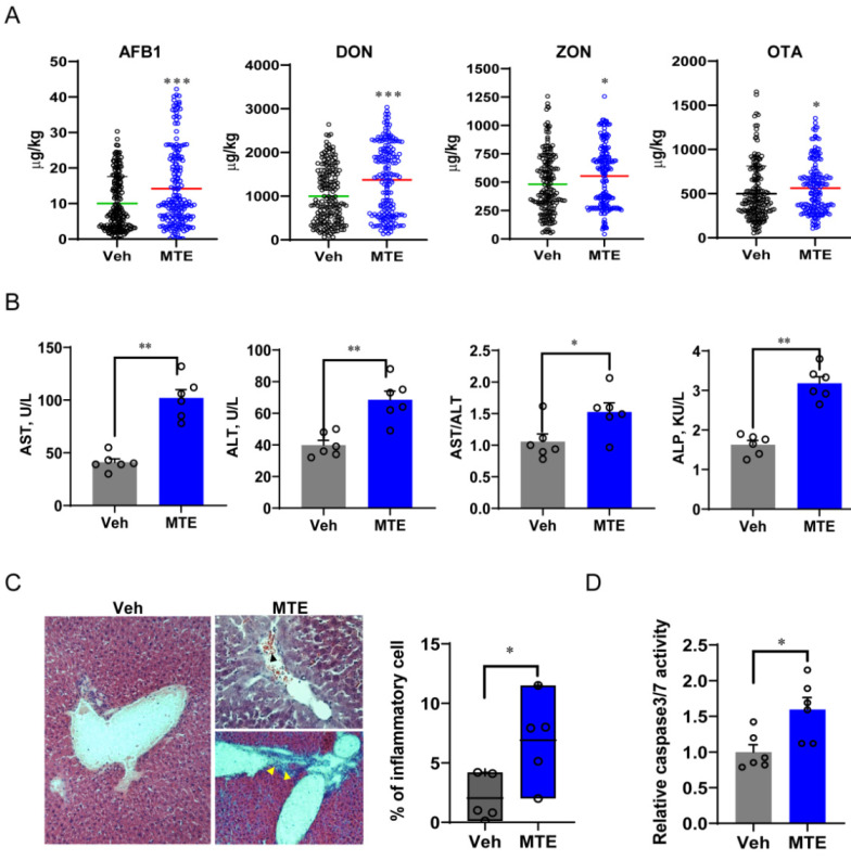
Figure 1.
Mycotoxin exposure is associated with liver injury in piglets. (A) Feed samples were collected and used to measure the content of mycotoxins by the competitive enzyme immunoassay using the kits from R-Biopharm AG. (B) Blood AST (aspartate transaminase), ALT (alanine aminotransferase), and ALP (alkaline phosphatase) concentrations, and the AST/ALT ratio. (C) Representative images of liver sections stained with hematoxylin and eosin from the Veh and MTE groups, respectively. The upper right panel shows diffused cell infiltration and blood cell accumulation (black arrow). Yellow arrows (bottom right panel) point to mononuclear cell infiltration at the portal area. (D) Analysis of the percentage of inflammatory cells (n = 5); hepatocyte apoptosis is reflected by activation of caspase 3/7. The data are shown as the means ± SEM, n = 6, * p < 0.05, ** p < 0.01, *** p < 0.001, using ANOVA with Tukey’s post hoc test.

