Abstract
In this study, we investigated aqueous extracts of three edible mushrooms: Agaricus bisporus (white button mushroom), Pleurotus columbinus (oyster mushroom), and Pleurotus sajor-caju (grey oyster mushroom). The extracts were biochemically characterized for total carbohydrate, phenolic, flavonoid, vitamin, and protein contents besides amino acid analysis. Triple TOF proteome analysis showed 30.1% similarity between proteomes of the two Pleurotus spp. All three extracts showed promising antiviral activities. While Pleurotus columbinus extract showed potent activity against adenovirus (Ad7, selectivity index (SI) = 4.2), Agaricus bisporus showed strong activity against herpes simplex II (HSV-2; SI = 3.7). The extracts showed low cytotoxicity against normal human peripheral blood mononuclear cells (PBMCs) and moderate cytotoxicity against prostate (PC3, DU-145); colorectal (Colo-205); cecum carcinoma (LS-513); liver carcinoma (HepG2); cervical cancer (HeLa); breast adenocarcinoma (MDA-MB-231 and MCF-7) as well as leukemia (CCRF-CEM); acute monocytic leukemia (THP1); acute promyelocytic leukemia (NB4); and lymphoma (U937) cell lines. Antioxidant activity was evaluated using 2,2-diphenyl-1-picryl-hydrazyl-hydrate (DPPH) radical scavenging, 2,2′-Azinobis-(3-Ethylbenzthiazolin-6-Sulfonic Acid) ABTS radical cation scavenging, and oxygen radical absorbance capacity (ORAC) assays. The three extracts showed potential antioxidant activities with the maximum activity recorded for Pleurotus columbinus (IC50 µg/mL) = 35.13 ± 3.27 for DPPH, 13.97 ± 4.91 for ABTS, and 29.42 ± 3.21 for ORAC assays.
Keywords: white rot fungus, Pleurotus columbinus, Pleurotus sajor-caju, Agaricus bisporus, medicinal mushrooms, antioxidant, cytotoxic, antiviral
1. Introduction
Mushrooms have been used in traditional ancient therapies since the Neolithic period. Humankind has valued mushrooms as an edible and medicinal food source. Most of the ancient knowledge about medicinal mushrooms has been confirmed and registered by modern science [1]. Current mushrooms with medicinal value are used in various fields such as dietary food, nutritive auxiliary products, and in the medicinal field called “pharmaceuticals of mushrooms” [2]. Remedial mushrooms are similar to “curing plants” in that they are observable fungi, mostly some Ascomycetes and higher Basidiomycetes, that are used in the extract formula or powder for disease avoidance, allegation or mending, and/or to provide an equitable healthy regimen [2]. About 130 therapeutic actions are thought to exist in medicinal mushroom and fungi, including antitumor, antiviral, antiparasitic, antibacterial, antifungal, antioxidant, radical scavenging, detoxicating, immunomodulating,, antidiabetic, and hepatoprotective effects [3]. Medicinal mushroom are used with patients under chemotherapeutic treatment or radiation therapy, in different forms of cancers, bloodborne chronic viral infections of hepatitis B, C, and D, and herpes simplex virus (HSV) [4].
Many edible mushrooms have been portrayed as therapeutic cures for a range of ailments. Both mycelia and fruiting bodies were used to extract fractions with antiviral activity [5]. The identified active compounds appeared to operate by directly inhibiting viral enzymes, viral replication, absorption of virus, and mammalian cells uptake. Small molecules were responsible for the direct antiviral activity, whereas the indirect effects may be elicited by polysaccharides or complex molecules [6].
Mushroom carbohydrates inhibit tumor genesis, have direct anticancer activities against a variety of synergetic tumors, and stop tumors from spreading. When used in combination with chemotherapy, their operation is particularly beneficial [7]. Polysaccharides’ antitumor activity is resolved through a thymus-dependent immune system, which requires an intact T cell portion [8]. In vitro and in vivo studies indicate that medicinal mushroom drugs besides medicinal mushroom polysaccharide preparations from various mushroom species are effective in treating cancer [9]. Biological reaction modifiers are a new class of antitumor medicinal mushrooms medications (BRMs) [10]. Along with surgery, chemotherapy, and radiotherapy, BRMs have been used as an advanced type of cancer treatment [10]. Moreover, enzyme therapies, particularly those produced by mushrooms, have been shown to be effective in the treatment of cancer by limiting oxidative stress and restricting cell growth [11].
Interestingly, mushrooms are rich in high-quality proteins that contain the essential amino acids, vitamins, carbohydrates, minerals, unsaturated fatty acids, and fibers. They are considered an excellent and nutritious food for people with high blood cholesterol levels or hypertension due to their low energy, fat, and sodium contents [12]. Aside from phenolic, mushrooms with relatively high levels of vitamins A, C, and β-carotene have been shown to be the key contributor to their antioxidant activity [13]. Researchers are interested in quantifying in vitro mushroom extracts antioxidant activities as a way to easily evaluate total antioxidant concentrations and possible effectiveness due to the potential benefits of antioxidant intake [14]. In addition, Barros et al. reported that flavonoids of mushroom can play an important role as free radical scavengers, stopping the chain reactions that occur during triglyceride oxidation in the food system [15]. Therefore, this study was conducted to assess the antioxidant, cytotoxic, and antiviral activities of the three edible mushroom aqueous extracts, namely Pleurotus columbinus, Pleurotus sajor-caju, and Agaricus bisporus followed by composition analysis of the respective extracts.
2. Materials and Methods
2.1. Collection and Culture Conditions of Fungal Isolates
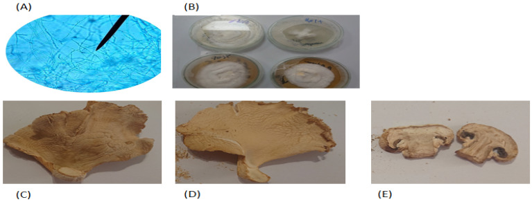
Preidentified Pleurotus columbinus, Pleurotus sajor-caju, and Agaricus bisporus mushrooms were obtained, as spawns and/or fruiting bodies (Figure 1), from the culture collection of Al-Orman botanical reservoir, Cairo, Egypt [16,17,18]. For subculturing from fungal fruits, sterile surgical blades were aseptically used to cut the fruit. The inner mycelia were picked by sterile needles and subcultured in sterile potato dextrose agar (PDA; Merck, Darmstadt, Germany) plates [19]. For subculturing of fungal spawns, sterile forceps were used to pick the seeds and aseptically transfer them to sterile PDA plates that were incubated at 28 °C for 7 days, and then the fruiting bodies of each isolate were separately washed. Aliquots (500 g) of each isolate were air dried at room temperature for 48 h, ground in a kitchen grinder and stored at room temperature in a well-aerated place until use.
Figure 1.
(A) Microscopic examination of the white rot fungus using light microscope, (B) growth of mushroom on potato dextrose agar post 7 days culture. Dried fruiting bodies of the collected mushroom isolates, (C) Pleurotus columbinus, (D) Pleurotus sajor-caju, and (E) Agaricus bisporus.
2.2. Molecular Identification of Mushroom Species
2.2.1. Extraction of Genomic DNA and PCR Amplification
Total genomic DNA was extracted from 7-day-old mycelial mats using Zymo Research (ZR) Fungal/Bacterial DNA kit™ (Zymo Research, Irvine, CA, USA) following the manufacturer instructions.
The internal transcribed spacer (ITS) region of the rDNA was amplified by PCR with previously described universal primers ITS1 (5′-TCC GTA GGT GAA CCT GCG G-3′) and ITS4 (5′-TCC TCC GCT TAT TGA TAT GC-3′) [20]. PCR reaction mixture was performed in a total volume of 50 μL containing 8 μL DNA, 1 μL (20 pico mol) of each primer, 25 μL My Taq Red mixture (Bioline GmbH, Luckenwalde, Germany) and 15 μL nuclease free water. The amplification reaction was done in a C1000 Touch thermal cycler (BioRad, Hercules, CA, USA) according to Das et al. [21] with slight modifications. Initial denaturation at 94 °C for 6 min, followed by 35 cycles of denaturation at 94 °C for 45 s, annealing at 56 °C for 45 s, extension at 72 °C for 1 min, and a final extension at 72 °C for 5 min.
2.2.2. DNA Sequencing and ITS Sequence Analysis
The pair of universal ITS primers ITS-1 (forward) and ITS-4 (reverse) was used for the Sanger sequencing of the purified PCR products using model ABI PRISM® 3500XL DNA Sequencer (Applied Biosystems, Foster City, CA, USA) following manufacturer’s instructions. For good quality sequence assurance, FinchTV version1.4.0 software was used for the analysis of sequences (sense and antisense) resulting from sequencing reaction (Geospiza, Inc.; Seattle, WA, USA; https://digitalworldbiology.com/FinchTV, http://www.geospiza.com (accessed on 5 August 2021)). The resulting sequences were edited using BioEdit Sequence Alignment Editor (Ibis Therapeutics, Carlsbad, CA, USA). Then, the resulting consensus ITS sequences were blasted in the NCBI (http://www.ncbi.nlm.nih.gov, accessed on 5 August 2021) database with the BLASTn for homology in order to identify the probable mushroom in question. The sequences were deposited in the GenBank®.
2.3. Mushroom Aqueous Extract Preparation
The dried fruit of each isolate was blended using a household blender to give 500 g powder, then macerated three times for three successive days in 7 L of distilled water at room temperature and filtered. Maceration and filtration were repeated until exhaustion. The yield was stored with ethanol. A total of 500 mL of each isolate was freeze-dried after ethanol evaporation at 45 °C to give approximately 20 g dry residue of each extract [22].
2.4. Biochemical Charactrization of the Crude Mushroom Extracts
2.4.1. Total Carbohydrate Content
Total soluble carbohydrate content was assayed using phenol sulfuric acid method as previously reported [23]. Data are presented as means ± standard deviation (SD) and experiments were performed in triplicate.
2.4.2. Total Phenolic and Total Falvonoid Content
Phenolic and flavonoid content was determined as reported by Ryan et al. [24]. Samples were prepared at a concentration of 20 mg/mL in water. Gallic acid (1 mg/mL stock solution) was prepared in methanol, and 9 serial dilutions were prepared (concentrations: 12.5, 25, 50, 100, 200, 400, 500, 800, and 1000 µg/mL) for the calibration curve. Rutin (1 mg/mL stock solution) was prepared in methanol, and 10 serial dilutions were prepared (concentrations 1000, 800, 500, 400, 200, 100, 50, 25, 12.5, and 6.25 µg/mL) used in the calibration curve. Gallic acid standards and samples were pipetted in the wells of the plate in six replicates and measured at 630 nm. Each of the 10 rutin standards and samples in six replicates were measured at 420 nm.
2.4.3. Analysis of Phenolics and Flavonoids Using High-Performance Liquid Chromatography (HPLC)
This was performed according to the method of Singh et al. [25]. Each of the samples and 10 different standards solutions were dissolved in methanol and filtered using 0.22 µm syringe filters; then, 100 µL of each sample and 10 µL of each standard were injected using HPLC column, Waters 2690 Alliance HPLC system equipped with a Waters 996 photodiode array detector. Column C18 Inertsil ODS 3: 4.6 × 250 mm, 5 µm, and mobile phase: Buffer (0.1% phosphoric acid in water) and methanol mode of elution was gradient with a flow rate of 1 mL/min at wavelength 280 nm.
2.4.4. Vitamin Content
Water Soluble Vitamins
The three tested mushroom extracts (50 mg/mL), and reference standards (10 mg in 10 mL 0.05 M sodium hydroxide) of each of the seven water soluble vitamins (thiamine HCl, ascorbic acid, riboflavin, nicotinic acid, nicotinamide, pyridoxine HCl, and folic acid) were diluted to 100 µg/mL and filtered using a 0.22 µm syringe filter; then, 100 µL were injected onto the HPLC column, Waters 2690 Alliance HPLC system (Milford, CT, USA) equipped with a Waters 996 photodiode array detector. Column Inertsil ODS 3: 4.6 × 250 mm, 5 µm, mobile phase: Buffer (0.85 g hexane sulphonic acid sodium salt in 1000 mL water and pH was adjusted to 3 with orthophosphoric acid): Methanol elution was gradient at a flow rate of 1 mL/min at wavelength 210 nm [26].
Fat Soluble Vitamins
Solutions (50 mg/mL) of the three mushroom extracts and a standard solution in methanol of three fat vitamins; E, D3, and A, at 806.2, 114, and 400 IU/mL, respectively, were filtered using 0.22 µm syringe filters; then, 10 µL of each was injected onto HPLC column, Waters 2690 Alliance HPLC system equipped with photodiode array detector. Column Inertsil ODS 3: 4.6 × 250 mm, 5 µ, mobile phase: Methanol 100% with isocratic elution of flow rate 1 mL/min at wavelength: 210 nm [27].
2.4.5. Total Protein Analysis
This was performed using the bicinchoninic acid (BCA) assay [28] using bovine serum albumin as a standard. One mg of each sample was dispersed in 1 mL phosphate buffer saline (pH 7.4), sonicated for 30 min to extract proteins among other water-soluble substances, filtrated using a 0.45 µm syringe filter, then tested using the BCA Protein Assay Kit (Novagen, Madison, WI, USA) as stated by the manufacturer’s instructions.
2.4.6. Amino Acids Analysis
Composition of amino acid of the mushroom extracts was performed using the Sykam amino acid analyzer (Sykam GmbH, Eresing, Germany) equipped with solvent delivery system S 2100 (Quaternary pump with flow range 0.01 to 10.00 mL/min and maximum pressure up to 400 bar), auto sampler S5200, amino acid reaction module S4300 (with built-in dual filter photometer at 570 nm), and refrigerated reagent organizer S4130. One g of each mushroom extract was mixed with 5 ml hexane, allowed to macerate for 24 h, and then the mixture was filtered using Whatman no. 1 filter paper. The residue was placed in a test tube containing 10 mL 6 M HCl and incubated in an oven at 110 °C for 24 h. After the incubation, filtration was done using Whatman no. 1 filter paper, followed by evaporation in a rotary evaporator. The residue was dissolved completely in 2 mL of dilution buffer (Tri-sodium citrate dehydrate 0.06 M; citric acid 0.03 M, phenol 0.02 M, Thiodiglycol 1.4%, HCl 32%), diluted 1000 folds in the same buffer, then loaded onto an ammonia filter column (LCA, K04/Na, 4.6 × 100 mm, Sykam, GmbH, Eresing, Germany) equipped with an automatic amino acid analyzer (Sykam GmbH, Eresing, Germany) [29].
2.5. TripleTOF Analysis of Proteomes
Proteomic analysis was conducted at the proteomics and metabolomics unit at Children’s Cancer Hospital Egypt 57,357 (CCHE), Cairo, Egypt. About 600 μL 8 M urea in 500 mM Tris (pH 8.5) and 60 μL complete ultraproteases (Roche, Mannheim, Germany) were added to each sample; then, samples were shaken vigorously and centrifuged at 10,000 RPM for 30 min. Supernatants were collected and fractions (1 μg/10 μL) were injected using the NanoLC system. The mass spectrometry triple TOF system (Sciex TripleTOF 5600+, AB SCIEX, Concord) was coupled with liquid chromatography (LC) (3 µm, ChromXP C18CL, 120A, 150 × 0.3 mm), consisting of the Eksigent nanoLC400 auto sampler attached to an Ekspert nanoLC425 pump with a flow rate of 10 µL/min for 55 min for each sample [30]. Data analysis was performed by Protein pilot (version 5.0.1.0, 4895) and paragon Algorithm (version 5.0.1.0, 4874). Protein sequences were aligned against sequences in the Swiss-Prot and TrEMBL databases (Pleurotus sp. containing 14,792 entries for Pleurotus columbinus and Pleurotus sajor-caju, and Agaricus sp. containing 29,258 entries for Agricus bisporus) [31]. Commonly identified proteins between the mushroom species were illustrated using Venny2.1.0.BioinfoGP software [32] (Kingston upon Hull, UK).
2.6. Cell Lines and Cultures
Primary peripheral blood mononuclear cells (PBMCs), normal, human (ATCC® PCS-800-011™) were purchased from Vascera, Cairo, Egypt. The cancer cell lines human leukemia (CCRF-CEM); acute promyelocytic leukemia (NB-4); human lymphoma (U937); prostate cancer (DU-145 and PC3); hepatocellular carcinoma (HepG2; breast adenocarcinoma (MCF-7 and MDA-MB-231); Vero; and Hep-2 cells were obtained from Nawah Scientific Inc., (Mokatam, Cairo, Egypt). Cervical cancer (HeLa), acute monocytic leukemia (Thp1), colorectal carcinoma (Colo-205); and cecum carcinoma (LS-513) cells were maintained in Roswell Park Memorial Institute (RPMI) medium (Lonza, Germany) supplemented with 100 mg/mL of streptomycin, 100 units/mL of penicillin, and 10% of heat-inactivated fetal bovine serum. For other cell lines, Dulbecco’s modified Eagle’s medium (DMEM; Invitrogen, Carlsbad, CA, USA) supplemented with 10% FBS (Hyclone, Marlborough, MA, USA), 10 µg/mL of insulin (Sigma-Aldrich, St. Louis, MO, USA), 100 mg/mL of streptomycin, and 100 U/mL of penicillin (Grand Island, NY, USA) was used for cell culture. Incubation was performed at 37 °C in a humidified atmosphere with 5% CO2.
2.7. Antiviral Activity
Antiviral activity was tested against adenovirus type 7 (Ad7) and herpes simplex virus type 2 (HSV-2) (Nawah Scientific, Mokattam, Egypt). Antiviral activity was assessed as previously described [33]. Vero and Hep-2 cells were seeded into a 96-well culture plate at a density of 2 × 10⁴ cells/well one day before infection. The cell culture medium (2.5) was removed the next day, and phosphate-buffered saline (PBS) was used for cells to be washed. The infectivity of human Ad7 and HSV2 was determined using the sulforhodamine B (SRB) method [34], which monitored CPE and calculated the percentage of cell viability. One hundred microliters of diluted virus suspension of Ad7 and HSV-2 containing 50% cell culture infective dose (CCID50) of virus stock was added to mammalian cells. To test the antiviral activity of mushroom extracts, 0.01 mL of medium containing each extract (10-fold serial dilution ranging from 0.1 to 100 µg/mL) was added to cells. Controls (virus-infected, non-extract-treated cells and noninfected, non-extract-treated cells) were included. After 4 days of incubation at 37 °C in 5% CO2, PBS was used to wash cells, then 0.01 mL of 70% (v/v) cold acetone was added and left for 30 min at −20 °C. After removing acetone, plates were dried at 60 °C for 30 min, then wells were filled with 0.01 mL of 0.4% (w/v) SRB solution in 1% acetic acid (v/v) and at room temperature incubated for 30 min. Unbound SRB was removed from the plates by washing 5 times using 1% acetic acid (v/v) and allowing them to dry. Fixed SRB in wells was dissolved in 100 µL of unbuffered Tris base solution (10 mM), and then plates were incubated for 30 min at room temperature. Finally, optical density (OD) was measured at 540 nm and a reference absorbance of 620 nm using a microplate reader (FluoStar Omega, BMG labtech, Ortenberg, Germany).
To assess cytotoxicity, cells were seeded at a density of 2 × 10⁴ cells/well in a 96-well culture plate. The next day, the cells were given culture medium containing serially diluted samples, which was cultured for 48 h before being withdrawn and the cells rinsed with PBS. The following step was carried out in the same manner as described above for the antiviral activity assay [35].The antiviral activity was calculated based on the extracts ability to inhibit the viral cytopathogenic effects where the 50% cytotoxic concentrations (CC50) and the 50% inhibitory concentration (IC50) were determined using GraphPad PRISM software (GraphPad Software, San Diego, CA, USA). Selectivity index (SI) was calculated as stated by Doğan et al. [36] according to the Equation:
| Selectivity index (SI) = CC50/IC50 | (1) |
2.8. Evaluation of Cytotoxic Effect against PBMCs
Cytotoxicity was assessed using MTT assay [37]. Aliquots (100 µL) of cell suspensions (cell density 1.2–1.8 × 10⁴ cells/well) were placed in 96-well plates, complete medium (2.5) was added, and cells were incubated for 24 h. Then cells were treated with serial dilutions of fungal extracts for 48 h. MTT (3-(4, 5-dimethylthiazol-2-yl)-2, 5-diphenyltetrazolium bromide, 5 mg/mL in PBS) was added to wells and incubated for 4 h. Formazan crystals were extracted using MTT solubilization solution (10% Triton X-100 plus, 0.1 M HCl in anhydrous isopropanol) and at 570 nm absorbance were read using microplate reader.
2.9. Evaluation of Cytotoxic Effect against Cancer Cell Lines
Cytotoxicity against prostate cancer (DU-145 and PC3); hepatocellular carcinoma (HepG2); colorectal carcinoma (Colo-205); cecum carcinoma (LS-513); cervical cancer (HeLa); and breast adenocarcinoma (MDA-MB-231 and MCF-7) cell lines was evaluated using a sulforhodamine B (SRB) colorimetric assay. Cell suspensions in complete media (2.5) were incubated in 96-well plates for 24 h. Then, various concentrations (0 to 100 μg/mL in complete media) of mushroom extracts were added and incubated for 72 h. Cell viability was measured using SRB method as described above (2.6).
2.10. Cytotoxcicty Assay against Leukemia and Lymphoma Cell Lines
Cytotoxicity against human leukemia (CCRF-CEM), acute promyelocytic leukemia (NB4), acute monocytic leukemia (THP1), and human lymphoma (U937) was determined using the Abcam® Water Soluble Tetrazolium Salts (WST-1) assay kit (ab155902 WST-1 Cell Proliferation Reagent, UK). Cell suspensions were incubated for 24 h, and then treated with different concentrations (0 to 100 μg/mL in complete media) of mushroom extracts for 48 h. Cells were then treated with 10 μL WST-1 reagent for 1 h, then A450 was read using a microplate reader [38].
2.11. Antioxidant Activity Assay
2.11.1. DPPH Radical Scavenging Activity
DPPH radical scavenging assay was carried out according to the method of [39]. Briefly, in 96-well plates, 100 µL of freshly prepared DPPH reagent (Sigma-Aldrich, Taufkirchen, Germany) (0.1% in methanol) were added to 100 µL of a range of concentrations (0 to 100 μg/mL) of the mushroom extracts, in 6 replicates each, and incubated in the dark at room temperature for 30 min. The resulting reduction in DPPH color was measured at 540 nm. Antioxidant activity was expressed as the inhibition percentage with reference to calibration curve (R2 = 0.9903). Data are represented as means ± SD according to Equation (2):
| Percentage inhibition% = (Average absorbance of blank − average absorbance of the extract) × 100 Average absorbance of blank |
(2) |
2.11.2. ABTS Radical Cation Scavenging Activity
The assay was carried out as previously stated [40]. Mushroom extract (10 µL of 0 to 100 μg/mL solutions) were mixed with 190 µL of freshly prepared ABTS (Sigma-Aldrich, Taufkirchen, Germany) in 96-well plates, incubated in the dark at room temperature for 2 h. Each sample was tested in 4 replicates and absorbance was measured at 734 nm. Antioxidant activity was expressed as percentage of inhibition (Equation (1)) with reference to calibration curve (R2 = 0.9948).
2.11.3. Oxygen Radical Absorbance Capacity (ORAC) Assay
The analysis was carried out as previously determined [41], with minor modifications. Briefly, 12.5 µL of the mushroom extracts, in triplicate, were incubated with 75 µL of fluorescein (10 nM) for 30 min at 37 °C. Fluorescence measurement (485 EX, 520 EM, nm) was carried out for three cycles (cycle time = 90 s). Afterwards, 12.5 µL of freshly prepared 240 mM solution of 2,2′-Azobis 2-amidinopropane dihydrochloride (AAPH) (Abcam, Cambridge, UK) were added to each well immediately and fluorescence measurement was continued for 2.5 h (85 cycles, each 90 sec). Antioxidant activity was expressed as percentage of inhibition (Equation (1)) with reference to calibration curve (R2 = 0.9957).
2.12. Statistical Analysis
For all experiments, results were presented as the mean ± standard deviation (SD) of, unless otherwise specified, three independent readings. Statistical analyses were performed using one-way ANOVA. The significance of differences between means was evaluated with the Tukey–Kramer multiple comparisons test. p ≤ 0.05 was considered statistically significant. Statistical evaluation was determined using GraphPad PRISM software (Graph-Pad Software, San Diego, CA, USA).
3. Results
3.1. Molecular Identification of Mushrooms
According to BLASTn results the mushrooms were identified as Pleurotus columbinus, Pleurotus sajor-caju (Lentinus sajor-caju), and Agaricus bisporus, and the sequences were deposited in GenBank® with accession numbers MZ642245, MZ642259, and MZ642282, respectively.
3.2. Biochemical Characterization of Mushroom Extract
The biochemical analysis (total carbohydrate, protein, phenolic, and flavonoid contents) showed distinctive differences between the three mushrooms (Table 1). Pleurotus columbinus extract had the highest carbohydrate content and the lowest phenolic content. It is worth noting that flavonoids were not detected in the Pleurotus columbinus extract; neither were carbohydrates in the Pleurotus sajor-caju extract.
Table 1.
Biochemical characterization of the three mushroom extracts.
| Mushroom | Glucose Content (mg/g Extract) | Total Phenolic Content (mg/g Extract) | Total Flavonoid Content (mg/g Extract) | Total ProteinContent (mg/g Extract) |
|---|---|---|---|---|
| Pleurotus columbinus | 82.24 ± 3.98 a | 18.16 ± 0.54 b | ND * | 0.08 ± 0.006 b |
| Pleurotus sajor-caju | ND * | 22.50 ± 1.53 b | 2.27 ± 0.18 b | 0.06 ± 0.005 b |
| Agaricus bisporus | 34.18 ± 1.48 b | 27.45 ± 0.8 a | 11.96 ± 1.81 a | 0.12 ± 0.004 a |
All results are expressed as mean ± SD from three experiments (n = 6). Values with different letters within the columns are significantly different (p ≤ 0.05), * ND: not detected in the mushroom.
3.2.1. Characterization of Total Phenolic and Total Falvonoid Contents by HPLC
Characterization of phenols and flavonoids in the three mushroom extracts was done by HPLC. The chromatogram is shown in Figure S1 and the TPC and TFC quantification is in Table 2. Catechin was found in all 3 mushroom extracts (retention time 26.48 min), cholorgenic acid was detected in Pleurotus sajor-caju and Agaricus bisporus extracts at retention time 29.61 min, and gallic acid was found in Pleurotus columbinus and Pleurotus sajor-caju extracts at retention time 12.22 min. Caffeic acid, hesperidin, rutin, ellagic acid, kampeferol, quercetin, and apigenin were not detected in any of the mushroom extracts. There results were confirmed by including a mixture of standard phenolics and flavonoids that has shown the same retention times as those of the detected in the mushroom extracts.
Table 2.
Phenolic and flavonoid composition of mushroom Sp. (µg/mL).
| Compounds | Pleurotus columbinus | Pleurotus sajor-caju | Agaricus bisporus |
|---|---|---|---|
| Gallic acid | 0.18 ± 0.05 a | 0.15 ± 0.03 | ND * |
| Catechin | 0.10 ± 0.03 a | 0.14 ± 0.02 a | 0.07 ± 0.01 b |
| Chlorogenic acid | ND * | 0.17 ± 0.04 a | 0.09 ± 0.02 b |
| Caffeic acid | ND * | ND * | ND * |
| Rutin | ND * | ND * | ND * |
| Ellagic acid | ND * | ND * | ND * |
| Hesperidin | ND * | ND * | ND * |
| Querceitin | ND * | ND * | ND * |
| Kampeferol | ND * | ND * | ND * |
| Apigenin | ND * | ND * | ND * |
All results are expressed as mean ± SD from three experiments (n = 3). Values with different letters within the rows are significantly different (p ≤ 0.05). ND * = Not detected.
3.2.2. Vitamin Content
The content of both water-soluble and fat-soluble vitamins was analyzed using HPLC (chromatograms in Figures S2 and S3). Vitamin C (at a high content), nicotinic acid, nictinamide, and vitamin D were detected in all three mushrooms, unlike folic acid, thiamine, vitamin A, and vitamin E (Table 3). Riboflavin was only detected in Agaricus bisporus (0.11 µg/100 g).
Table 3.
Total vitamin content of the test mushroom species.
| Vitamins | Pleurotus columbinus | Pleurotus sajor-caju | Agaricus bisporus | |
|---|---|---|---|---|
| Ascorbic acid (Vitamin C) (mg/100 g) | 1.40 ± 0.39 a | 0.75 ± 0.23 a | 0.96 ± 0.08 a | |
| Nicotinic acid (µg/100 g) | 0.21 ± 0.02 b | 0.22 ± 0.03 b | 0.18 ± 0.02 c | |
| Nicotinamide (µg/100 g) | 0.05 ± 0.03 c | 0.03 ± 0.01 c | 0.07 ± 0.02 d | |
| Water soluble Vitamins | Pyridoxine (Vitamin B6) (µg/100 g) | 0.25 ± 0.15 b | 0.27 ± 0.20 b | 0.30 ± 0.23 b |
| Folic acid (Vitamin B9) (µg/100 g) | ND * | ND * | ND * | |
| Thiamine (Vitamin B1) (µg/100 g) | ND * | ND * | ND * | |
| Riboflavin (Vitamin B2) (µg/100 g) | ND * | ND * | 0.11 ± 0.02 c | |
| Fat soluble vitamins | Retinol (Vitamin A) (µg/100 g) | ND * | ND * | ND * |
| Cholecalciferol (Vitamin D) (µg/100 g) | 0.04 ± 0.02 c | 0.03 ± 0.01 c | 0.29 ± 0.03 b | |
| Tocopherol (Vitamin E) (mg/100 g) | ND * | ND * | ND * |
Each value is presented as the mean ± standard deviation (n = 3). Data with different superscript letters in the same column of variety indicate a significant difference (p ≤ 0.05). ND *: Not detected.
3.2.3. Amino Acids Analysis
The composition of amino acids of the mushroom extracts as detected using the Sykam Amino Acid Analyzer is shown in Table 4 and Figure S4. A wide range of amino acids was detected in the mushroom extracts, with the exceptions of histidine and aspartic acid, which were not detected. It is worth noting that glutamic acid was the major amino acid in all three mushroom extracts.
Table 4.
Amino acids composition in three mushroom species.
| Amino Acids Content (g/100 g Protein) | |||
|---|---|---|---|
| Amino Acids | Pleurotus columbinus | Pleurotus sajor-caju | Agaricus bisporus |
| Aspartic acid | ND * | ND * | ND * |
| Threonine | 0.266 | 0.315 | 0.321 |
| Serine | 0.367 | 0.446 | 0.412 |
| Glutamic acid | 1.118 | 1.209 | 1.277 |
| Proline | 0.209 | 0.599 | 0.839 |
| Glycine | 0.365 | 0.388 | 0.302 |
| Alanine | 0.49 | 0.496 | 0.523 |
| Cystine | 0.206 | 0.201 | 0.223 |
| Valine | 0.241 | 0.257 | 0.268 |
| Methionine | 0.281 | 0.276 | 0.353 |
| Isoleucine | 0.117 | 0.124 | 0.127 |
| Leucine | 0.381 | 0.417 | 0.337 |
| Tyrosine | 0.167 | 0.176 | 0.197 |
| Phenylalanine | 0.202 | 0.217 | 0.161 |
| Histadine | ND * | ND * | ND * |
| Lysine | 0.343 | 0.347 | 0.299 |
| Arginine | 0.286 | 0.326 | 0.215 |
| Total | 5.04 | 5.75 | 5.58 |
ND *: Not detected
3.2.4. Triple TOF Analysis of Proteomes
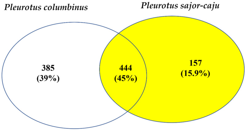
Protein and peptide identification and quantification of the three mushroom extracts were performed using triple TOF. A total of 541 proteins were identified in Pleurotus columbinus, 402 in Pleurotus sajor-caju, and 408 in Agaricus bisporus (Tables S1–S3, respectively). The Venn diagram in Figure 2 summarizes the common proteins between the two tested Pleurotus species. Whereas 444 proteins with the same accession codes were found in both Pleurotus columbinus and Pleurotus sajor-caju, no proteins with the same accession codes were found in common between the two Pleurotus mushrooms and Agaricus bisporus. A multitude of proteins and enzymes were detected, including S-adenosyl-L-homocysteine hydrolase, intradiol dioxygenase, alpha- and beta-form tubulin, thioredoxin reductase, haloacid dyhydrogenase, serine proteinase, ubiquitin-like protein, prohibitin, tubulin, septin, and chaperone. Pleurotolysin and lectin were also identified in both Pleurotus species.
Figure 2.
Venn diagram showing the common identified proteins between the three mushroom species Pleurotus columbinus, Pleurotus sajor-caju, and Agaricus bisporus.
3.3. Cytopatheic Effect against Viral Cell Lines
The antiviral activities of the aqueous extracts of Pleurotus columbinus, Pleurotus sajor-caju, and Agaricus bisporus were examined in vitro against human adenovirus type 7 and herpes simplex virus type II. The dose–response curves are shown in Figures S5 and S6, and the CC50, IC50, and selectivity indices (SI) in Table 5. Potent antiviral activities with relatively low toxicities were recorded for Pleurotus columbinus against Ad7 and Agaricus bisporus against HSV2.
Table 5.
Antiviral activities of the mushroom extracts.
| Virus | Mushroom | CC50 (µg/mL) | IC50 (µg/mL) | SI |
|---|---|---|---|---|
| Pleurotus columbinus | 185.5 | 40.29 | 4.60 | |
| Ad7 | Pleurotus sajor-caju | 60.96 | 43.61 | 1.39 |
| Agaricus bisporus | 8.339 | 15.28 | 0.54 | |
| Pleurotus columbinus | 17.615 | 20.04 | 0.87 | |
| HSV2 | Pleurotus sajor-caju | 25.43 | 34.34 | 0.74 |
| Agaricus bisporus | 59.07 | 15.9 | 3.7 |
CC50, half-maximal cytotoxic concentration; IC50, half-maximal inhibitory concentration; SI, selectivity index = CC50/IC50.
3.4. Cytotoxic Activity against Normal Human PBMCs
All three mushroom extracts (Pleurotus columbinus, Pleurotus sajor-caju, and Agaricus bisporus) showed low cytotoxicity against the normal human PBMCs (IC50 µg/mL; 75.03 ± 0.62, 70.42 ± 1.89, and 57.72 ± 0.95, respectively).
3.4.1. Cytotoxic Activity against Cancer Cell Lines
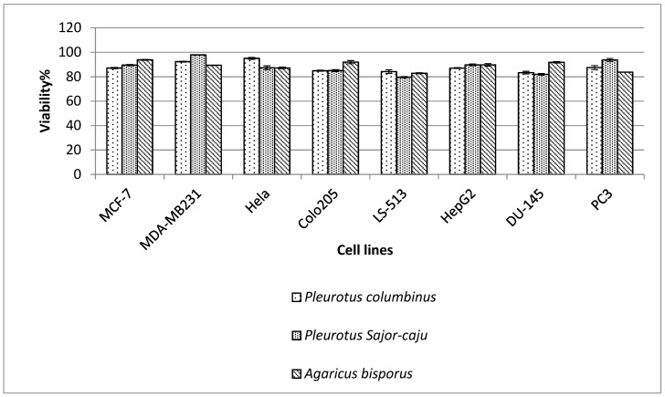
Cytotoxicity of the three mushroom extracts was tested at five different concentrations (0.01, 0.1, 1, 10, and 100 µM) on a range of cancer cell lines including prostate cancer (DU-145 and PC3); hepatocellular carcinoma (HepG2); colorectal carcinoma (Colo-205); cecum carcinoma (LS-513); cervical cancer (HeLa); and breast adenocarcinoma (MDA-MB-231 and MCF-7). Results are shown in Figure 3. Pleurotus sajor-caju and Agaricus bisporus reported the highest cytotoxicity against LS-513 cell line with cell viabilities 79.60 ± 0.53% and 82.83 ± 0.42%, respectively.
Figure 3.
Cytotoxic effect of the three mushrooms extracts Pleurotus columbinus, Pleurotus sajor-caju, and Agaricus bisporus against cancer cell lines MCF-7, MDA-MBA-231, Hela, Colo-205, LS-513, HepG2, Du-145, and PC-3.
3.4.2. Cytotoxic Activity against Leukemia and Lymphoma
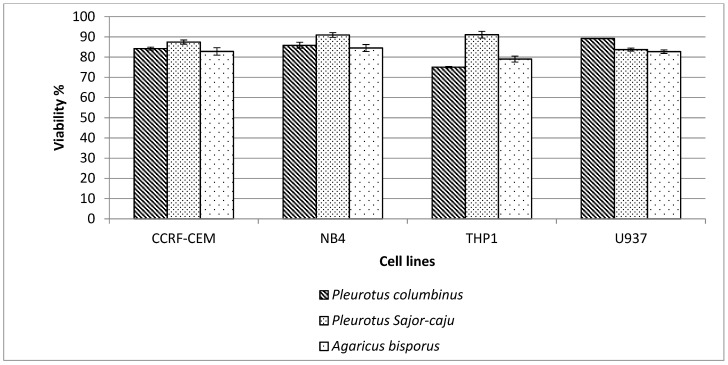
Mushroom extracts of Pleurotus columbinus, Pleurotus sajor-caju, and Agaricus bisporus were tested for cytotoxicity against leukemia and lymphoma cells at a concentration of 100 µM. As shown in Figure 4, comparable results were obtained for the three mushroom extracts.
Figure 4.
Cell viability of the three mushrooms extracts Pleurotus columbinus, Pleurotus sajor-caju, and Agaricus bisporus against leukemic cells (CCRF-CEM, NB4, and THP1) and lymphoma U937.
3.5. Antioxidant Activity
Antioxidant activity was investigated through three different evaluation methods (Table 6) against the antioxidant standard, Trolox. The DPPH assay showed that the free radicle scavenging ability of Pleurotus columbinus was significantly higher than Pleurotus sajor-caju and much higher than that of Agaricus bisporus. The ABTS scavenging activity showed that all three extracts showed antioxidant activities higher than that of Trolox. Results of the ORAC assay are shown in Table 6, Figures S7a,b and S8 with results of various concentrations of Trolox. ORAC assay results show that Pleurotus columbinus and Pleurotus sajor-caju have significantly higher antioxidant capacities than Agaricus bisporus.
Table 6.
IC50 values of Pleurotus columbinus, Pleurotus sajor-caju, and Agaricus bisporus in the DPPH, ABTS radical scavenging, and ORAC assays.
| IC50(µg/mL) | |||
|---|---|---|---|
| Mushroom | DPPH Radical Scavenging | ABTS Radical Cation Scavenging | ORAC Assay |
| Pleurotus columbinus | 35.13 ± 3.27 c | 13.97 ± 4.91 c | 29.42 ± 3.21 b |
| Pleurotus sajor-caju | 40.91 ± 2.27 b | 16.89 ± 5.77 b | 32.00± 2.17 b |
| Agaricus bisporus | 83.93 ± 0.62 a | 29.96 ± 7.03 a | 75.64 ± 4.65 a |
| Trolox | 24.00 ± 0.87 | 40.00 ± 0.03 | 55.51 ± 0.06 |
All results are expressed as mean ± SD from three experiments (n = 6). Values with different letters within the columns are significantly different (p ≤ 0.05).
4. Discussion
The three mushrooms Pleurotus columbinus, Pleurotus sajor-caju, and Agaricus bisporus are edible mushrooms with high consumption in many countries [42]. In this study, we biochemically characterized the aqueous extracts of the respective three mushrooms. Our results revealed that they were all protein-rich and that Pleurotus sajor-caju contained the least carbohydrate content [43]. Similar data were reported by Agarwal et al. (2017), who recommended Pleurotus sajor-caju for diabetics and weight watchers [44]. It is well documented that phenolics are ubiquitous in mushrooms at approximately 2 to more than 30 mg/g concentration for Pleurotus spp. [45,46]. Our data reported phenolic concentrations of 22.50 ± 1.53 and 18.16 ± 0.54 for P. columbinus and P. sajor-caju, respectively. The phenol content of Agaricus bisporus was 27.45 ± 0.8 mg/g. Gąsecka et al. [47] studied the properties of seven Agaricus spp. mushrooms and reported a wide range of total phenolic content (132.7 to 1154.7 mg GAE/100 g DW). The presence of flavonoids in Pleurotus spp. is reported to be species specific [45,48]. In this study, flavonoids were not detected in Pleurotus columbinus. Similarly, Vieira et al. [49] reported the absence of flavonoids in P. ostreatus. The highest flavonoid concentration was found in A. campestris (15.63 mg GAE/g extract). Catechin was detected in the three mushrooms. Likewise, Butkhup et al. [14] reported the presence of catechin in 25 edible mushrooms. Catechin has often been linked to antioxidant activity of natural extracts [50,51,52].
Since all three mushroom species belong to Agaricomycetes, their amino acids profiles were remarkably similar. While overall amino acid contents were similar in the three mushrooms (5.04%, 5.78%, and 5.58% for Pleurotus columbinus, Pleurotus sajor-caju, and Agaricus bisporus, respectively), specific amino acids were found at different concentrations. Glutamic acid was the most prominent amino acid in the three mushroom extracts. Tagkouli et al. [53] also reported that glutamic acid was the most abundant amino acid in the studied three Pleurotus extracts.
The vitamin profiles of the three mushrooms were relatively close; this may be because they belong to the same family. Regarding water soluble vitamins, vitamin C was detected in high amounts (1.40, 0.75, and 0.96 mg/100 g in Pleurotus columbinus, Pleurotus sajor-caju, and Agaricus bisporus, respectively) compared to other vitamins. Sánchez et al. [54] reported that mushrooms contain vitamin C at concentrations in the range of 0.15–0.06 mg/mL. Several mushrooms have also been shown to contain vitamin C including Boletus edulis [55], B. pseudosulphureus [56], Lactarius deliciosus [57], Pleurotus ostreatus [58], and Suillus luteus [59]. Vitamin D was the only fat-soluble vitamin detected in the extracts.
Pleurotus columbinus, Pleurotus sajor-caju, and Agaricus bisporus showed effective antiviral activities against the Adv7 and HSV2. Selectivity index (SI) is commonly used parameters for measuring how safe it is to use a compound as an antiviral agent. SI estimates the gap between the cytotoxic and antiviral activity. Higher SI values indicate higher efficacy and safety of drug use. A perfect drug would affect the virus at low concentration and the cells at a high one; thus, it would eradicate the virus at a concentration that will not harm host cells [60,61].
The antiviral properties of edible mushrooms have been attributed to water extracts and are typically linked to the presence of water-soluble polysaccharides. β glucans isolated from the Pleurotus tuber-regium sclerotium have antiviral action against HSV-1 [62]. Huang et al. [63] discovered that an acidic polysaccharide bound to protein isolated from water-soluble extracts of Ganoderma lucidum fruiting bodies has ant herpetic action. Sulphates of Lentinus edodes (lentinan) β-(1-3)-D-glucan have also been shown to have significant anti-HSV-1 action. Antiviral activity may also be attributed to other molecules. Two phenolic compounds, for example, were extracted from the fruiting bodies of Inonotus hispidus and found to have exceptional antiviral activity in face of influenza viruses. [64]
Mushroom components such as peptide RC28, polysaccharide, proteoglycan, sulfated polysaccharide, and triterpenoid (lucialdehyde B, ganoderone A, and ganodermadiol) have exhibited pre- and post-treatment antiviral effects [65]. They could work on all stages of viral replication. The activity of the peptide RC28 against herpes viruses was comparable to the activity of ganciclovir [66]. Peptide RC28 and sulfated polysaccharide from mushrooms have antiviral activity against HSV in vitro as well as in vivo, suggesting that they could be used as therapeutic treatments [66].
The aqueous extracts of the mushrooms showed inhibiting growth effects on the cell lines prostate cancer (DU-145 and PC3); hepatocellular carcinoma (HepG2); colorectal carcinoma (Colo-205); cecum carcinoma (LS-513); cervical cancer (HeLa); and breast adenocarcinoma (MCF-7 and MDA-MB-231) at concentration 100 μg/mL. Pleurotus columbinus and Pleurotus sajor-caju demonstrated a decrease in the cell viability of MCF-7, Hela, Colo-205, LS-513, HepG2, Du-145, PC-3 and a slight reduction in viability of MDA-MD-231 cells. Agaricus bisporus showed the same effect as the other two mushroom isolates but with a little more reduction in the cell viability of MDA-MD-231. Agaricus bisporus conjugated linoleic acids have been proved to constrain prostate cancer cell types in vitro [67]. In a similar way, Adams et al. [68] reported that this extract suppresses breast cancer cell proliferation, and drinking white button mushroom powder with green tea may help to prevent breast cancer. Abdalla et al. [69] proposed that Agaricus blazei extracts suppressed breast cancer cell proliferation by inhibiting aromatase activity. Gu and Leonard discovered that edible mushrooms Coprinellus sp., Coprinus comatus, and Flammulina velutipes had strong antiproliferative activity in human breast cancer cell lines (MCF-7) and (MDA-MB-231) [70].
Though the antiproliferative effect of polysaccharides on tumor cell lines in vitro is unknown, certain studies have shown that incubating polysaccharides with tumor cells can cause changes in signal expression inside the tumor cells. Such modifications could cause cell cycle arrest and apoptosis, which would explain polysaccharides’ antiproliferative activity in vitro [71]. Inonotus obliquus mushroom water extract reported to have antitumor activity towards colon cancer cells HT-29. The stimulation of apoptosis, which causes cancer cells to die, was used to restrict cell growth. The mushroom Inonotu obliquus has been shown to have anticancer properties in several research studies. Sclerotia extracts from Inonotu obliquus, for example, have been shown to decrease tumor cell proliferation and protein synthesis [72]. Suillin, a tetraprenylphenol derivative separated from Suillus placidus, Suillin was reported to destroy human liver cancer cells preferentially. Suillin could cause apoptotic death in HepG2 cells in addition to inhibiting proliferation [73]. Lau et al. [74] found that an ethanol–water extract of Coriolus versicolor, a commonly used Chinese medicinal mushroom, may dramatically reduce the development of human promyelocytic leukemia in its natural form. HL-60 and NB-4 cells, as well as Raji cells from B-cell lymphoma, were tested in vitro using the MTT assay. The present study found out that the three mushrooms have some cytotoxic effect on the leukemia and lymphoma cell lines. Pleurotus columbinus, Pleurotus sajor-caju, and Agaricus bisporus declared some decline in cell viability of the CCRF-CEM, NB-4, THP1, and human lymphoma (U937). However, Pleurotus sajor-caju showed no significant effect against NB4 and THP1 cell lines.
In this study, the protein expression profiles of fruiting bodies were investigated using Triple TOF protein identification. A myriad of proteins and enzymes were detected including catalase, S-adenosyl-L-homocysteine hydrolase, intradiol dioxygenase, alpha- and beta-form tubulin, thioredoxin reductase, haloacid dyhydrogenase, prohibitin, septin, chaperone, serine proteinase, and ubiquitin-like protein. Tubulin was reported to contribute to the cytotoxic activity of mushroom extracts [75,76,77]. Similarly, thioredoxin reductase has shown high cytotoxic activity against a range of cell lines vis-à-vis low systemic toxicity [78,79,80]. Pleurotolysin was detected in the Pleurotus species. It is a sphingomyelin-specific cytolysin composed of two subunits (17 and 59 kDa) that integrate to cause an outflow of K ions leading to selective lysis of cancer cells [81,82]. Serine proteinase plays an important role in the antiviral properties of mushrooms [83]. Wang et al. proposed that the anti-HIV effect of the aqueous extracts of some mushroom species may be caused by inhibition of reverse transcriptase enzyme by ubiquitin-like protein [84]. The antioxidant properties of mushroom extracts was attributed to the presence of enzymes including superoxide dismutase, catalase, and glutathione peroxidase, which quench free radicals and detoxify reactive oxygen species [85].
The antioxidant activity of the mushroom extracts was investigated using three different activity measurements. This is because antioxidants have multiple mechanisms of action, and no single approach can capture all of them. The two major mechanisms of antioxidant activity are HAT (hydrogen atom transfer) and SET (single electron transfer). whereas DPPH is classified as SET, ABTS, and ORAC assays are HAT reactions [86]. The measurements of the ABTS test showed slightly lower scavenging capacity compared to the DPPH method. The IC50 values of the ABTS scavenging method for Pleurotus columbinus, Pleurotus sajor-caju, and Agaricus bisporus were 13.97 ± 4.91, 16.89 ± 5.77, and 29.96 ± 7.03 µg/mL, respectively.
Our findings revealed that, based on the different antioxidant assays, DPPH radical scavenging activity, ABTS radical cation scavenging activity, and the ORAC assay, the mushroom aqueous extracts had strong antioxidant activities, comparable to those of the standard antioxidant, Trolox. Bakir et al. [87] compared the DPPH scavenging ability of Pleurotus ostreatus stored at different temperatures and found that the highest antioxidant activity IC50 = 0.321 mg/mL) was when the mushroom was stored at 20 °C. In the present study, we report lower IC50. Highly effective antioxidant capacities were recorded for Pleurotus columbinus (IC50 = 35.13 ± 3.2 µg/mL) and Pleurotus sajor-caju (IC50 = 40.91 ± 2.27 µg/mL). That of Agaricus bisporus was significantly lower (83.93 ± 0.62 µg/mL) (p ≤ 0.05).
In the ORAC assay, oxidative degradation or quenching of fluorescence probe by the proxy radicals is assessed. Antioxidants can prevent surrogate radicals from quenching the fluorescence probe. As a result, the time required to quench the fluorescent probe may vary depending on the sample’s antioxidant potential [88,89]. The IC50 values by ORAC assay for Pleurotus sajor-caju and Pleurotus columbinus (29.42 ± 3.21 and 32.00 ± 2.17 µg/mL, respectively) were also significantly higher than those of Agaricus bisporus (75.64 ± 4.65 µg/mL).
The strong fungal extracts’ antioxidant activity is commonly linked to high content of total phenols. However, our findings report that Pleurotus columbinus having the lowest phenolic content exerted the strongest antioxidant activity compared to the other tested mushrooms. This agrees with the previous study by Matuszewska et al. [5], in which it was reported that the fractions of the mushroom Cerrena unicolor with lowest phenol content had the strongest antioxidant effect and suggested that phenols may not be the key player in their antioxidant activity.
The antioxidant effect of the test extracts may be attributed to more than one bioactive compound. All three mushroom extracts were rich in vitamin C (1.40, 0.75 and 0.96 mg/100 g for Pleurotus sajor-caju, Pleurotus columbinus, and Agaricus bisporus, respectively). Antioxidants, including vitamins C and E, are nonenzymatic scavengers. Since L-ascorbic acid (vitamin C) is water-soluble, it can fight free radical damage both within and outside the cell [90]. Vitamin C is most commonly linked to antioxidant effect; however, vitamin D’s antioxidant impact is one of the most recent noncalcemic activities proposed for this molecule [91]. It may initiate an antioxidant by inhibiting nitric oxide synthase (iNOS) or boosting glutamate concentrations [92]. The mushrooms also contain catechin, which has been linked to antioxidant effect. They may exert their effect through breaking chains and inhibiting lipid peroxidation of low-density lipoprotein (LDL) [93].
5. Conclusions
The three mushrooms Pleurotus columbinus, Pleurotus sajor-caju, and Agaricus bisporus contain a myriad of bioactive compounds. Aqueous extracts of the mushrooms have promising antiviral activities against Ad7 and HSV2 viruses. Cytotoxic effects were detected against cancer cell lines but not against normal human PBMCs. The extracts show potent antioxidant effects. Pleurotus columbinus, Pleurotus sajor-caju and Agaricus bisporus mushrooms offer significant medicinal potential for the prohibition and treatment of a variety of ailments.
Acknowledgments
Authors extend their gratefulness to Microbiology and immunology department, Faculty of Pharmacy, Ain Shams University, and Ahram Canadian University (ACU) Cairo, Egypt for the great help, and support in the current study. Authors also acknowledge Taif University, Taif, Saudi Arabia, for funding current work by Taif University Researchers Supporting Project number (TURSP-2020/111) at Taif University. Permission have been taken from all acknowledged individuals and institutions.
Supplementary Materials
The following are available online at https://www.mdpi.com/article/10.3390/jof7080645/s1. Figure S1: HPLC chromatogram (λ280 nm) elution profile of pure phenolics and flavonoids standards (a) mixture (gallic acid, catechin, chlorogenic acid, caffeic acid, hespiridin, rutin, ellagic acid, quercetin, kampeferol, and apigenin) and the three mushroom extracts Pleurotus columbinus, Pleurotus sajor-caju, and Agaricus bisporus (b–d), Figure S2. HPLC chromatogram the standard mixture of water-soluble vitamins (a) and the three mushroom extracts Pleurotus columbinus, Pleurotus sajor-caju, and Agaricus bisporus (b–d), Figure S3: HPLC chromatogram of the standard mixture of fat-soluble vitamins (a), and the three mushroom extracts Pleurotus columbinus, Pleurotus sajor-caju, and Agaricus bisporus (b–d), Figure S4: Chromatogram of amino acids analysis of the mushroom isolates, Figure S5. Cytotoxicity concentration 50 (CC50) and inhibitory concentration 50 (IC50) of Hep 2 cells and Adv7, Figure S6. Cytotoxicity concentration (CC50) and 50% inhibitory concentration (IC50) on Vero cells and HSV 2, Figure S7. Effect of Trolox on the decay of fluorescein in ORAC assay. (A) Blank corrected linear regression curve of Trolox. (B) Signal curves of different Trolox concentrations and blank indicating the decay of fluorescein with different concentrations of Trolox, Figure S8. Signal curves of the three mushroom extracts and blank indicating the decay of fluorescein upon applying the samples. (1) Pleurotus columbinus, (2) Pleurotus sajor-caju, and (3) Agaricus bisporus. Table S1: Protein identification of Pleurotus columbinus by Uniprot database; Table S2: Protein identification of Pleurotus sajor caju by Uniprot database; Table S3: Protein identification of Agricus bisporus by Uniprot database.
Author Contributions
All persons who meet authorship criteria are listed as authors, and all authors certify that they have participated sufficiently in the work to take public responsibility for the content, including participation in the concept, design, analysis, writing, or revision of the manuscript. Furthermore, each author certifies that this material or similar material has not been submitted or published in any other journals before and all authors have approved the final article. Study conception and design: S.M.E., M.A.Y., K.M.A., M.M.S.F., T.S.E.-M. and N.S.E. Acquisition of data: S.M.E., M.A.Y., K.M.A., M.M.S.F., T.S.E.-M., M.F.A. and N.S.E. Analysis and interpretation of data: S.M.E., M.A.Y., K.M.A., M.M.S.F., T.S.E.-M., M.F.A. and N.S.E. Drafting of manuscript: S.M.E., M.A.Y., K.M.A., M.M.S.F., T.S.E.-M., M.F.A. and N.S.E. Critical revision: S.M.E., K.M.A., M.M.S.F., T.S.E.-M. and N.S.E. All authors have read and agreed to the published version of the manuscript.
Funding
The current work was funded by Taif University Researchers Supporting Project number (TURSP-2020/111), Taif University, Taif, Saudi Arabia.
Informed Consent Statement
Not applicable.
Conflicts of Interest
The authors declare no conflict of interest.
Footnotes
Publisher’s Note: MDPI stays neutral with regard to jurisdictional claims in published maps and institutional affiliations.
References
- 1.Chang S.T., Wasser S.P. The Role of Culinary-Medicinal Mushrooms on Human Welfare with a Pyramid Model for Human Health. Int. J. Med. Mushrooms. 2012;14:95–134. doi: 10.1615/IntJMedMushr.v14.i2.10. [DOI] [PubMed] [Google Scholar]
- 2.Lindequist U. The Merit of Medicinal Mushrooms from a Pharmaceutical Point of View. Int. J. Med. Mushrooms. 2013;15:517–523. doi: 10.1615/IntJMedMushr.v15.i6.10. [DOI] [PubMed] [Google Scholar]
- 3.Wasser S.P., Didukh M.Y. Medicinal Value of Species of the Family Agaricaceae Cohn (Higher Basidiomycetes): Current Stage of Knowledge and Future Perspectives. Int. J. Med. Mushrooms. 2003;5:20. doi: 10.1615/InterJMedicMush.v5.i2.30. [DOI] [Google Scholar]
- 4.Dai Y.-C., Yang Z.-L., Cui B.-K., Yu C.-J., Zhou L.-W. Species Diversity and Utilization of Medicinal Mushrooms and Fungi in China. Int. J. Med. Mushrooms. 2009;11:287–302. doi: 10.1615/IntJMedMushr.v11.i3.80. [DOI] [Google Scholar]
- 5.Matuszewska A., Jaszek M., Stefaniuk D., Ciszewski T., Matuszewski Ł. Anticancer, Antioxidant, and Antibacterial Activities of Low Molecular Weight Bioactive Subfractions Isolated from Cultures of Wood Degrading Fungus Cerrena Unicolor. PLoS ONE. 2018;13:e0197044. doi: 10.1371/journal.pone.0197044. [DOI] [PMC free article] [PubMed] [Google Scholar]
- 6.Mensah-Agyei G.O., Ayeni K.I., Ezeamagu C.O. GC-MS Analysis of Bioactive Compounds and Evaluation of Antimicrobial Activity of the Extracts of Daedalea Elegans: A Nigerian Mushroom. Afr. J. Microbiol. Res. 2020;14:204–210. [Google Scholar]
- 7.Khan A.A., Gani A., Khanday F.A., Masoodi F. Biological and Pharmaceutical Activities of Mushroom β-Glucan Discussed as a Potential Functional Food Ingredient. Bioact. Carbohydr. Diet. Fibre. 2018;16:1–13. doi: 10.1016/j.bcdf.2017.12.002. [DOI] [Google Scholar]
- 8.Lee D.H., Kim H.W. Innate Immunity Induced by Fungal β-Glucans via Dectin-1 Signaling Pathway. Int. J. Med. Mushrooms. 2014;16:1–16. doi: 10.1615/IntJMedMushr.v16.i1.10. [DOI] [PubMed] [Google Scholar]
- 9.Chaturvedi V.K., Agarwal S., Gupta K.K., Ramteke P.W., Singh M. Medicinal Mushroom: Boon for Therapeutic Applications. 3 Biotech. 2018;8:1–20. doi: 10.1007/s13205-018-1358-0. [DOI] [PMC free article] [PubMed] [Google Scholar]
- 10.Zhang Y., Kong H., Fang Y., Nishinari K., Phillips G.O. Schizophyllan: A Review on Its Structure, Properties, Bioactivities and Recent Developments. Bioact. Carbohydr. Diet. Fibre. 2013;1:53–71. doi: 10.1016/j.bcdf.2013.01.002. [DOI] [Google Scholar]
- 11.Zaidman B.-Z., Yassin M., Mahajna J., Wasser S.P. Medicinal Mushroom Modulators of Molecular Targets as Cancer Therapeutics. Appl. Microbiol. Biotechnol. 2005;67:453–468. doi: 10.1007/s00253-004-1787-z. [DOI] [PubMed] [Google Scholar]
- 12.Rathore H., Prasad S., Sharma S. Mushroom Nutraceuticals for Improved Nutrition and Better Human Health: A Review. PharmaNutrition. 2017;5:35–46. doi: 10.1016/j.phanu.2017.02.001. [DOI] [Google Scholar]
- 13.Reis F.S., Martins A., Barros L., Ferreira I.C. Antioxidant Properties and Phenolic Profile of the Most Widely Appreciated Cultivated Mushrooms: A Comparative Study between in Vivo and in Vitro Samples. Food Chem. Toxicol. 2012;50:1201–1207. doi: 10.1016/j.fct.2012.02.013. [DOI] [PubMed] [Google Scholar]
- 14.Butkhup L., Samappito W., Jorjong S. Evaluation of Bioactivities and Phenolic Contents of Wild Edible Mushrooms from Northeastern Thailand. Food Sci. Biotechnol. 2018;27:193–202. doi: 10.1007/s10068-017-0237-5. [DOI] [PMC free article] [PubMed] [Google Scholar]
- 15.Barros L., Cruz T., Baptista P., Estevinho L.M., Ferreira I.C. Wild and Commercial Mushrooms as Source of Nutrients and Nutraceuticals. Food Chem. Toxicol. 2008;46:2742–2747. doi: 10.1016/j.fct.2008.04.030. [DOI] [PubMed] [Google Scholar]
- 16.Mohamed M.F., Refaei E.F., Abdalla M.M., Abdelgalil S.H. Fruiting Bodies Yield of Oyster Mushroom (Pleurotus Columbinus) as Affected by Different Portions of Compost in the Substrate. Int. J. Recycl. Org. Waste Agricult. 2016;5:281–288. doi: 10.1007/s40093-016-0138-2. [DOI] [Google Scholar]
- 17.Mostafa D.M., Allah S.F.A., Awad-Allah E.F. Potential of Pleurotus Sajor-Caju Compost for Controlling Meloidogyne Incognita and Improve Nutritional Status of Tomato Plants. Power. 2019;2:4. [Google Scholar]
- 18.Abou Raya M., Shalaby M., Hafez S., Hamouda A.M. Chemical Composition and Nutritional Potential of Some Mushroom Varieties Cultivated in Egypt. J. Food Dairy Sci. 2014;5:421–434. doi: 10.21608/jfds.2014.52999. [DOI] [Google Scholar]
- 19.Komura D.L., Ruthes A.C., Carbonero E.R., Gorin P.A.J., Iacomini M. Water-Soluble Polysaccharides from Pleurotus Ostreatus Var. Florida Mycelial Biomass. Int. J. Biol. Macromol. 2014;70:354–359. doi: 10.1016/j.ijbiomac.2014.06.007. [DOI] [PubMed] [Google Scholar]
- 20.White T.J., Bruns T., Lee S., Taylor J. Amplification and Direct Sequencing of Fungal Ribosomal RNA Genes for Phylogenetics. PCR Protoc. A Guide Methods Appl. 1990;18:315–322. [Google Scholar]
- 21.Das S.K., Mandal A., Datta A.K., Gupta S., Paul R., Saha A., Sengupta S., Dubey P.K. Nucleotide Sequencing and Identification of Some Wild Mushrooms. Sci. World J. 2013;2013:403191. doi: 10.1155/2013/403191. [DOI] [PMC free article] [PubMed] [Google Scholar]
- 22.Zhang M., Zhu L., Cui S.W., Wang Q., Zhou T., Shen H. Fractionation, Partial Characterization and Bioactivity of Water-Soluble Polysaccharides and Polysaccharide-Protein Complexes from Pleurotus Geesteranus. Int. J. Biol. Macromol. 2011;48:5–12. doi: 10.1016/j.ijbiomac.2010.09.003. [DOI] [PubMed] [Google Scholar]
- 23.Masuko T., Minami A., Iwasaki N., Majima T., Nishimura S.-I., Lee Y.C. Carbohydrate Analysis by a Phenol–Sulfuric Acid Method in Microplate Format. Anal. Biochem. 2005;339:69–72. doi: 10.1016/j.ab.2004.12.001. [DOI] [PubMed] [Google Scholar]
- 24.Ryan C.M., Khoo W., Ye L., Lambert J.D., O’Keefe S.F., Neilson A.P. Loss of Native Flavanols during Fermentation and Roasting Does Not Necessarily Reduce Digestive Enzyme-Inhibiting Bioactivities of Cocoa. J. Agric. Food Chem. 2016;64:3616–3625. doi: 10.1021/acs.jafc.6b01725. [DOI] [PubMed] [Google Scholar]
- 25.Singh A., Sharma S., Singh B. Influence of Grain Activation Conditions on Functional Characteristics of Brown Rice Flour. Food Sci. Technol. Int. 2017;23:500–512. doi: 10.1177/1082013217704327. [DOI] [PubMed] [Google Scholar]
- 26.Tsai S.-Y., Mau J.-L., Huang S.-J. Enhancement of Antioxidant Properties and Increase of Content of Vitamin D2 and Non-Volatile Components in Fresh Button Mushroom, Agaricus Bisporus (Higher Basidiomycetes) by γ-Irradiation. Int. J. Med. Mushrooms. 2014;16:137–147. doi: 10.1615/IntJMedMushr.v16.i2.40. [DOI] [PubMed] [Google Scholar]
- 27.Duffy S.K., Kelly A.K., Rajauria G., Jakobsen J., Clarke L.C., Monahan F.J., Dowling K.G., Hull G., Galvin K., Cashman K.D., et al. The Use of Synthetic and Natural Vitamin D Sources in Pig Diets to Improve Meat Quality and Vitamin D Content. Meat Sci. 2018;143:60–68. doi: 10.1016/j.meatsci.2018.04.014. [DOI] [PubMed] [Google Scholar]
- 28.González A., Nobre C., Simões L.S., Cruz M., Loredo A., Rodríguez-Jasso R.M., Contreras J., Texeira J., Belmares R. Evaluation of Functional and Nutritional Potential of a Protein Concentrate from Pleurotus Ostreatus Mushroom. Food Chem. 2021;346:128884. doi: 10.1016/j.foodchem.2020.128884. [DOI] [PubMed] [Google Scholar]
- 29.Zakaria Z., Mat Jais A., Goh Y., Sulaiman M., Somchit M. Amino Acid and Fatty Acid Composition of an Aqueous Extract of Channa Striatus (Haruan) That Exhibits Antinociceptive Activity. Clin. Exp. Pharmacol. Physiol. 2007;34:198–204. doi: 10.1111/j.1440-1681.2007.04572.x. [DOI] [PubMed] [Google Scholar]
- 30.Saadeldin I.M., Swelum A.A.-A., Elsafadi M., Mahmood A., Osama A., Shikshaky H., Alfayez M., Alowaimer A.N., Magdeldin S. Thermotolerance and Plasticity of Camel Somatic Cells Exposed to Acute and Chronic Heat Stress. J. Adv. Res. 2020;22:105–118. doi: 10.1016/j.jare.2019.11.009. [DOI] [PMC free article] [PubMed] [Google Scholar]
- 31.Wiśniewski J.R., Gaugaz F.Z. Fast and Sensitive Total Protein and Peptide Assays for Proteomic Analysis. Anal. Chem. 2015;87:4110–4116. doi: 10.1021/ac504689z. [DOI] [PubMed] [Google Scholar]
- 32.Oliveros J.C. VENNY. An Interactive Tool for Comparing Lists with Venn Diagrams. [(accessed on 5 July 2021)]; Available online: http://bioinfogp.cnb.csic.es/tools/venny/index.html.
- 33.Donalisio M., Nana H.M., Ngane R.A.N., Gatsing D., Tchinda A.T., Rovito R., Cagno V., Cagliero C., Boyom F.F., Rubiolo P., et al. In Vitro Anti-Herpes Simplex Virus Activity of Crude Extract of the Roots of Nauclea Latifolia Smith (Rubiaceae) BMC Complement. Altern. Med. 2013;13:1–8. doi: 10.1186/1472-6882-13-266. [DOI] [PMC free article] [PubMed] [Google Scholar]
- 34.Allam R.M., Al-Abd A.M., Khedr A., Sharaf O.A., Nofal S.M., Khalifa A.E., Mosli H.A., Abdel-Naim A.B. Fingolimod Interrupts the Cross Talk between Estrogen Metabolism and Sphingolipid Metabolism within Prostate Cancer Cells. Toxicol. Lett. 2018;291:77–85. doi: 10.1016/j.toxlet.2018.04.008. [DOI] [PubMed] [Google Scholar]
- 35.Vichai V., Kirtikara K. Sulforhodamine B Colorimetric Assay for Cytotoxicity Screening. Nat. Protoc. 2006;1:1112. doi: 10.1038/nprot.2006.179. [DOI] [PubMed] [Google Scholar]
- 36.Doğan H.H., Karagöz S., Duman R. In Vitro Evaluation of the Antiviral Activity of Some Mushrooms from Turkey. Int. J. Med. Mushrooms. 2018;20:201–212. doi: 10.1615/IntJMedMushrooms.2018025468. [DOI] [PubMed] [Google Scholar]
- 37.El Sayed O.H., Asker M.M.S., Swelim M.A., Abbas I.H., Attwa A.I., El Awady M.E. Production of Hydroxy Marilone C as a Bioactive Compound FromStreptomyces Badius. J. Genet. Eng. Biotechnol. 2016;14:161–168. doi: 10.1016/j.jgeb.2016.04.001. [DOI] [PMC free article] [PubMed] [Google Scholar]
- 38.Alaufi O.M., Noorwali A., Zahran F., Al-Abd A.M., Al-Attas S. Cytotoxicity of Thymoquinone Alone or in Combination with Cisplatin (CDDP) against Oral Squamous Cell Carcinoma in Vitro. Sci. Rep. 2017;7:13131. doi: 10.1038/s41598-017-13357-5. [DOI] [PMC free article] [PubMed] [Google Scholar]
- 39.Lu C., Li H., Li C., Chen B., Shen Y. Chemical Composition and Radical Scavenging Activity of Amygdalus Pedunculata Pall Leaves’ Essential Oil. Food Chem. Toxicol. 2018;119:368–374. doi: 10.1016/j.fct.2018.02.012. [DOI] [PubMed] [Google Scholar]
- 40.Hao J., Zhang W., Wang H., Ziya N., Luo Y., Jia P., Zhang G., Ng T. Purification and Properties of a Laccase from the Mushroom Agaricus Sinodeliciosus. Biotechnol. Appl. Biochem. 2020;68:297–306. doi: 10.1002/bab.1926. [DOI] [PubMed] [Google Scholar]
- 41.Liang Z., Cheng L., Zhong G.-Y., Liu R.H. Antioxidant and Antiproliferative Activities of Twenty-Four Vitis Vinifera Grapes. PLoS ONE. 2014;9:e105146. doi: 10.1371/journal.pone.0105146. [DOI] [PMC free article] [PubMed] [Google Scholar]
- 42.Llauradó Maury G., Morris-Quevedo H.J., Heykers A., Lanckacker E., Cappoen D., Delputte P., Vanden Berghe W., Salgueiro Z., Cos P. Differential Induction Pattern Towards Classically Activated Macrophages in Response to an Immunomodulatory Extract from Pleurotus Ostreatus Mycelium. J. Fungi. 2021;7:206. doi: 10.3390/jof7030206. [DOI] [PMC free article] [PubMed] [Google Scholar]
- 43.Pennerman K.K., Yin G., Bennett J.W. Health Effects of Small Volatile Compounds from East Asian Medicinal Mushrooms. Mycobiology. 2015;43:9–13. doi: 10.5941/MYCO.2015.43.1.9. [DOI] [PMC free article] [PubMed] [Google Scholar]
- 44.Agarwal S., Kushwaha A., Verma V., Singh M. Nutritional Attributes of Pleurotus Mushroom. In: Singh M.P., Verma V., Singh A.K., editors. Incredible World of Biotechnology. Nova Science Publishers, Inc.; Hauppauge, NY, USA: 2017. pp. 13–24. [Google Scholar]
- 45.Babu D.R., Pandey M., Rao G.N. Antioxidant and Electrochemical Properties of Cultivated Pleurotus Spp. and Their Sporeless/Low Sporing Mutants. J. Food Sci. Technol. 2014;51:3317–3324. doi: 10.1007/s13197-012-0822-9. [DOI] [PMC free article] [PubMed] [Google Scholar]
- 46.Sudha G., Vadivukkarasi S., Shree R.B.I., Lakshmanan P. Antioxidant Activity of Various Extracts from an Edible Mushroom Pleurotus Eous. Food Sci. Biotechnol. 2012;21:661–668. doi: 10.1007/s10068-012-0086-1. [DOI] [Google Scholar]
- 47.Gąsecka M., Magdziak Z., Siwulski M., Mleczek M. Profile of Phenolic and Organic Acids, Antioxidant Properties and Ergosterol Content in Cultivated and Wild Growing Species of Agaricus. Eur. Food Res. Technol. 2018;244:259–268. doi: 10.1007/s00217-017-2952-9. [DOI] [Google Scholar]
- 48.Singdevsachan S.K., Patra J.K., Thatoi H. Nutritional and Bioactive Potential of Two Wild Edible Mushrooms (Lentinus Sajor-Caju and Lentinus Torulosus) from Similipal Biosphere Reserve, India. Food Sci. Biotechnol. 2013;22:137–145. doi: 10.1007/s10068-013-0019-7. [DOI] [Google Scholar]
- 49.Vieira P.A.F., Gontijo D.C., Vieira B.C., Fontes E.A.F., de Assunção L.S., Leite J.P.V., Oliveira M.G.d.A., Kasuya M.C.M. Antioxidant Activities, Total Phenolics and Metal Contents in Pleurotus Ostreatus Mushrooms Enriched with Iron, Zinc or Lithium. LWT-Food Sci. Technol. 2013;54:421–425. doi: 10.1016/j.lwt.2013.06.016. [DOI] [Google Scholar]
- 50.Wang X.-M., Zhang J., Wu L.-H., Zhao Y.-L., Li T., Li J.-Q., Wang Y.-Z., Liu H.-G. A Mini-Review of Chemical Composition and Nutritional Value of Edible Wild-Grown Mushroom from China. Food Chem. 2014;151:279–285. doi: 10.1016/j.foodchem.2013.11.062. [DOI] [PubMed] [Google Scholar]
- 51.Shirakami Y., Shimizu M. Possible Mechanisms of Green Tea and Its Constituents against Cancer. Molecules. 2018;23:2284. doi: 10.3390/molecules23092284. [DOI] [PMC free article] [PubMed] [Google Scholar]
- 52.Maeda H., Fukuda S., Izumi H., Saga N. Anti-Oxidant and Fucoxanthin Contents of Brown Alga Ishimozuku (Sphaerotrichia Divaricata) from the West Coast of Aomori, Japan. Mar. Drugs. 2018;16:255. doi: 10.3390/md16080255. [DOI] [PMC free article] [PubMed] [Google Scholar]
- 53.Tagkouli D., Kaliora A., Bekiaris G., Koutrotsios G., Christea M., Zervakis G.I., Kalogeropoulos N. Free Amino Acids in Three Pleurotus Species Cultivated on Agricultural and Agro-Industrial By-Products. Molecules. 2020;25:4015. doi: 10.3390/molecules25174015. [DOI] [PMC free article] [PubMed] [Google Scholar]
- 54.Sánchez C. Reactive Oxygen Species and Antioxidant Properties from Mushrooms. Synth. Syst. Biotechnol. 2017;2:13–22. doi: 10.1016/j.synbio.2016.12.001. [DOI] [PMC free article] [PubMed] [Google Scholar]
- 55.Olfati J., Peyvast G., Mami Y. Identification and Chemical Properties of Popular Wild Edible Mushrooms from Northern Iran. J. Horticult. For. 2009;1:48–51. [Google Scholar]
- 56.Islam T., Ganesan K., Xu B.B. New Insight into Mycochemical Profiles and Antioxidant Potential of Edible and Medicinal Mushrooms: A Review. Int. J. Med. Mushrooms. 2019;21:237–251. doi: 10.1615/IntJMedMushrooms.2019030079. [DOI] [PubMed] [Google Scholar]
- 57.Rani E.P., Fernando R.R.S. Effect of Cooking on Total Antioxidant Activity in Selected Vegetables. Int. J. Home Sci. 2016;2:218–222. [Google Scholar]
- 58.Proserpio C., Lavelli V., Gallotti F., Laureati M., Pagliarini E. Effect of Vitamin D2 Fortification Using Pleurotus Ostreatus in a Whole-Grain Cereal Product on Child Acceptability. Nutrients. 2019;11:2441. doi: 10.3390/nu11102441. [DOI] [PMC free article] [PubMed] [Google Scholar]
- 59.Mirończuk-Chodakowska I., Socha K., Zujko M.E., Terlikowska K.M., Borawska M.H., Witkowska A.M. Copper, Manganese, Selenium and Zinc in Wild-Growing Edible Mushrooms from the Eastern Territory of “Green Lungs of Poland”: Nutritional and Toxicological Implications. Int. J. Environ. Res. Public Health. 2019;16:3614. doi: 10.3390/ijerph16193614. [DOI] [PMC free article] [PubMed] [Google Scholar]
- 60.Anderson O., Beckett J., Briggs C.C., Natrass L.A., Cranston C.F., Wilkinson E.J., Owen J.H., Williams R.M., Loukaidis A., Bouillon M.E., et al. An Investigation of the Antileishmanial Properties of Semi-Synthetic Saponins. RSC Med. Chem. 2020;11:833–842. doi: 10.1039/D0MD00123F. [DOI] [PMC free article] [PubMed] [Google Scholar]
- 61.Zubair M.S., Khairunisa S.Q., Widodo A., Pitopang R. Antiviral Screening on Alpinia Eremochlamys, Etlingera Flexuosa, and Etlingera Acanthoides Extracts against HIV-Infected MT-4 Cells. Heliyon. 2021;7:e06710. doi: 10.1016/j.heliyon.2021.e06710. [DOI] [PMC free article] [PubMed] [Google Scholar]
- 62.Zhang M., Cheung P.C., Ooi V.E., Zhang L. Evaluation of Sulfated Fungal β-Glucans from the Sclerotium of Pleurotus Tuber-Regium as a Potential Water-Soluble Anti-Viral Agent. Carbohydr. Res. 2004;339:2297–2301. doi: 10.1016/j.carres.2004.07.003. [DOI] [PubMed] [Google Scholar]
- 63.Huang C.-H., Lin W.-K., Chang S.-H., Tsai G.-J. Evaluation of the Hypoglycaemic and Antioxidant Effects of Submerged Ganoderma Lucidum Cultures in Type 2 Diabetic Rats. Mycology. 2021;12:82–93. doi: 10.1080/21501203.2020.1733119. [DOI] [PMC free article] [PubMed] [Google Scholar]
- 64.Ali N.A., Mothana R., Lesnau A., Pilgrim H., Lindequist U. Antiviral Activity of Inonotus Hispidus. Fitoterapia. 2003;74:483–485. doi: 10.1016/s0367-326x(03)00119-9. [DOI] [PubMed] [Google Scholar]
- 65.Shahzad F., Anderson D., Najafzadeh M. The Antiviral, Anti-Inflammatory Effects of Natural Medicinal Herbs and Mushrooms and SARS-CoV-2 Infection. Nutrients. 2020;12:2573. doi: 10.3390/nu12092573. [DOI] [PMC free article] [PubMed] [Google Scholar]
- 66.Seo D.J., Choi C. Antiviral Bioactive Compounds of Mushrooms and Their Antiviral Mechanisms: A Review. Viruses. 2021;13:350. doi: 10.3390/v13020350. [DOI] [PMC free article] [PubMed] [Google Scholar]
- 67.Zhang M., Huang J., Xie X., Holman C.D.J. Dietary Intakes of Mushrooms and Green Tea Combine to Reduce the Risk of Breast Cancer in Chinese Women. Int. J. Cancer. 2009;124:1404–1408. doi: 10.1002/ijc.24047. [DOI] [PubMed] [Google Scholar]
- 68.Adams L.S., Chen S., Phung S., Wu X., Ki L. White Button Mushroom (Agaricus Bisporus) Exhibits Antiproliferative and Proapoptotic Properties and Inhibits Prostate Tumor Growth in Athymic Mice. Nutr. Cancer. 2008;60:744–756. doi: 10.1080/01635580802192866. [DOI] [PubMed] [Google Scholar]
- 69.Abdalla M.M., Al-Omar M.A., Bhat M.A., Amr A.-G.E., Al-Mohizea A.M. Steroidal Pyrazolines Evaluated as Aromatase and Quinone Reductase-2 Inhibitors for Chemoprevention of Cancer. Int. J. Biol. Macromol. 2012;50:1127–1132. doi: 10.1016/j.ijbiomac.2012.02.006. [DOI] [PubMed] [Google Scholar]
- 70.Gu Y.-H., Leonard J. In Vitro Effects on Proliferation, Apoptosis and Colony Inhibition in ER-Dependent and ER-Independent Human Breast Cancer Cells by Selected Mushroom Species. Oncol. Rep. 2006;15:417–423. doi: 10.3892/or.15.2.417. [DOI] [PubMed] [Google Scholar]
- 71.Wang Y.-Y., Khoo K.-H., Chen S.-T., Lin C.-C., Wong C.-H., Lin C.-H. Studies on the Immuno-Modulating and Antitumor Activities of Ganoderma Lucidum (Reishi) Polysaccharides: Functional and Proteomic Analyses of a Fucose-Containing Glycoprotein Fraction Responsible for the Activities. Bioorg. Med. Chem. 2002;10:1057–1062. doi: 10.1016/S0968-0896(01)00377-7. [DOI] [PubMed] [Google Scholar]
- 72.Lee S.H., Hwang H.S., Yun J.W. Antitumor Activity of Water Extract of a Mushroom, Inonotus Obliquus, against HT-29 Human Colon Cancer Cells. Phytother. Res. 2009;23:1784–1789. doi: 10.1002/ptr.2836. [DOI] [PubMed] [Google Scholar]
- 73.Liu F., Luo K., Yu Z., Co N., Wu S., Wu P., Fung K., Kwok T. Suillin from the Mushroom Suillus Placidus as Potent Apoptosis Inducer in Human Hepatoma HepG2 Cells. Chem. Biol. Interact. 2009;181:168–174. doi: 10.1016/j.cbi.2009.07.008. [DOI] [PubMed] [Google Scholar]
- 74.Lau C., Ho C., Kim C., Leung K., Fung K., Tse T., Chan H., Chow M. Cytotoxic Activities of Coriolus Versicolor (Yunzhi) Extract on Human Leukemia and Lymphoma Cells by Induction of Apoptosis. Life Sci. 2004;75:797–808. doi: 10.1016/j.lfs.2004.04.001. [DOI] [PubMed] [Google Scholar]
- 75.Khattab M., Al-Karmalawy A.A. Revisiting Activity of Some Nocodazole Analogues as a Potential Anticancer Drugs Using Molecular Docking and DFT Calculations. Front. Chem. 2021;9:628398. doi: 10.3389/fchem.2021.628398. [DOI] [PMC free article] [PubMed] [Google Scholar]
- 76.Majcher U., Klejborowska G., Kaik M., Maj E., Wietrzyk J., Moshari M., Preto J., Tuszynski J.A., Huczyński A. Synthesis and Biological Evaluation of Novel Triple-Modified Colchicine Derivatives as Potent Tubulin-Targeting Anticancer Agents. Cells. 2018;7:216. doi: 10.3390/cells7110216. [DOI] [PMC free article] [PubMed] [Google Scholar]
- 77.Mirzaei S., Hadizadeh F., Eisvand F., Mosaffa F., Ghodsi R. Synthesis, Structure-Activity Relationship and Molecular Docking Studies of Novel Quinoline-Chalcone Hybrids as Potential Anticancer Agents and Tubulin Inhibitors. J. Mol. Struct. 2020;1202:127310. doi: 10.1016/j.molstruc.2019.127310. [DOI] [Google Scholar]
- 78.Stafford W.C., Peng X., Olofsson M.H., Zhang X., Luci D.K., Lu L., Cheng Q., Trésaugues L., Dexheimer T.S., Coussens N.P., et al. Irreversible Inhibition of Cytosolic Thioredoxin Reductase 1 as a Mechanistic Basis for Anticancer Therapy. Sci. Transl. Med. 2018;10:eaaf7444. doi: 10.1126/scitranslmed.aaf7444. [DOI] [PMC free article] [PubMed] [Google Scholar]
- 79.Zhang J., Zhang B., Li X., Han X., Liu R., Fang J. Small Molecule Inhibitors of Mammalian Thioredoxin Reductase as Potential Anticancer Agents: An Update. Med. Res. Rev. 2019;39:5–39. doi: 10.1002/med.21507. [DOI] [PubMed] [Google Scholar]
- 80.Li X., Zhang B., Yan C., Li J., Wang S., Wei X., Jiang X., Zhou P., Fang J. A Fast and Specific Fluorescent Probe for Thioredoxin Reductase That Works via Disulphide Bond Cleavage. Nat. Commun. 2019;10:2745. doi: 10.1038/s41467-019-10807-8. [DOI] [PMC free article] [PubMed] [Google Scholar]
- 81.Frangež R., Šuput D., Molgó J., Benoit E. Ostreolysin A/Pleurotolysin B and Equinatoxins: Structure, Function and Pathophysiological Effects of These Pore-Forming Proteins. Toxins. 2017;9:128. doi: 10.3390/toxins9040128. [DOI] [PMC free article] [PubMed] [Google Scholar]
- 82.Rebolj K., Batista U., Sepčić K., Cestnik V., Maček P., Frangež R. Ostreolysin Affects Rat Aorta Ring Tension and Endothelial Cell Viability in Vitro. Toxicon. 2007;49:1211–1213. doi: 10.1016/j.toxicon.2007.01.016. [DOI] [PubMed] [Google Scholar]
- 83.Gallego P., Rojas Á., Falcón G., Carbonero P., García-Lozano M.R., Gil A., Grande L., Cremades O., Romero-Gómez M., Bautista J.D., et al. Water-Soluble Extracts from Edible Mushrooms (Agaricus Bisporus) as Inhibitors of Hepatitis C Viral Replication. Food Funct. 2019;10:3758–3767. doi: 10.1039/C9FO00733D. [DOI] [PubMed] [Google Scholar]
- 84.Wang H.X., Ng T.B. Isolation of a Novel Ubiquitin-like Protein from Pleurotus Ostreatus Mushroom with Anti-Human Immunodeficiency Virus, Translation-Inhibitory, and Ribonuclease Activities. Biochem. Biophys. Res. Commun. 2000;276:587–593. doi: 10.1006/bbrc.2000.3540. [DOI] [PubMed] [Google Scholar]
- 85.Kedare S.B., Singh R. Genesis and Development of DPPH Method of Antioxidant Assay. J. Food Sci. Technol. 2011;48:412–422. doi: 10.1007/s13197-011-0251-1. [DOI] [PMC free article] [PubMed] [Google Scholar]
- 86.Priya R.R., Bhaduhsha N., Manivannan V., Gunasekaran T. Evaluation of In-Vitro Antioxidant Activity in Senna Alata of Hydroalcoholic Extract and Methanolic Extract. Ann. Rom. Soc. Cell Biol. 2021;25:5291–5303. [Google Scholar]
- 87.Bakir T., Karadeniz M., Unal S. Investigation of Antioxidant Activities of Pleurotus Ostreatus Stored at Different Temperatures. Food Sci. Nutr. 2018;6:1040–1044. doi: 10.1002/fsn3.644. [DOI] [PMC free article] [PubMed] [Google Scholar]
- 88.Ilyasov I.R., Beloborodov V.L., Selivanova I.A., Terekhov R.P. ABTS/PP Decolorization Assay of Antioxidant Capacity Reaction Pathways. Int. J. Mol. Sci. 2020;21:1131. doi: 10.3390/ijms21031131. [DOI] [PMC free article] [PubMed] [Google Scholar]
- 89.Gunawardena H., Silva R., Ranasinghe P. Human Plasma Dynamically Quenches the Fluorescein at the Initial Point of Oxygen Radical Absorption Capacity (ORAC) Assay. BMC Res. Notes. 2019;12:809. doi: 10.1186/s13104-019-4845-4. [DOI] [PMC free article] [PubMed] [Google Scholar]
- 90.Salehi B., Martorell M., Arbiser J.L., Sureda A., Martins N., Maurya P.K., Sharifi-Rad M., Kumar P., Sharifi-Rad J. Antioxidants: Positive or Negative Actors? Biomolecules. 2018;8:124. doi: 10.3390/biom8040124. [DOI] [PMC free article] [PubMed] [Google Scholar]
- 91.Pittas A.G., Lau J., Hu F.B., Dawson-Hughes B. The Role of Vitamin D and Calcium in Type 2 Diabetes. A Systematic Review and Meta-Analysis. J. Clin. Endocrinol. Metab. 2007;92:2017–2029. doi: 10.1210/jc.2007-0298. [DOI] [PMC free article] [PubMed] [Google Scholar]
- 92.Mokhtari Z., Hekmatdoost A., Nourian M. Antioxidant Efficacy of Vitamin D. J. Parathyr. Dis. 2016;5:11–16. [Google Scholar]
- 93.Lambert J.D., Elias R.J. The Antioxidant and Pro-Oxidant Activities of Green Tea Polyphenols: A Role in Cancer Prevention. Arch. Biochem. Biophys. 2010;501:65–72. doi: 10.1016/j.abb.2010.06.013. [DOI] [PMC free article] [PubMed] [Google Scholar]
Associated Data
This section collects any data citations, data availability statements, or supplementary materials included in this article.


