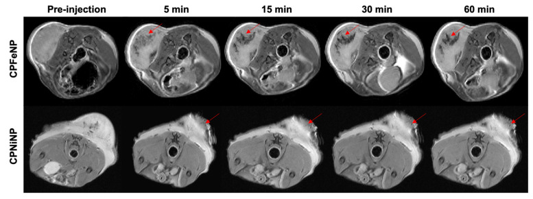
Figure 8.
MRI monitoring after i.t. injection of IONP-doped CPNs in the flank. T2W axial images of the mouse body were acquired at 5, 15, 30 and 60 min after the i.t. nanoparticles injection. Red arrows point to the tumor region where injected nanoparticles (CPFeNP: upper panels, CPNiNP: lower panels) can be identified as a hypointense signal.

