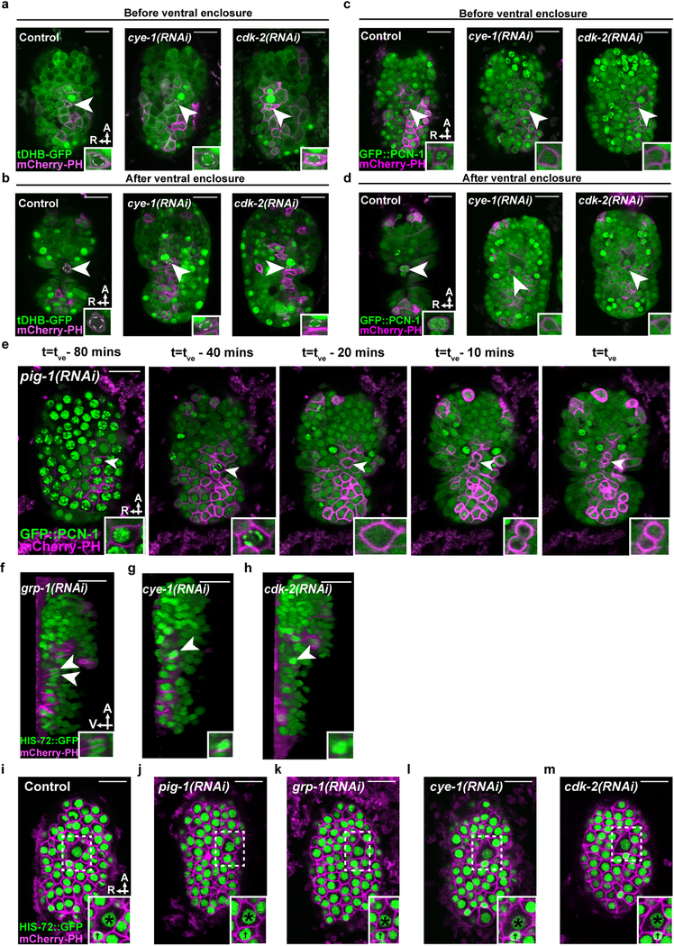
Extended Data Figure 3. ABplpappap, which is generated by an unequal cell division, arrests in S phase and is extruded.
a, b, Confocal fluorescence micrographs of tDHB-GFP fluorescence in ABplpappap (arrowhead) (a) before and (b) after ventral enclosure in heSi192[Peft-3::tDHB-GFP]; ced-3(lf); nIs861[Pegl-1::mCherry::PH] embryos after the indicated RNAi treatment. Dotted line, ABplpappap nucleus, as identified by Nomarski optics. c, d, Confocal fluorescence micrographs of GFP::PCN-1 fluorescence in ABplpappap (arrowhead) (c) before and (d) after ventral enclosure in ced-3(lf); isIs17[Ppie-1::GFP::pcn-1]; nIs861 embryos after the indicated RNAi treatment. e, Time-lapse confocal fluorescence micrographs of GFP::PCN-1 fluorescence in ABplpappap (arrowhead) in a ced-3(lf); isIs17; nIs861; pig-1(RNAi) embryo at the indicated times. tve- time point of ventral enclosure. f-h, Micrographs of virtual lateral section of ced-3(lf); nIs861; stIs10026 embryos showing either ABplpappap (arrowhead) or its daughters (arrowheads) after indicated RNAi treatment. i-m, Confocal fluorescence micrographs of ced-3(lf); ltIs44[Ppie-1::mCherry::PH]; stIs10026 embryos showing the relative sizes of ABplpappap and its sister, ABplpappaa, in embryos after the indicated RNAi treatment. Insets, ABplpappap (a-d); ABplpappap or its daughters (e); magnified view of the region indicated, which includes ABplpappap (†) and ABplpappaa (∗) (i-m). A, anterior; R, right; V, ventral. Scale bars, 10 μm.

