Abstract
Objectives:
To evaluate the accuracy of the measurements of the maxillary sinus (MS) and frontal sinus (FS) in sex estimation among Brazilian adults using multislice computed tomography (MCT) and to develop and cross-validate a new formula for sex estimation.
Methods:
The present cross-sectional research was conducted in two phases: (1) development of a formula on the basis of the measurements of both the sinuses (50 males and 50 females); and (2) validation study (20 males and 20 females). The linear measurements (height, width and diameter) were assessed using the RadiAnt DICOM software. A new formula for sex estimation was developed (multivariate statistical approach) and validated. Receiver operating characteristic curves, area under the curve, sensitivity, specificity, positive and negative predictive values, accuracy and likelihood ratio were estimated.
Results:
Males displayed higher mean values (width, height and diameter) of the FS and MS (p < 0.05). The MS was a better predictor in sex estimation (males vs females), compared to the FS (accuracy between 61–74% and 58–69%, respectively). The distance between the right and left MS displayed the highest accuracy (74%). The sensitivity, specificity and accuracy of the new formula were 80%, 95.5% and 87.5%, respectively. 63.1% reduction was observed in the number of predictive values for sex estimation (individuals older than 30 years).
Conclusions:
The present MCT measurements showed a higher accuracy in the estimation of sex in males. The highest accuracy was associated with the distance between the right and left MS. The new formula displayed high precision for sex estimation.
Keywords: Paranasal sinuses, Sexual dimorphism, Maxillary sinus, Frontal sinus, Computed tomography
Introduction
The process of identification of human remains one of the most relevant aspects of forensic sciences. The aforementioned process has received considerable attention in the scenarios concerning individuals involved in natural calamities or mass disasters, where the body of the deceased person is decomposed, dismembered, skeletonized or burnt, as well as in criminal situations associated with the attempts to conceal an individual’s identity.1,2
It is noteworthy that the tools for sex determination are useful in the reconstruction of biological profiles, especially in unidentified individuals. In forensic odontology, human teeth have been commonly used to distinguish between males and females, in view of the fact that dental units can resist postmortem deterioration. Sexual dimorphism is a well-recognized and prominent area of interest in the fields of forensic sciences and anthropology, and is associated with the assessment of several anatomical structures, including the skull, pelvis, long bones, foramen magnum and paranasal sinuses.3,4 Among human bones, the skull components are considered to be the second best part of the skeleton, after the pelvis, which can be used for the estimation of sex.5
Osteometry is a preferable approach that can be used to differentiate between males and females, owing to the high accuracy reported in literature, such as 77–92%6 and 72–95.5%.7 In extreme situations, such as explosions, war and different types of mass disasters, the skull and other bones might be severely disfigured. However, it has been reported that the maxillary sinus (MS) remains intact in incinerated victims. Hence, this anatomic structure can be used for the purpose of identification.8 Similarly, previous literature has reported that the frontal sinus (FS) was observed to be preserved in dismembered or carbonized corpses, mainly due to its high resistance to traumatic injuries.9
Skull-related morphological aspects have been reported as a potential method that can be used to study sexual dimorphism. However, literature does not give emphasis to any specific feature regarding the same.5 Accordingly, the estimation of sex on the basis of craniofacial structures, such as the paranasal sinuses, should be considered as a topic of interest in forensic research, as it may address the lack of population-specific parameters in the current literature. In view of the fact that the paranasal sinuses display significant interindividual variations,10 imaging studies may provide substantial scientific evidence regarding sex estimation using paranasal sinuses.
Currently, CT is a common imaging modality used by forensic institutes across the world, and its application on skeletal bones for the purpose of sex estimation has received continuous attention of the forensic community.11 Additional aspects that favour the application of the aforementioned radiographical examination in postmortem investigations are the common clinical applications for the preoperative assessment of paranasal sinuses and adjacent structures, as well as the high accuracy regarding details, even in severely disfigured corpses.12 In addition, CT assessment has been validated by several forensic science groups for postmortem assessment.13–15
Few in vivo studies on sexual dimorphism have evaluated the accuracy of the parameters of linear measurements of the FS9,16 or MS16–20 on multislice computed tomography (MCT). Hence, the primary goal of the present study was to evaluate the specificity, sensitivity and accuracy of the linear measurements pertaining to the MS and FS in sex estimation among Brazilian adults on the basis of MCT. Furthermore, the present research aimed to develop and cross-validate a new formula for the purpose of distinguishing between males and females.
Methods and materials
Study design and ethics statement
The present cross-sectional investigation was performed after obtaining approval from the Ethics and Research Committee (number 2.253.923) of the Dr José Frota Hospital (Ceará, Brazil) and followed the principles of the Declaration of Helsinki. The current study was conducted and reported in accordance with the guidelines in the Strengthening the Reporting of Observational Studies in Epidemiology (STROBE) Statement.21
Sample
The MCT data pertaining to the individuals who were referred to the Dr José Frota Hospital for radiographical examination during the time period from May to June 2017 (two months) were evaluated. The sample was selected during the period of maximum CT scan requisitions at the imaging centre. Initially, two investigators analysed the hospital image database, in order to obtain the necessary image samples, as the database included all the MCT images that were acquired for various clinical purposes (e.g., assessment of cranio-maxillofacial trauma).
The present two-phase study involved 140 MCT images. The phases are stated as follows: Phase 1: development of a formula on the basis of the measurements of the MS and FS (n = 100 images from 50 males and 50 females); Phase 2: validation of the new formula using a random sample of MCTs of Brazilian individuals that were not used in Phase 1 (n = 40 images from 20 males and 20 females). All the MCT scans were evaluated by two investigators (DSM and ASWA), in accordance with the eligibility criteria. The inclusion criteria of the first phase of the current study were: (1) radiographical examinations of the individuals between the age of 18 and 40 years; (2) images that clearly show at least the MS and FS; and (3) the presence of posterior maxillary teeth (at least first premolar to the second upper molar). The exclusion criteria were: (1) duplicate examination data; (2) images that revealed signs of pathology or fracture; (3) signs suggestive of facial growth disorders or craniofacial syndromes; (4) any metallic artefacts that could impair accurate measurements (e.g., those from dental implants, metallic restorations and osteosynthesis materials; (5) movement artefacts; and 6) low-quality images.
The present study employed the student’s t-test to obtain a representative sample, which was estimated to be 50 MCT scans per group (90% power; assuming a 95% CI). Regarding the sample size estimation, a previous study by Sherif et al22 reported a statistically significant difference between the males and females with regard to the anteroposterior measurement of the FS (6.90 ± 2.30 vs 8.76 ± 3.25 mm, respectively).
Variables
The dichotomous variable analysed in the present study was sex. The quantitative data were the linear measurements.
Image acquisition process
MCT scans were obtained using a single scanner (Somatom Emotion 6, Siemens, Forchheim, Medical Solutions, Germany) under the following acquisition protocols: 1 mm of table increment, 130 kVp, milliamperage ranging from 80 to 120 mA, cross-sectional image thickness up to 2.0 mm, 180 mm field of view (FOV), and 0.6 s of rotation time. All the analyses were performed using the same computer system (Dell Inc., model G3 3590, Intel® Core ™ processor i5-9300H CPU @ 2.40 GHz, 2400 MHz, four colours, eight logic processors and LED HD backlight screen), and the Digital Imaging and Communications in Medicine (DICOM) files were imported into the free RadiAnt software (Medixant, Poznan, Poland), v.4.6.9.18463 (64 bit).
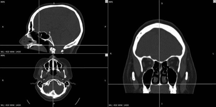
A trained observer (DSM) performed all the evaluations in a dedicated room with dimmed light. The observer was free to modify the brightness of the screen during analysis. The evaluation was performed in shifts and a maximum of two scans were evaluated per shift, in order to prevent visual fatigue, on account of the fact that a time period of approximately 45 min was required to perform all the measurements in a single scan. Initially, head orientation and tomographic alignment were performed to ensure that all the measurements were perpendicular to the horizontal plane. Subsequently, axial, sagittal and coronal sections were obtained to guide the observer. During this process, an axial plane parallel to hard palate in the coincident sagittal section with anterior and posterior nasal spine was used as a reference landmark in the present study (Figure 1).
Figure 1.
Definition of the positioning parameters to measure the frontal and maxillary sinuses in the sagittal, axial and coronal planes.
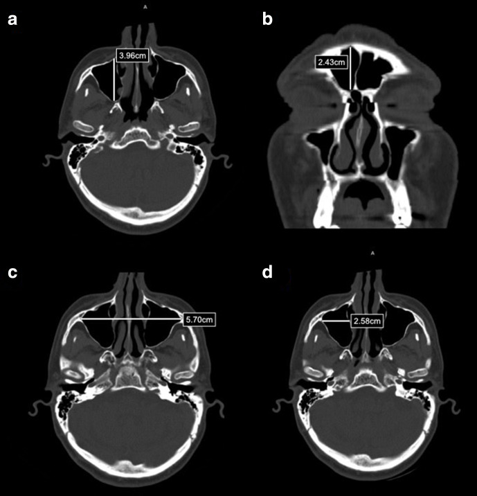
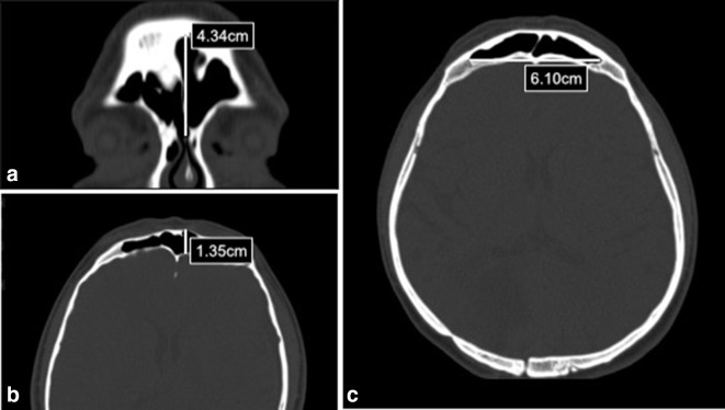
The greatest linear measurements were estimated by means of the axial and coronal images (Figures 2 and 3, Table 1). Moreover, subsequent to the detection of the section with the largest measurement (main image), all the linear assessments were repeated using two slices above and two sections below the main segment, in order to improve the accuracy of measurements. Subsequently, the mean of these five values was adopted as the value of each variable under evaluation in the current study (Figures 1 and 2).
Figure 2.
(a) Maximum distance between the anterior and posterior sinus walls (diameter); (b) maximum distance between the upper and lower sinus borders (height); (c) maximum distance between the external limits of the MS (maximum width); (d) maximum distance between the medial and lateral sinus walls of the MS (individual width).
Figure 3.
(a) Maximum distance between upper and lower sinus borders (height); (b) maximum distance between anterior and posterior sinus walls (diameter); (c) maximum distance between the external limits of the FS (maximum width).
Table 1.
Definition of the adopted morphometric parameters
| Spatial view | Parameter | Paranasal sinus | |
|---|---|---|---|
| Maximum distance between upper and lower sinus borders | Coronal | Height | Frontal and maxillary |
| Maximum distance between anterior and posterior sinus walls | Axial | Diameter | Frontal and maxillary |
| Maximum distance between medial and lateral sinus walls | Axial | Individual width | Maxillary |
| Maximum distance between external limits of the sinus walls | Axial | Maximum width | Frontal and maxillary |
Measurement training and study error
In order to minimize the occurrence of measurement bias, the observer (DSM) who performed the analysis in the present study underwent prior training under senior investigators (FWGC and LMK) who were experienced in the field of oral and maxillofacial radiology. First, an image dataset comprised of randomly selected MCT images was evaluated in a blind process. After a time interval of 15 days, the same procedure was repeated with the same image set, in order to estimate the intrarater agreement. The current study used the SPSS v.20.0 (IBM Corporation, Sommers, NY, USA) for Windows (Microsoft Corporation, Redmond, WA, USA) for data analysis. The analysis of systematic errors was performed using the paired t-test, Pearson‘s correlation, and intraclass correlation coefficient (ICC). The ICC was estimated using a random bidirectional effect model with a 95% CI and p-value less than 0.05. The ICC assessed the intraobserver reproducibility, as per the Koo and Li23 reliability criteria: poor (<0.5), moderate (0.5–0.74), good (0.75–0.9) and excellent (>0.9).
Development and validation of a formula for sex estimation
A linear regression model was estimated for each measurement, in order to predict the sex accurately. The correlation coefficients were used to devise a mathematical formula using the measurements of the MS and FS (isolated or combined). The present study assessed a random sample of MCT scans of Brazilian individuals (20 males and 20 females), in order to validate the formula. The second phase of the current study (validation of the formula) followed the eligibility criteria stated by Farias-Gomes et al24. Accordingly, the current study selected tomographic images of individuals in a broader age range that clearly showed the MS and FS, regardless of the presence or absence of posterior maxillary teeth.
Bias
The current study took certain aspects into consideration to minimize the occurrence of bias20: (1) selection bias: the sample size calculation was planned to estimate adequate and equally divided samples between males and females; (2) sample selection: the dental status was standardized and potential confounding factors (i.e., signs suggestive of pathological changes or bone fractures) were avoided; (3) measurement errors: images were evaluated by a trained observer who was blinded to the gender of the subject in each image, and the reliability of the measurements was determined.
Statistical methods
All the statistical analyses were performed by an investigator (PGBS) using the SPSS v.20.0 (IBM Corporation, Armonk, NY, USA) with a 95% confidence level. Regarding the validation of the five measurements per MCT scan, Cronbach’s α was used as a measure of internal consistency, the ICC was obtained to evaluate systematic error, and the Hotelling’s T-squared analysis (multivariate counterpart of the t-test) was used to estimate the random error.
The Kolmogorov-Smirnov test was used to assess the normality of the data. Linear measurements are expressed as mean and standard deviation (SD) and categorical data are expressed as absolute and relative frequencies. Bivariate analysis was performed using the Student’s t-test (linear measurements between males and females). In addition, the coefficient of variation was estimated and the Levene test was used to compare the variance regarding sex. The measurements of the right and left MSs were compared using the paired t-test.
Receiver operating characteristic (ROC) curves were plotted to identify the cutoff points associated with sexual dimorphism and to estimate the area under the curve (AUC), sensitivity, specificity, positive- and negative-predictive values, accuracy and likelihood ratio (Supplementary Material 1). Moreover, the present study performed an age-related subgroup analysis and estimated the sensitivity, specificity, positive- and negative-predictive values, accuracy and likelihood ratio in relation to two subgroups: the individuals below the age of 30 years and those above the age of 30 years.
Results
Reliability
The reliability with regard to the method was observed to be significant for the linear measurements relating to the sinuses, varying from satisfactory (r = 0.822) to highly satisfactory (r = 0.997). The paired t-test did not reveal any statistically significant difference between the initial set of measurements and the one repeated after 15 days. The ICC showed satisfactory values ranging from 0.896 to 0.998.
Reproducibility analysis
The validation analysis (Table 2) of the FS measurements and the maximum height and width of bilateral MSs showed excellent values with regard to the Cronbach’s α (>0.800) and ICC (>0.800) values, and significant Hotelling’s T-squared correlation (p < 0.001). Moreover, two observers assessed all the linear measurements twice within a 15-day time interval, in order to estimate the inter-observer reproducibility.
Table 2.
Parameters related to the reproducibility analysis
| Validation coefficientsa | Mean ± SD (cm) | Variation coefficientb | ||||||||
|---|---|---|---|---|---|---|---|---|---|---|
| Cronbach’s α | Hotelling’s T-Squared | ICC | Total | Females | Males | p valuec | Females | Males | p valued | |
| FS height | 0.999 | <0.001 | 0.997 | 2.92 ± 1.13 | 2.65 ± 0.93 | 3.19 ± 1.26 | 0.018 | 35.1% | 39.5% | 0.035 |
| FS width | 0,993 | <0.001 | 0.967 | 5.21 ± 1.73 | 4.99 ± 1.59 | 5.43 ± 1.85 | 0.201 | 31.9% | 34.1% | 0.406 |
| FS diameter | 0.993 | <0.001 | 0.967 | 1.12 ± 0.39 | 0.96 ± 0.26 | 1.28 ± 0.43 | <0.001 | 27.1% | 33.6% | 0.001 |
| RMS height | 0.999 | <0.001 | 0.994 | 3.65 ± 0.49 | 3.48 ± 0.45 | 3.82 ± 0.47 | <0.001 | 12.9% | 12.3% | 0.699 |
| RMS width | 0.999 | <0.001 | 0.994 | 2.83 ± 0.51 | 2.70 ± 0.42 | 2.95 ± 0.56 | 0.011 | 15.6% | 19.0% | 0.337 |
| RMS diameter | 0.998 | 0.074 | 0.998 | 3.92 ± 0.38 | 3.83 ± 0.25 | 4.02 ± 0.46 | 0.011 | 6.5% | 11.4% | 0.015 |
| LMS height | 0.999 | <0.001 | 0.999 | 3.67 ± 0.50 | 3.51 ± 0.45 | 3.84 ± 0.49 | 0.001 | 12.8% | 12.8% | 0.966 |
| LMS width | 0.999 | <0.001 | 0.994 | 2.82 ± 0.49 | 2.66 ± 0.41 | 2.98 ± 0.51 | 0.001 | 15.4% | 17.1% | 0.287 |
| LMS diameter | 0.947 | 0.351 | 0.781 | 3.92 ± 0.36 | 3.82 ± 0.27 | 4.02 ± 0.41 | 0.004 | 7.1% | 10.2% | 0.048 |
| MS(x--[R;L]) | 1.000 | 0.028 | 0.998 | 8.59 ± 0.94 | 8.21 ± 0.97 | 8.96 ± 0.74 | <0.001 | 11.8% | 8.3% | 0.467 |
FS, Frontal sinus; ICC, Intraclass correlation coefficient; L, Left; MS, Maxillary sinus; R, Right; SD, Standard deviation; (x--[R;L]), Arithmetic mean between R and L measurements.
Intra observer reproducibility.
Inter observer reproducibility.
Student t-test.
Levene’s test.
Males showed higher mean values of the measurements pertaining to the paranasal sinuses
The mean maximum height (p = 0.018) and width (p = 0.201), as well as the diameter (p < 0.001) of the FS (both sides) were observed to be significantly higher in males compared to females (Table 3). In addition, males showed increased values of the following measurements of the MS, compared to the females: maximum height (right side, p < 0.001; left side, p = 0.001), maximum width (right side, p = 0.011; left side, p = 0.001) and diameters (right side, p = 0.011; left side, p = 0.004).
Table 3.
Summary of the sensitivity, specificity, positive-/negative-predictive values, accuracy and likelihood ratio pertaining to the study variables (cm) to estimate sex
| Estimated sex | Sens. (M) | Spec. (F) | PPV (M) | PNV (F) | Accuracy | LR (95% CI) | ||
|---|---|---|---|---|---|---|---|---|
| F (n = 50) | M (n = 50) | |||||||
| FS height | 26 | 35 | 70.0% | 52.0% | 59.3% | 63.4% | 61.0% | 2.53 (1.11–5.74) |
| FS width | 29 | 29 | 58.0% | 58.0% | 58.0% | 58.0% | 58.0% | 1.91 (0.86–4.22) |
| FS diameter | 34 | 35 | 70.0% | 68.0% | 68.6% | 69.4% | 69.0% | 4.96 (2.12–11.58) |
| RMS height | 34 | 34 | 68.0% | 68.0% | 68.0% | 68.0% | 68.0% | 4.52 (1.95–10.46) |
| RMS width | 34 | 32 | 64.0% | 68.0% | 66.7% | 65.4% | 66.0% | 3.78 (1.65–8.65) |
| RMS diameter | 39 | 33 | 66.0% | 78.0% | 75.0% | 69.6% | 72.0% | 6.88 (2.83–16.74) |
| LMS height | 32 | 29 | 58.0% | 64.0% | 61.7% | 60.4% | 61.0% | 2.46 (1.10–5.49) |
| LMS width | 37 | 26 | 52.0% | 74.0% | 66.7% | 60.7% | 63.0% | 3,08 (1.33–7.15) |
| LMS diameter | 41 | 30 | 60.0% | 82.0% | 76.9% | 67.2% | 71.0% | 6.83 (2.73–17.09) |
| MS height(x--[R;L]) | 34 | 31 | 62.0% | 68.0% | 66.0% | 64.2% | 65.0% | 3.47 (1.52–7.90) |
| MS width(x--[R;L]) | 35 | 30 | 60.0% | 70.0% | 66.7% | 63.6% | 65.0% | 3.50 (1.53–8.01) |
| MS diameter(x--[R;L]) | 40 | 30 | 60.0% | 80.0% | 75.0% | 66.7% | 70.0% | 6.00 (2.45–14.68) |
| Width between both MS | 37 | 37 | 74.0% | 74.0% | 74.0% | 74.0% | 74.0% | 8.10 (3.31–19.80) |
CI, Confidence interval; F, Female; FS, Frontal sinus; L, Left; LR, Likelihood;M, Male; MS, Maxillary sinus; NPV, Negative predictive value; PPV, Positive predictive value; R, Right; Sens., Sensibility; spec., Specificity;(x-- [R;L]), Arithmeticmean between R and L measurements.
The data variance, which is a measure of statistical dispersion, was observed to be significantly higher for the maximum height (p = 0.035) and diameter (p = 0.001) of the FS. The evaluation of the right (p = 0.015) and left (p = 0.048) MSs revealed that the diameter was significantly higher in males, compared to females (Table 2).
The measurements of the MS were significant predictors of sexual dimorphism
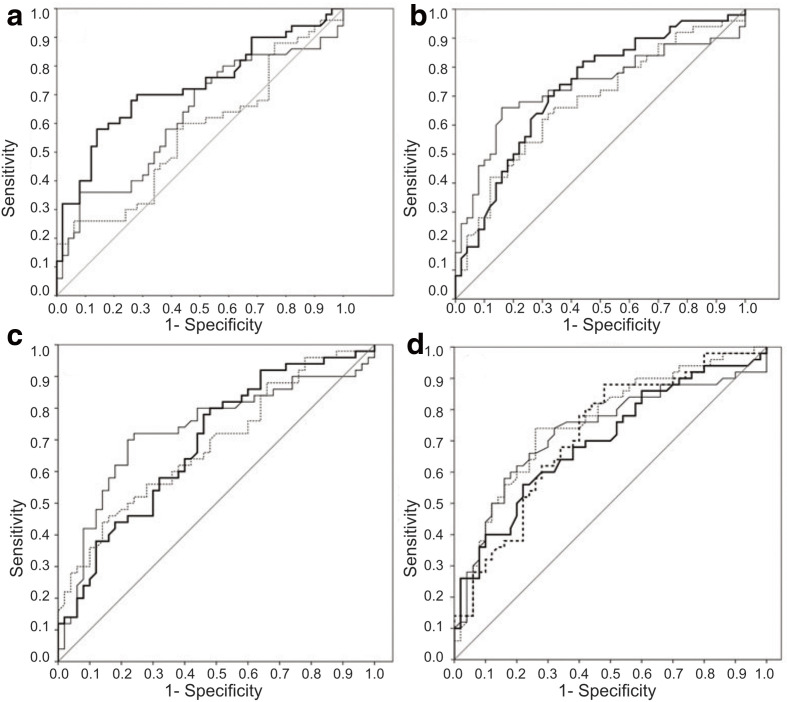
The ROC curve-based (Figure 4) cutoff points for the estimation of sexual dimorphism are shown in Table 4. Most of the AUC values were significantly higher, compared to the null axis of the ROC curve (>0.500); higher AUC values were observed in relation to the maximum width of the MS.
Figure 4.
ROC curves demonstrating the cutoff values, sensitivities and specificities of the measurements of the frontal sinus (a), right (b) and left (c) maxillary sinuses and bilateral maxillary sinuses (d). Continuous thick line = maximum height; continuous thin line = diameter; discontinuous thin line = individual width; discontinuous thick line = maximum width.
Table 4.
Variation of the predictive values with reference to sex estimation, in accordance with the age groups
| Up to 30 years | >30 years | ||||||||||
|---|---|---|---|---|---|---|---|---|---|---|---|
| Sens. (M) |
Spec. (F) |
PPV (M) |
PNV (F) |
Accuracy | Sens. (M) |
Spec. (F) |
PPV (M) |
PNV (F) |
Accuracy | ||
| Frontal sinus height | −5.9% | −0.4% | 3.2% | −10.1% | −2.4% | 20.9% | 0.6% | −6.7% | 27.5% | 5.7% | |
| Frontal sinus width | 3.5% | −9.6% | 2.0% | −8.0% | −2.3% | −12.5% | 15.7% | −8.0% | 12.0% | 5.3% | |
| Frontal sinus diameter | −0.8% | −9.9% | −1.1% | −9.4% | −4.7% | 2.7% | 16.2% | 4.1% | 14.8% | 11.0% | |
| R maxillary sinus height | 1.2% | 3.0% | 7.0% | −3.3% | 2.0% | −4.4% | −4.8% | −18.0% | 7.0% | −4.7% | |
| R maxillary sinus width | 2.7% | 6.2% | 9.8% | −1.5% | 4.0% | −9.5% | −10.1% | −23.8% | 3.4% | −9.3% | |
| R maxillary sinus diameter | 0.7% | −0.6% | 3.8% | −4.7% | −0.6% | −2.4% | 0.9% | −11.4% | 9.3% | 1.3% | |
| L maxillary sinus height | 3.5% | 3.7% | 8.9% | −2.1% | 3.3% | −12.5% | −6.1% | −23.2% | 4.3% | −7.7% | |
| L maxillary sinus width | 4.4% | 3.4% | 9.2% | −2.2% | 2.7% | −15.6% | −5.6% | −26.7% | 4.3% | −6.3% | |
| L maxillary sinus diameter | 4.1% | 1.9% | 6.4% | −2.2% | 1.9% | −14.5% | −3.1% | −21.3% | 4.2% | −4.3% | |
| Maxillary sinus height (x--[R;L]) | 2.1% | 6.2% | 9.8% | −2.0% | 3.6% | −7.5% | −10.1% | −23.1% | 4.6% | −8.3% | |
| Maxillary sinus width (x--[R;L]) | 4.1% | 10.6% | 13.9% | 0.5% | 6.4% | −14.5% | −17.4% | −31.0% | −1.1% | −15.0% | |
| Maxillary sinus diameter (x--[R;L]) | 1.5% | 0.6% | 5.0% | −4.2% | 0.0% | −5.5% | −1.1% | −15.0% | 8.3% | 0.0% | |
| Width between both maxillary sinuses | 2.9% | 6.6% | 9.3% | −0.5% | 4.6% | −10.4% | −10.8% | −24.0% | 1.0% | −10.7% | |
CI, Confidence interval; F, Female; FS, Frontal sinus; L, Left; LR, Likelihood;M, Male; MS, Maxillary sinus; NPV, Negative predictive value; PPV, Positive predictive value; R, Right; Sens., Sensibility; spec., Specificity.
x-- = arithmetic mean between left and right measurements. Bold numbers represent reduced values of accuracy measures.
The maximum width of the MS displayed the maximum sensitivity with regard to the identification of the male sex (74.0%), as shown in Table 3. Among females, the diameters of the left (82.0%) and right (78.0%) MSs, and the mean diameters of both sides (80.0%) presented the most significant values of specificity. The mean maximum width of MS (74.0%) displayed the highest accuracy with the highest likelihood ratio (8.10; 95% confidence interval (CI)=3.31–19.80).
Age-related subgroup analysis
The frequency distribution of the age on the basis of gender is shown in Table 4. The current study observed that 23 of the 65 (35.4 %) measurements of the patients below the age of 30 years showed lower values, compared to the total sample. Moreover, 41 of the 65 measurements (63.1%) of the patients above the age of 30 years showed lower values, compared to the total sample.
In the subgroup comprising the subjects below the age of 30 year, the distance between the right and left MSs (76.9%) displayed the highest sensitivity with reference to the identification of the male sex. Conversely, the diameter of the left MS (83.9%) displayed the highest specificity with regard to the identification of the female sex. The highest accuracy was observed in relation to the distance between the right and left MSs (78.6%). Analysis of the CT scans of the patients above the age of 30 years revealed that the height of the FS (90.9%) displayed the highest sensitivity with reference to the identification of the male sex. Furthermore, the diameter of the FS (84.2%) displayed the highest specificity in relation to the identification of the female sex. The highest accuracy was observed in relation to the diameter of the FS (80.0%).
Formula for sex estimation and external validation
A multiple linear regression model was designed to obtain the adjusted β values, which were inserted in a linear formula to estimate the sex.24 On the basis of the coefficients of collinearity with sex, the following formula was constructed to distinguish between females (value <0) and males (value >0):
where A, FS height; B, FS width; C, FS diameter; D, MS width; E, arithmetic mean of the right and left MS height; F, arithmetic mean of the right and left MS width; G, arithmetic mean of the right and left MS diameter.
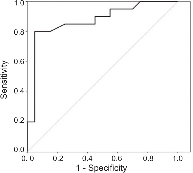
Subsequently, a ROC curve with a statistically significant AUC (0.757 ± 0.002; [CI 95% = 0.619–0.894]; p = 0.002) was plotted, and an optimal cutoff point value of 2.23 was estimated (Figure 5). In addition, the current study performed a sample size calculation, in order to estimate the sample size for external validation. On the basis of the best measurements of this study (FS diameter: females = 0.96 ± 0.26, males = 1.28 ± 0.43), the estimated sample size required to perform the validation of the formula, in accordance with the statistical approach, was 20 patients per group (power 80% and CI 95%; t-test).
Figure 5.
ROC to establish an optimal cutoff point for the estimation of sex.
In an independent sample comprised of 40 CT scans (20 females and 20 males) that were not part of the original study sample, the estimated cutoff point showed a sensitivity of 80.0% (males), a specificity of 95.5% (females), a positive-predictive value of 94.1% (males), a negative-predictive value of 82.6% (females) and a likelihood ratio of 76.00 (CI05% = 7.70–750.49), as shown in Table 5.
Table 5.
Summary of the sensitivity, specificity, positive-/negative-predictive values, accuracy and likelihood ratio pertaining to the external validation of the formula developed for sex estimation
| Estimated sex | Sens. (M) | Spec. (F) | PPV (M) | PNV (F) | Accuracy | LR (95% CI) | |
|---|---|---|---|---|---|---|---|
| F (n = 20) | M (n = 20) | ||||||
| 19 | 16 | 80.0% | 95.5% | 94.1% | 82.6% | 87.5% | 76.00 (7.70–750.49) |
CI, Confidence interval; F, Female; LR, Likelihood; M, Male; NPV, Negative predictive value; PPV, Positive predictive value; Sens., Sensibility;spec., Specificity.
Sens., sensibility; spec., specificity; PPV, positive predictive value; NPV, negative predictive value; LR, likelihood; CI, confidence interval; M, male; F, female.
Power of the sample size
On the basis of the sensitivity (80.0%) to determine the male sex and the specificity (95.5%) to identify the female sex, the present sample showed a power of 89.4 and 99.9%, respectively, thereby rejecting the null hypothesis (50.0%) with reference to sexual dimorphism in relation to the linear measurements of the MS (chi-squared test).
Discussion
The present research evaluated the MCT images of the MS and FS, in order to investigate sexual dimorphism. Previous studies have used conventional radiographs of the craniofacial complex,5,23,25 cone-beam CT (CBCT),12 and magnetic resonance imaging to estimate determine the sex. There are few studies involving MCT that have developed and validated the formula for estimating the sex by combining the morphometric (linear distances) measurements of the MS and FS.22 To the best of our knowledge, no Brazilian studies with similar methodological aspects have been published to date.
Besides the application of MCT in surgical procedures, exploration of the utility of this method in the estimation of sex by means of the measurements of MS and FS has been limited over the years. In fact, very few diagnostic accuracy studies that evaluated both paranasal sinuses using MCT images have been published to date (Supplementary Material 2). In view of the fact that this imaging modality requires standardized measurement methods to obtain reliable results, the present investigation used validated coefficients (Cronbach´s α, Hotelling’s T-squared, and ICC) to assess the intraobserver agreement. The current study observed an almost perfect agreement, which was concurrent with the results reported by Sahlstrand-Johnson et al10. In addition, it should be highlighted that a mean value was obtained after the evaluation of five consecutive MCT slices, which guaranteed a higher fidelity of the measurements.
The present, cross-sectional investigation adopted the method of convenience sampling, in order to obtain the MCT scans that were used to compare the tridimensional measurements of the MS and FS in males and females. A dataset of MCT scans with a large FOV was used, as the tomographic investigations that follow this acquisition parameter were necessary and justified for the purpose of clinical and surgical planning. Although MCT is a radiographic investigation that emits a higher dose of radiation, compared to CBCT,26 the investigation is frequently used in hospitals to assess patients with cranio-maxillofacial trauma. A previous study has reported similar precision and accuracy of the linear measurements obtained using MCT (0.6 mm resolution) and CBCT (0.25 mm resolution). An additional advantage of MCT, compared to CBCT, is the high accuracy of soft tissue measurements, which plays a substantial role during the preoperative planning in orthognathic surgery.27
In the context of forensic sciences, the paranasal sinuses are highly individualistic, like fingerprints. The aforementioned observation supports the use of the FS measurements in scenarios that warrant quantitative reasoning for forensic identification.16 Regarding the MS, although literature includes previous studies that focused on their anatomical aspects, Sahlstrand-Johnson et al10 mentioned that there are dimensions related to these anatomical structures that need to be investigated further. Hence, both the paranasal sinuses that were assessed in the present study are recommended to be used as reliable indicators in the current literature on sexual dimorphism. Moreover, the development of a formula that combines the measurements of two paranasal sinuses has not been reported to date, which highlights the relevance of the current research. Further studies involving other population-based samples and similar study designs may be conducted to evaluate the accuracy of the measurements of MS and FS in distinguishing between males and females.
Both the paranasal sinuses displayed significantly higher values of measurements in males, compared to females, which is concurrent with the FS-related data reported by previous studies from Brazil28 and Iraq,29 and the MS-related findings reported by the studies involving the individuals from Iraq2,17 and Turkey.30 In addition, the present results were obtained from a miscegenated population, as the ancestry was shown to be a mixture of predominantly European and Amerindian population,31 which partially explains the dissimilar values, compared to other studies. Farias-Gomes et al24 highlighted certain factors that can influence these differences, namely, the sample size, age, type of radiographic investigation, methods used for measurement and statistical protocol.
The height of the right MS displayed a sensitivity of 68% in the identification of the male sex, which was higher, compared to females. Furthermore, previous studies involving non-Brazilian populations9,10,17,18,30,32–34 showed that height is a useful variable in sex estimation, owing to the ability to provide a faster method of identification, thereby facilitating the work done by anthropologists.35 In addition, the other linear measurements of the sinuses that were evaluated in the present study (width and diameter) are considered to be significant for gender differentiation, as reported by previous studies that involved computed tomography evaluations.36,37
The FS was assessed for sexual dimorphism in 2014 by Belaldavar et al38, who evaluated the width, height and sinus area on posteroanterior plain radiographs from an Indian population. The authors reported a sex estimation index of 64.6%, which is concurrent with the results of the present study (range of linear measurements: 61 to 69%). Regarding the FS measurements, Luo et al39 reported medium significance for sex estimation while employing the area and an index calculated using the height and width. The accuracy of the height and width of the FS in the current study was 61 and 58%, respectively. The aforementioned results confirm the significance of the parameters in sex estimation.
In the present study, the most significant values of accuracy for the estimation of sex with reference to the type of paranasal sinus were 74% (MS) and 69% (FS). In a study that involved the Turkish population,30 the accuracy rate in relation to the MS was 69.3%. A previous study by Ekizoglu et al40, which involved comparable population and methodology, reported an accuracy of 77.1%. Uthman et al16 employed CT evaluation to assess the measurements of the MS in an Iraqi population, and reported an overall accuracy of 73.9% with regard to sex estimation. Akhlaghi and colleagues conducted two separate investigations involving Iraqi individuals. The first study9 did not recommend the use of FS parameters to estimate sex among Persian adults. The second study performed in 201717 reported that in young Iranian adults, the width of the MS displayed considerable accuracy (65.6%) with reference to the identification of the correct sex.
The mean distance between the right and left MSs showed an accuracy of 74% and a likelihood ratio of 8.10 with regard to the accurate identification of males, thereby emphasizing the role of this measurement using MCT scans in the field of sexual dimorphism research. Moreover, the aforementioned finding was concurrent with the results reported by previous studies involving the Iranian population,9,16,17 which reported a high sensitivity with regard to the identification of the male sex using the measurements of the MS.
In the current study, the estimated accuracy pertaining to the diameters of the right and left MSs were 72 and 71%, respectively; the accuracy of the width of the right and left MSs and the height of the right MS were 66%, 63%, and 68%, respectively. Regarding males, Ekizoglu et al40 obtained the highest precision rates while assessing the diameter of the right MS (75.7%) and the mean height of the right and left MSs (71.4%). Uthman et al16 reported an accuracy of 60.5 and 72.1% with regard to the width of the right and left MSs.
The maximum distance between the right and left MSs (74%) and the FS diameter (69%) showed high precision with reference to sex estimation, whereas the width of the left MS and FS displayed low accuracy rates. In a study that involved the evaluation of MS, the best variables for sex estimation were observed to be the height and anteroposterior diameter of the left MS.16 Teke et al30 reported low accuracy (67.7%) for the linear measurements of the left MS and the distance between the right and left MSs, which is in contrast with the results of the present study. Regarding the FS, Akhlaghi et al17 reported a precision of 61.3% with regard to the height of the left FS and a low precision rate (51.3%) with regard to the width of the right FS. Furthermore, the present study evaluated the maximum width of the MS, as previous studies have shown that it is a valuable parameter in the assessment of sexual dimorphism using MCT.16,19 In addition, in 2010, a previous study by Uthman et al16 reported that the maximum width of MS displayed an accuracy of 73.9% with regard to the estimation of sex.
Furthermore, the current study developed and validated a mathematical equation to estimate the sex in a sample of adult individuals from Northeast Brazil. Owing to the differences in methodological approaches, comparison with published data pertaining to the Brazilian population is limited. Farias-Gomes et al24 used a formula based on the three-dimensional volume of the MS that was assessed using the CBCT images of a south-west Brazilian sample population and reported an accuracy of 84% for sex estimation. However, the aforementioned study used a single paranasal sinus, whereas the formula developed in the present study used a multivariate statistical approach, based on the linear measurements pertaining to the FS and MS. Wanzeler et al41 reported that the probabilities of accurate estimation of sex by means of CBCT-based volumetric analyses of the male and female subjects from North Brazil were 96.2 and 92.7%, respectively. Nevertheless, the authors included the measurements of the sphenoidal sinus in their formula, which was not included in the present study.
On the topic of the validation study, the reliability of the formula was tested in another sample of the Brazilian population, which was selected without the restrictions of the selection criteria, such as the age or the presence of posterior teeth, as recommended in a previous investigation on sexual dimorphism.24 The cross-validated formula showed moderate accuracy (74%) and low error. Although the formula developed by the present study used simple and relatively easy-to-apply anthropometric measures of selected paranasal sinuses, it should be validated prior to the application in other ethnic groups by means of further investigations.
In the current investigation, with advancing age, the accuracy in sex estimation of the measurements pertaining to the MS were significantly reduced, which may be attributed to the bone resorption and age-related degenerative changes. Akhlaghi et al17 reported that age influences the reduction of predictive anthropometric indices of the MS with reference to sex estimation. Regarding the individuals above the age of 30 years, the FS provided better measures of accuracy regarding the estimation of sex, which was concurrent with the previous reports in literature.17
The present study has certain limitations. On account of the rigorous inclusion criteria, a sample of 100 MCT scans were included in the study. Although the sample size in the present study was not large, it was similar to that in the study reported by Sherif et al22, which evaluated the MCT scans of 100 adult Egyptians. The current study performed a sample size calculation, in order to minimize the selection bias. There was a relatively subjective selection of the slice with the highest measurement. However, a substantial intrarater agreement was observed and the methodology used in the present study involved the estimation of the mean of five consecutive measurements (thickness of 0.6–2.0 mm). The formula that was developed in the present study was based on a linear relationship between specific measurements and a category (0: female; 1: male), which is not constantly encountered in sex estimation. Hence, intersection ranges between the variables can influence the final sex estimation. However, it is worth mentioning that the present study adopted a multivariate model that was able to determine the different weights with respect to each measurement by means of an interassociated approach. After this adjustment, it was possible to develop a formula with a sensitivity of 80% and specificity of 95.5%, which are significant values in forensic research. Another limitation of the present study was that the MCT scans that were included were obtained from symmetrical individuals, which probably represent a minority of the subjects or bodies that are discovered. Hence, tomographic studies involving sexual dimorphism should include samples of asymmetrical patients, in order to cover the aforementioned aspect.
The development of a formula to estimate sex using MCT-based linear measurements is a topic that is rarely addressed by accuracy studies. Consequently, it was the primary aim of the present study. An advantage of the linear measurements is that the data assessment and volume procurement do not require any specific software, which could enhance the reproducibility of the methodology. Hence, the authors believe that a multivariate statistics-based formula may provide better precision in relation to the differentiation between males and females. The current study recommends that this approach can be used in future investigations involving other imaging modalities or the evaluation of other bones for the purpose of studying sexual dimorphism. Furthermore, future studies could address the volumetric analysis of the paranasal sinuses.
Conclusion
The present study observed that higher mean values of measurements of the paranasal sinuses were chiefly observed in males. The most significant values of accuracy for sex estimation were observed in relation to the MS, followed by the FS. High precision was observed in relation to the distance between the right and left MSs. In addition, the present study developed and validated a formula that showed high precision for sex estimation. The data obtained through the current study highlight the importance of MCT-based morphometric assessment of FS and MS as a complementary tool for sex estimation in the field of forensic sciences.
Footnotes
Acknowledgements: The authors would like to thank the Brazilian National Council for Scientific and Technological Development (CNPq) for providing Dr. Fábio Costa with a PQ fellowship in category 2.
Contributor Information
Diego Santiago de Mendonça, Email: diego.santiago@alu.ufc.br.
Lúcio Mitsuo Kurita, Email: luciokurita@gmail.com.
Francisco Samuel Rodrigues Carvalho, Email: samuelcarvalho@ufc.br.
Fabrício Mesquita Tuji, Email: fmtuji@gmail.com.
Paulo Goberlânio de Barros Silva, Email: paulo_goberlanio@yahoo.com.br.
Tácio Pinheiro Bezerra, Email: taciopb@gmail.com.
Andréa Silvia Walter de Aguiar, Email: aswaguiar@gmail.com.
Fábio Wildson Gurgel Costa, Email: fwildson@yahoo.com.br.
REFERENCES
- 1.Mathur H, Mathur A, Ahmed J, Ahmed J. Conventional frontal sinus imaging in identification of sex: original study in population of Udaipur City, India. J Med Sci Clin Res 2013; 1: 33–7. [Google Scholar]
- 2.Tsuchiya MJ, Gomes EM, Abe DM, et al. Human identification through the analysis of dental records registered in the context of a dental institution. Rev Gaúcha Odontol 2013; 61: 389–93. [Google Scholar]
- 3.Papaloucas C, Fiska A, Demetriou T. Sexual dimorphism of the hip joint in Greeks. Forensic Sci Int 2008; 179: 83.e1–83.e3. doi: 10.1016/j.forsciint.2008.03.007 [DOI] [PubMed] [Google Scholar]
- 4.Zorba E, Moraitis K, Manolis SK. Sexual dimorphism in permanent teeth of modern Greeks. Forensic Sci Int 2011; 210(1-3): 74–81. doi: 10.1016/j.forsciint.2011.02.001 [DOI] [PubMed] [Google Scholar]
- 5.Naikmasur VG, Shrivastava R, Mutalik S. Determination of sex in South Indians and immigrant Tibetans from cephalometric analysis and discriminant functions. Forensic Sci Int 2010; 197(1-3): 122.e1–122.e6. doi: 10.1016/j.forsciint.2009.12.052 [DOI] [PubMed] [Google Scholar]
- 6.Biggerstaff RH. Craniofacial characteristics as determinants of age, sex, and race in forensic dentistry. Dent Clin North Am 1977; 21: 85–97. [PubMed] [Google Scholar]
- 7.Robinson MS, Bidmos MA. The skull and humerus in the determination of sex: reliability of discriminant function equations. Forensic Sci Int 2009; 186(1-3): 86.e1–86.e5. doi: 10.1016/j.forsciint.2009.01.003 [DOI] [PubMed] [Google Scholar]
- 8.Kajoak SA, Ayad CE, Abdalla EA, Mohammed MN, Yousif MO, Mohammed AM. Characterization of sphenoid sinuses for Sudanese population using computed tomography. Glob J Health Sci 2013; 6: 135–41. doi: 10.5539/gjhs.v6n1p135 [DOI] [PMC free article] [PubMed] [Google Scholar]
- 9.Akhlaghi M, Bakhtavar K, Moarefdoost J, Kamali A, Rafeifar S. Frontal sinus parameters in computed tomography and sex determination. Leg Med 2016; 19: 22–7. doi: 10.1016/j.legalmed.2016.01.008 [DOI] [PubMed] [Google Scholar]
- 10.Sahlstrand-Johnson P, Jannert M, Strömbeck A, Abul-Kasim K. Computed tomography measurements of different dimensions of maxillary and frontal sinuses. BMC Med Imaging 2011; 11: 8. doi: 10.1186/1471-2342-11-8 [DOI] [PMC free article] [PubMed] [Google Scholar]
- 11.Tunis TS, Sarig R, Cohen H, Medlej B, Peled N, May H. Sex estimation using computed tomography of the mandible. Int J Legal Med 2017; 131: 1691–700. doi: 10.1007/s00414-017-1554-1 [DOI] [PubMed] [Google Scholar]
- 12.Gamba TdeO, Yamasaki MC, Groppo FC, da Silveira HLD, Boscolo SMdeA, Sanderink GCH, et al. Validation study of a new method for sexual prediction based on CBCT analysis of maxillary sinus and mandibular canal. Arch Oral Biol 2017; 83: 118–23. doi: 10.1016/j.archoralbio.2017.07.010 [DOI] [PubMed] [Google Scholar]
- 13.Ramsthaler F, Kettner M, Gehl A, Verhoff MA. Digital forensic osteology: morphological sexing of skeletal remains using volume-rendered cranial CT scans. Forensic Sci Int 2010; 195(1-3): 148–52. doi: 10.1016/j.forsciint.2009.12.010 [DOI] [PubMed] [Google Scholar]
- 14.Roberts ISD, Benamore RE, Benbow EW, Lee SH, Harris JN, Jackson A, et al. Post-Mortem imaging as an alternative to autopsy in the diagnosis of adult deaths: a validation study. The Lancet 2012; 379: 136–42. doi: 10.1016/S0140-6736(11)61483-9 [DOI] [PMC free article] [PubMed] [Google Scholar]
- 15.Thali MJ, Yen K, Plattner T, Schweitzer W, Vock P, Ozdoba C, et al. Charred body: virtual autopsy with multi-slice computed tomography and magnetic resonance imaging. J Forensic Sci 2002; 47: 15569J–31. doi: 10.1520/JFS15569J [DOI] [PubMed] [Google Scholar]
- 16.Uthman AT, Al-Rawi NH, Al-Naaimi AS, Tawfeeq AS, Suhail EH. Evaluation of frontal sinus and skull measurements using spiral CT scanning: an aid in unknown person identification. Forensic Sci Int 2010; 197(1-3): 124.e1–124.e7. doi: 10.1016/j.forsciint.2009.12.064 [DOI] [PubMed] [Google Scholar]
- 17.Akhlaghi M, Bakhtavar K, Kamali A, Maarefdoost J, Sheikhazadi A, Mousavi F, et al. The diagnostic value of anthropometric indices of maxillary sinuses for sex determination using CT-scan images in Iranian adults: a cross-sectional study. J Forensic Leg Med 2017; 49: 94–100. doi: 10.1016/j.jflm.2017.05.017 [DOI] [PubMed] [Google Scholar]
- 18.Amin MF, Hassan EI. Sex identification in Egyptian population using multidetector computed tomography of the maxillary sinus. J Forensic Leg Med 2012; 19: 65–9. doi: 10.1016/j.jflm.2011.10.005 [DOI] [PubMed] [Google Scholar]
- 19.Attia A, El-Badrawy A, Shebel H. Gender identification from maxillary sinus using Multi-Detector computed tomography. Mansoura Journal of Forensic Medicine and Clinical Toxicology 2012; 20: 17–28. doi: 10.21608/mjfmct.2012.47769 [DOI] [Google Scholar]
- 20.Emirzeoglu M, Sahin B, Bilgic S, Celebi M, Uzun A. Volumetric evaluation of the paranasal sinuses in normal subjects using computer tomography images: a stereological study. Auris Nasus Larynx 2007; 34: 191–5. doi: 10.1016/j.anl.2006.09.003 [DOI] [PubMed] [Google Scholar]
- 21.von Elm E, Altman DG, Egger M, Pocock SJ, Gøtzsche PC, Vandenbroucke JP, et al. Strengthening the reporting of observational studies in epidemiology (STROBE) statement: guidelines for reporting observational studies. BMJ 2007; 335: 806–8. doi: 10.1136/bmj.39335.541782.AD [DOI] [PMC free article] [PubMed] [Google Scholar]
- 22.Sherif NAE-H, Sheta AAE-M, Ibrahim ME, Kaka RAE-M, Henaidy MF, et al. Evaluation of the paranasal sinuses dimensions in sex estimation among a sample of adult egyptians using multidetector computed tomography. Journal of Forensic Radiology and Imaging 2017; 11: 33–9. doi: 10.1016/j.jofri.2017.11.001 [DOI] [Google Scholar]
- 23.Krishan K. Anthropometry in forensic medicine and forensic sciences- ‘Forensic Anthropometry’. Internet J Forensic Sci 2006; 2: 1–8. [Google Scholar]
- 24.Farias Gomes A, de Oliveira Gamba T, Yamasaki MC, Groppo FC, Haiter Neto F, Possobon RdeF. Development and validation of a formula based on maxillary sinus measurements as a tool for sex estimation: a cone beam computed tomography study. Int J Legal Med 2019; 133: 1241–9. doi: 10.1007/s00414-018-1869-6 [DOI] [PubMed] [Google Scholar]
- 25.Hsiao T-H, Tsai S-M, Chou S-T, Pan J-Y, Tseng Y-C, Chang H-P, et al. Sex determination using discriminant function analysis in children and adolescents: a lateral cephalometric study. Int J Legal Med 2010; 124: 155–60. doi: 10.1007/s00414-009-0412-1 [DOI] [PubMed] [Google Scholar]
- 26.Pauwels R. Cone beam CT for dental and maxillofacial imaging: dose matters. Radiat Prot Dosimetry 2015; 165(1-4): 156–61. doi: 10.1093/rpd/ncv057 [DOI] [PubMed] [Google Scholar]
- 27.Gaia BF, Pinheiro LR, Umetsubo OS, Costa FF, Cavalcanti MGP. Validity of three-dimensional computed tomography measurements for Le Fort I osteotomy. Int J Oral Maxillofac Surg 2014; 43: 197–203. doi: 10.1016/j.ijom.2013.06.005 [DOI] [PubMed] [Google Scholar]
- 28.Camargo JR, Daruge E, Prado FB, et al. The frontal sinus morphology in radiographs of Brazilian subjects: its forensic importance. J Morphol Sci 2007; 24: 239–43. [Google Scholar]
- 29.Raoof TM, Saeed KA, Mahmood KA. Anatomical variation of frontal sinuses evaluated by CT scan in relation to age and sex in SULAIMANI City. J Sulaimani Med Col 2013; 3: 33–45. doi: 10.17656/jsmc.10029 [DOI] [Google Scholar]
- 30.Teke HY, Duran S, Canturk N, Canturk G. Determination of gender by measuring the size of the maxillary sinuses in computerized tomography scans. Surg Radiol Anat 2007; 29: 9–13. doi: 10.1007/s00276-006-0157-1 [DOI] [PubMed] [Google Scholar]
- 31.Magalhães da Silva T, Sandhya Rani MR, de Oliveira Costa GN, Figueiredo MA, Melo PS, Nascimento JF, et al. The correlation between ancestry and color in two cities of northeast Brazil with contrasting ethnic compositions. Eur J Hum Genet 2015; 23: 984–9. doi: 10.1038/ejhg.2014.215 [DOI] [PMC free article] [PubMed] [Google Scholar]
- 32.Jehan M, Bhadkaria V, Trivedi A, Sharma SK, et al. Sexual Dimorphism of Bizygomatic distance & Maxillary sinus using CT Scan. IOSRJDMS 2014; 13: 91–5. doi: 10.9790/0853-13329195 [DOI] [Google Scholar]
- 33.Kanthem RK, Guttikonda VR, Yeluri S, Kumari G. Sex determination using maxillary sinus. J Forensic Dent Sci 2015; 7: 163–7. doi: 10.4103/0975-1475.154595 [DOI] [PMC free article] [PubMed] [Google Scholar]
- 34.Paknahad M, Shahidi S, Zarei Z, Tambawala SS, Karjodkar FR, Sansare K. Sexual dimorphism of maxillary sinus dimensions using cone-beam computed tomography. J Forensic Sci 2017; 62: 120–5. doi: 10.1016/j.ejfs.2015.08.002 [DOI] [PubMed] [Google Scholar]
- 35.Khanpetch P, Prasitwattanseree S, Case DT, Mahakkanukrauh P, et al. Determination of sex from the metacarpals in a Thai population. Forensic Sci Int 2012; 217(1-3): 229.e1–229.e8. doi: 10.1016/j.forsciint.2011.10.044 [DOI] [PubMed] [Google Scholar]
- 36.Angel JS, Mincer HH, Chaudhry J, Scarbecz M. Cone-Beam computed tomography for analyzing variations in inferior alveolar canal location in adults in relation to age and sex. J Forensic Sci 2011; 56: 216–9. doi: 10.1111/j.1556-4029.2010.01508.x [DOI] [PubMed] [Google Scholar]
- 37.von See C, Bormann K-H, Schumann P, Goetz F, Gellrich N-C, Rücker M. Forensic imaging of projectiles using cone-beam computed tomography. Forensic Sci Int 2009; 190(1-3): 38–41. doi: 10.1016/j.forsciint.2009.05.009 [DOI] [PubMed] [Google Scholar]
- 38.Belaldavar C, Kotrashetti VS, Hallikerimath SR, Kale AD. Assessment of frontal sinus dimensions to determine sexual dimorphism among Indian adults. J Forensic Dent Sci 2014; 6: 25–30. doi: 10.4103/0975-1475.127766 [DOI] [PMC free article] [PubMed] [Google Scholar]
- 39.Luo H, Wang J, Zhang S, Mi C. The application of frontal sinus index and frontal sinus area in sex estimation based on lateral cephalograms among Han nationality adults in Xinjiang. J Forensic Leg Med 2018; 56: 1–4. doi: 10.1016/j.jflm.2017.12.014 [DOI] [PubMed] [Google Scholar]
- 40.Ekizoglu O, Inci E, Hocaoglu E, Sayin I, Kayhan FT, Can IO. The use of maxillary sinus dimensions in gender determination: a thin-slice multidetector computed tomography assisted morphometric study. J Craniofac Surg 2014; 25: 957–60. doi: 10.1097/SCS.0000000000000734 [DOI] [PubMed] [Google Scholar]
- 41.Wanzeler AMV, Alves-Júnior SM, Ayres L, da Costa Prestes MC, Gomes JT, Tuji FM. Sex estimation using paranasal sinus discriminant analysis: a new approach via cone beam computerized tomography volume analysis. Int J Legal Med 2019; 133: 1977–84. doi: 10.1007/s00414-019-02100-6 [DOI] [PubMed] [Google Scholar]
Associated Data
This section collects any data citations, data availability statements, or supplementary materials included in this article.


