Abstract
Background:
The margin of crown is a significant area for plaque accumulations. Therefore, the ability of the cement to seal the margin is very important. The aim of the present study was to evaluate the bond (retentive) strength, microleakage, and failure mode of four different types of cements in stainless steel crown (SSC) of primary molar teeth.
Materials and Methods:
In this experimental study, eighty extracted primary molar teeth were divided into two groups of forty teeth to test the microleakage and bond strength. The crowns were cemented according to the manufacturer guidelines with four cement types including self-cure glass ionomer, resin-modified glass ionomer, polycarboxylate, and resin cements. Stereomicroscope and universal testing machine were used to measure the microleakage and bond strength, respectively. For calculating the surface area of crowns, three-dimensional scanning was used. Furthermore, the failure mode was examined after the bond strength test. The cements surfaces and the tooth–cement interfaces were evaluated using scanning electron microscopy (SEM). The obtained values were analyzed using SPSS-23 software through Shapiro–Wilk and one-way analysis of variance tests. Means, standard deviations, medians, and interquartile ranges were calculated. P < 0.05 was considered as statistically significant in all analyses.
Results:
Significant differences between microleakage (P = 0.001) and failure mode (P = 0.041) of the four types of cements were obtained. However, the mean bond strengths of the four groups did not differ significantly (P = 0.124). The obtained SEM images confirmed the results of bond strength and microleakage.
Conclusion:
Resin cement and resin-modified glass ionomer, respectively, showed superior properties and are recommended for use in SSCs of primary molar teeth.
Keywords: Deciduous tooth, dental cement, stainless steel, tooth crown
INTRODUCTION
Primary teeth affect the nutrition and development of children and are influential in creating proper occlusion. Therefore, their preservation is of importance, and their early extraction can lead to chewing problems, occlusal inconsistencies, loss of interdental spaces, crowding, and misplaced growing of permanent teeth. There are a variety of methods for preserving these teeth including the use of amalgams, resin composites, glass ionomers, and stainless steel crowns (SSCs) to restore their function.[1,2] Several studies have compared the durability of multisurface amalgam restoration and SSC in primary molar teeth, which indicated the greater longevity of SSC.[3] SSCs, also known as chrome steel crowns, were introduced by Humphrey in 1950 to solve the problems in the restoration of severely degraded primary teeth and to preserve and maintain them until the growth of permanent teeth.[4,5,6] The advantages of these crowns are adequate bonding, easy replacement, and favorable durability. SSC is a selective restoration for primary teeth with multiple or extensive caries, treated teeth with pulpotomy or pulpectomy, primary teeth with developmental defects such as amelogenesis imperfecta and dentinogenesis imperfecta, as well as restoration of primary teeth used for the base of appliances.[5,7] Although SSC is more successful in restoring the teeth with extensive degradation than other restorations, failure in cementing is one of the main reasons for the clinical failure of this restoration.[8,9] Therefore, selecting the cement material is highly important in SSC bonding.[10] Adhesive cements are used to fill the space between the tooth structure and restorations or appliance made outside the mouth and to keep them together and act as a barrier against microleakage.[5,11] Ideal features of cement for adhering an indirect restoration such as SSC include low solubility, strong bonding with tooth structure, and adequate durability to withstand displacements during function. One of the important factors for the crown durability is the proper alignment of the crown edge with the tooth.[12,13] If there is no alignment between the edges of the crown and the tooth walls, the periodontal tissues will be affected and eventually the tooth health may be compromised, especially in cases of prefabricated crowns of primary teeth where the complete alignment with the edges of the tooth is not possible.[14,15] The margin of the crown is a significant area for plaque accumulations. Therefore, the ability of the cement to seal the margin is very important.[16] If there is no bonding between the cement and the tooth, or if debonding occurs, an inadequate marginal seal will result in microleakage.[17] Factors such as the restorative material used, the type of cement, the preparation of the tooth, and the selection of a suitable crown affect the amount of microleakage.[18,19,20,21,22] Another factor in the success of restoration is the bonding strength of the crown to the tooth structure, the main characteristic of which is the proper alignment of the crown margin with the tooth surface in the undercut areas of the cut tooth.[23] According to laboratory and clinical studies, adhesive cements also play an important role in crown bonding.
Considering the various nonconclusive results of the previous studies, and very few investigations on the mode of failure of the cements, the aim of the current article is to compare microleakage, bond strength, and mode of failure of four different types of cements including polycarboxylate, self-cure glass ionomer, resin-modified glass ionomer, and resin cement in SSC for primary molar teeth. In this study, primary molar teeth are examined due to the extensive use of SSC for their preservation.
MATERIALS AND METHODS
In this experimental study, a total of eighty first molar primary teeth of humans were collected. The teeth were cleaned by bistouri, brushes, and pumice and stored in distilled water at room temperature during collection. They were then stored in 0.1% timol solution for disinfection for 2 weeks. Each tooth was placed in an acrylic block individually where the crown was completely exposed. A 691-L bur was used to cut the occlusal surface of the tooth by 1–1.5 mm[24] and to cut interproximal regions parallel to the longitudinal axis of the tooth. The ledges and undercuts were removed, and the sharp angles were rounded. The cutting of buccal surface was performed only in teeth with large buccal bulges (especially the mandibular first molar). SSCs were aligned to the teeth after crimping and contouring to provide a good marginal alignment. The teeth were randomly divided into two groups of forty teeth for measuring the bond strength and microleakage of four different types of cements. The cements include self-cure glass ionomer (Glass Ionomer-Luting and lining cement-GC), resin-modified glass ionomer (GC Fuji Plus), polycarboxylate (Zinc Polycarboxylate Cement-Hoffmanns), and resin cement (G-CEM LinkAce-Self-adhesive resin cement-G).
Evaluation of microleakage
Forty teeth were used to test microleakage (four groups of ten teeth for four types of cements) based on previous studies.[16,25] In each group, the cement was mixed according to the manufacturer guidelines. Then, a small amount of cement was loaded inside the crown. The crown was placed on the top of the tooth and pressed by hand, and the excess cement was subsequently removed from the crown–tooth interface. Then, to apply a constant force on the tooth, a constant weight of 5 kg was put on each tooth for 10 min and extra cement around the crown was removed again. One hour after mixing the cement, the restored teeth with crown were transferred to a distilled water container and stored at ambient temperature for 4 weeks. Then, the samples were thermocycled to simulate the temperature changes inside the oral cavity. That is, they underwent 500 times of thermocycling between 5 ± 2 and 55°C ± 2°C for 30 s at each temperature with the transmission time of 20 s. Afterward, the samples were immersed in 1% methylene blue solution for 24 h. After removing the samples and washing them completely, they were cut in the mid-section in the buccolingual direction with a longitudinal cutting apparatus. Finally, the dye penetration was evaluated by a stereo-optical-microscope (SZX9, OLYMPUS, Tokyo, Japan) with ×60 magnification. The method of measurement was based on the level of linear penetration of dye from the edge of the crown inward and along the interface of the tooth and the cement. In the present study, the microleakage was measured in millimeters, which resulted in more accurate and reliable results. The results obtained were analyzed by SPSS-23 software (IBM, Chicago, United States) through Shapiro–Wilk and one-way analysis of variance (ANOVA) tests.
Evaluation of bond strength
Forty teeth were used to evaluate the bond strength of the cements, similar to studies of Reddy et al. and Veerabadhran et al.[26,27] The teeth were divided into four groups of ten teeth for four types of cements. As described in the previous section, the cement was mixed according to the manufacturer guidelines in each group and a small amount of cement was put in the crown. The crown was seated on the tooth by hand pressure of a single operator, and it was tried to apply equal steady pressure in the same direction on the crowns. When complete seating was ensured, marginal adaptation was checked and extra cement was removed from the crown–tooth interface after initial set. One hour after mixing the cement, the samples were stored in artificial saliva and stored in an incubator at 37°C for 24 h. Before testing the bond strength, SSCs were embedded in acrylic block. In order to prevent acrylic adhesion to the root, a small piece of plastic spacer was used around the cervical area of the tooth on the acrylic block around the root. A hole was made on both acrylic blocks of the crown and the root to provide a passage for a few twisted wire strands to be fixed in the jaws of the universal testing machine (UTM, SANTAM, Iran) [Figure 1]. In order to investigate the bond strength of cements, the samples were subjected to tension at the longitudinal axis of the tooth with the Instron universal testing machine. After placing and fixing the samples in the machine, a tensile force was applied on the teeth at a speed of 2 mm/min. The force was increased from 0 gradually until the first detachment of the crown from the tooth was observed. Here, the machine was stopped, and the corresponding force was recorded. This was carried out for all samples, and the bond strength was calculated by dividing the force to the surface of the crown. The surface area of crown can be obtained by cut opening of the crowns and developing the surface on a graph sheet.[26] However, this approach is not as precise as three-dimensional (3D) scanning and the use of computer modeling tools, which provides a detailed shape of an object.[28] For obtaining the area of the crowns, therefore, 3D scanning was conducted. This study used four groups of crowns of different sizes (D5, E2, E3, and E4) based on the size of the collected teeth. This does not affect the bond strength because it is obtained by dividing the force to the crown surface (normalized by surface area). One crown of each group was 3D scanned, and its point cloud file was generated [Figure 2]. The file was converted by SolidWorks software into the surface, and then, its area was determined. The results of bond strength were analyzed similarly to microleakage by SPSS-23 software using Shapiro–Wilk and one-way ANOVA tests.
Figure 1.
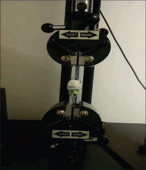
Specimen undergoing bond strength testing in the universal testing machine.
Figure 2.
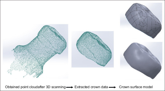
Steps of converting the point cloud to surface by SolidWorks software.
Evaluation of mode of failure and microstructure
The mode of failure was investigated after separating the crowns by bond strength test. The mode of failure was divided into four different categories according to previous studies.[29,30] These categories are presented in Table 1. The number of failure in each category was obtained and presented as percentage for each group. The data were analyzed using Shapiro–Wilk, one-way ANOVA (Kruskal–Wallis), and Chi-square tests.
Table 1.
Different groups of the mode of failure after removing the crowns in bond strength test
| Category | Description |
|---|---|
| 1 | Cement mostly remained on the teeth(>75%) |
| 2 | Cement remained on both the teeth and the crown(between 25% and 75%) |
| 3 | Cement mostly remained on crown(>75%) |
| 4 | Tooth root fracture without separation of the crown |
In addition, four samples (one from each group) tested under tensile bond strength were examined under scanning electron microscopy (SEM; FEI QUANTA 200 ESEM, USA), and their mode of failure was investigated. SEM images were taken with different magnifications of ×1300 and ×2500 using backscattered electron mode and the applied voltage of 25 kV. These images were provided from both the cement surface and the tooth–cement interface.
RESULTS
In this study, microleakage, bond strength, and mode of failure of four types of cements were studied. The results are presented in the following sections.
Microleakage
The mean, standard error, median, and interquartile range of microleakage obtained for four types of cements are presented in Table 2. Our results on microleakage did not support null hypothesis; it means there was not enough evidence to conclude that the four groups are equal. In other words, there was a significant difference (P = 0.001) between the microleakage of the four types of cements. The resin cement microleakage was lower than polycarboxylate cement (P = 0.008) and self-cure glass ionomer (P = 0.001) and did not differ significantly from resin-modified glass ionomer cement (P = 0.092). Microleakage of resin-modified glass ionomer cement was also significantly lower than polycarboxylate (P = 0.036) and self-cure glass ionomer (P = 0.005) cements. Furthermore, microleakage of polycarboxylate and self-cure glass ionomer cements did not differ significantly (P = 0.888). Figure 3 shows the dye penetration between the tooth and the cement.
Table 2.
Mean, standard error, median, and interquartile range of microleakage (in millimeters) in the four types of cement
| Cement type | Number of samples | Mean±SE | Median | P |
|---|---|---|---|---|
| Resin cement | 9 | 2.00±0.23 | 1.85 | 0.001 |
| Resin-modified glass ionomer | 8 | 2.40±0.19 | 2.15 | |
| Polycarboxylate | 9 | 3.31±0.31 | 3.53 | |
| Self-cure glass ionomer | 8 | 3.53±0.22 | 3.89 |
SE: Standard error; IQR: Interquartile range
Figure 3.
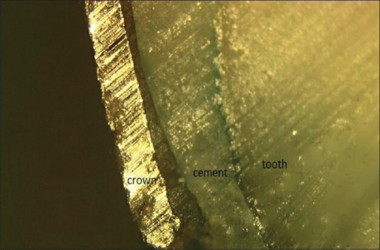
Methylene blue penetration between the tooth and the cement.
Bond strength
The mean, standard error, median, and interquartile range of bond strength in four types of cements are presented in Table 3. As Table 3 shows, the highest to the lowest bond strengths were those of resin cement, resin-modified glass ionomer, glass ionomer, and polycarboxylate, respectively. However, there was no significant difference (P = 0.124 and F (3,33) = 2.07) between the mean bond strengths of the four types of cements. According to the sampling (n = 10), there was no evidence for rejecting null hypothesis on bond strength. Therefore, more sampling is suggested for future research.
Table 3.
Mean, standard error, median, and interquartile range of bond strength (Newtons per square centimeter) in the four types of cement
| Cement type | Number of samples | Mean±SE | Median | IQR | P |
|---|---|---|---|---|---|
| Resin cement | 7 | 235.6±27.0 | 242.1 | 84.9 | 0.124 |
| Resin-modified glass ionomer | 10 | 220.1±25.2 | 233.4 | 157.5 | |
| Self-cure glass ionomer | 10 | 200.4±24.4 | 203.7 | 144.3 | |
| Polycarboxylate | 10 | 152.1±23.4 | 150.2 | 137.9 |
SE: Standard error; IQR: Interquartile range
Mode of failure and microstructure
The results of the mode of failure are presented in Table 4. As Table 4 shows, mode of failure in category 3 was not found in any of the cement, and mode of failure in category 4 was only observed in the resin cement. Furthermore, mode of failure in 50% of resin cement, 70% of each of the resin-modified glass ionomer and glass ionomer cements, and 100% of polycarboxylate cement was in the way that cement was remained on both the tooth and the crown. The distribution of failure in the four types of cements was significantly different (P = 0.041).
Table 4.
Percentage of different groups of the mode of failure in the cements
| Cement type | Mode of failure | ||
|---|---|---|---|
|
| |||
| >75% of cements remained on the tooth(category 1), n(%) | Cement remained on both the tooth and the crown(category 2), n(%) | Tooth root fracture without separation of the crown(category 4), n(%) | |
| Resin | 2(20.0) | 5(50.0) | 3(30.0) |
| Resin-modified glass ionomer | 3(30.0) | 7(70.0) | 0(0.0) |
| Glass ionomer | 3(30.0) | 7(70.0) | 0(0.0) |
| Polycarboxylate | 0(0.0) | 10(100) | 0(0.0) |
Figures 4 and 5 show the SEM images from the surface of the four types of cements, and the tooth–cement interface after the bond strength tests, respectively.
Figure 4.
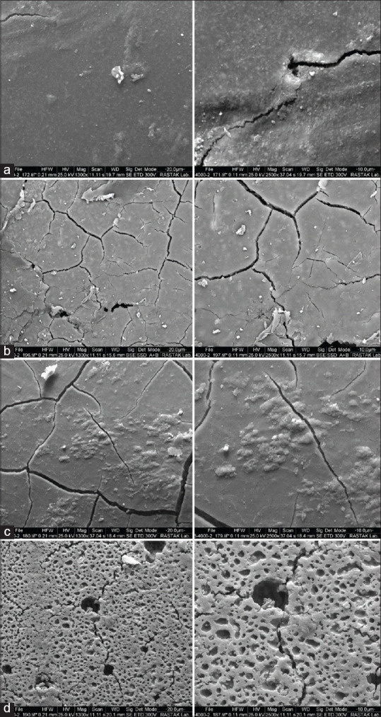
Scanning electron microscopy images of the cement surface (a) resin cement, (b) resin-modified glass ionomer, (c) glass ionomer, and (d) polycarboxylate at magnifications of ×1300 (left) and ×2500 (right).
Figure 5.
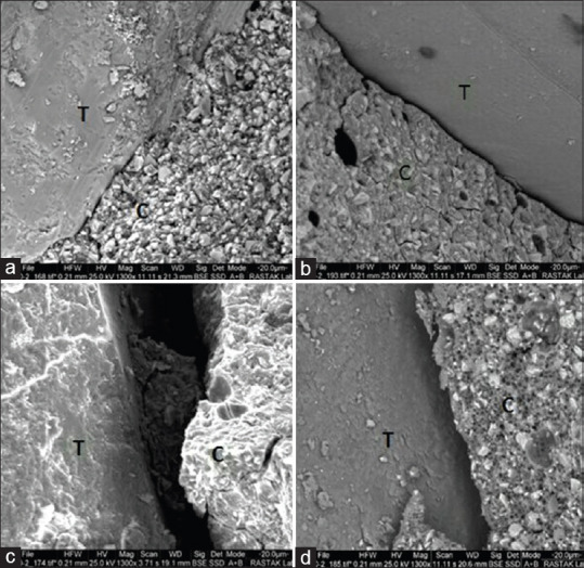
Scanning electron microscopy images from the tooth–cement interface in (a) resin cement, (b) RMGI, (c) glass ionomer, and (d) polycarboxylate at a magnification of ×1300.
DISCUSSION
SSC provides a complete cover for primary tooth crown. Although SSCs are more successful in restoring the teeth with extensive degradation than other restorations, failure in cementing is one of the main leading causes for the clinical failure of this restoration.[8,9] Factors such as the dissolution of cement, contraction during setting, and the absence of the acceptable bonding to the tooth structure and restoration can cause microleakage.[31] The presence of leakage in the margin of the crown allows bacterial penetration into the tooth structure and can lead to recurrent caries, inflammation of the tooth pulp, or re-infection of root-treated teeth.[32] One of the important factors that affects the microleakage is the type of cement used to bond SSCs.[18] The results of this study showed that the microleakage of resin cement and resin-modified glass ionomer cement was less than self-curing glass ionomer and polycarboxylate cements. Furthermore, microleakage of resin cement and resin-modified glass ionomer cement was not statistically significantly different, neither it was between self-cure glass ionomer and polycarboxylate cements. The results of microleakage on resin-modified glass ionomer and polycarboxylate cements obtained here are consistent with Memarpour et al.[33] Erdemci et al. showed that resin cement had the smallest amount of microleakage, followed by glass ionomer and polycarboxylate cement.[34] Their results on resin cement are similar to the present study. However, they showed that the microleakage of glass ionomer cement was less than that of polycarboxylate cement, while the current study identified that the microleakage of glass ionomer and polycarboxylate cements did not significantly differ statistically. The difference in the results obtained can be due to the different brands of cements used. The point that should be born in mind is that the rate of bacteria and their product penetration between the teeth and the cements is clinically less than the degree of penetration of the dye in the laboratory tests. This is because of easier diffusion of color than bacteria and their products. As a result, the cements are clinically more effective against microleakage than the results of laboratory penetration tests.[35,36]
The results of studies comparing SSC bonding fixed with and without cement showed that the crown fixed with cement to the tooth had considerably higher bonding strength than that fixed without cement.[26] Therefore, in the present study, four different types of cements were evaluated for their bond strengths. The results of our study showed that there was no significant difference between the bond strengths of the resin cement, resin-modified glass ionomer, self-cure glass ionomer, and polycarboxylate. However, the highest strength was related to the resin cement, followed by resin-modified glass ionomer, self-cure glass ionomer, and polycarboxylate, respectively. The results of Reddy et al. showed that the bond strength of zinc phosphate and glass ionomer cements was higher than that of the polycarboxylate cement, and there was little difference in bonding strength of zinc phosphate and glass ionomer.[26] The authors recommended the use of glass ionomer cement due to its easier use and release of fluoride for cementing SSC in children.[26] In the present study, the mean bond strength of the glass ionomer was higher than that of polycarboxylate, but the difference was not statistically significant. Subramaniam et al. also showed that the bond strength of resin cement and resin-modified glass ionomer was significantly higher than that of conventional glass ionomer.[37] Similarly, in the current study, the mean bond strengths of resin and resin-modified glass ionomer cements were higher than the mean bond strength of glass ionomer cement. In a study conducted in 2004, Yilmaz et al.[24] compared the tensile strength, microleakage, and SEM images of SSCs fixed by glass ionomer (Aqua Meron), resin-modified glass ionomer (RelyX luting), and resin (Panavia F) cements on primary molar teeth. They showed that the bond strength of resin cement was significantly higher than that of resin-modified glass ionomer, and there was no significant difference between the bond strengths of glass ionomer and resin cements. The resin cement had a significantly lower microleakage than resin-modified glass ionomer. However, there was no significant difference between the microleakage of resin and glass ionomer cements. Their results showed that the cement with the highest bond strength has the lowest microleakage. Their results regarding no significant difference between the bond strengths of resin and glass ionomer cements are consistent with our findings. In the present study, ten samples were considered for each group in the tests (based on power analysis), where significant differences were obtained for microleakage. However, there were no differences between the groups for bond strength, thus increasing the number of samples in each group for bond strength test is suggested for future research. The main limitation of bonding strength experimentation is that the results of various studies are not comparable. This is due to lack of standardization between studied groups. The differences in the results obtained in various researches may also be attributed to the different crown materials, different brands of cements used, and the method of testing as well. For instance, the bond strength test can be done using different methods for attaching the crown to the device applying tensile force (spot welding the brackets and buccal tubes to the buccal and lingual surface of the crown, using nail through a hole in the central fossa or clamps to the occlusal surface of the crown). The other difference between the studies conducted on bond strength is related to seating pressure for initial placement of crown[38] which is either finger/hand force[26,27] or applied controlled force.[39] Differences in pressure applied during the cementation may influence the cement film thickness uniformity, which subsequently can affect the micromechanics of retention, and marginal adaptation of restoration.[40] Meanwhile, because of the heterogeneous distribution of stress in the bonded interface, the mean value of bond strength cannot be indicative of the actual stress initiating debonding solely.[41]
In the present study, the mode of failure after detachment of the crowns in the bond strength tests was evaluated in the four cement groups. The failure mode in the four types of cements had significant differences (P = 0.041). Category 3 of mode of failure was not observed in any of the cements, i.e., the category in which the cement remained mostly on the crown. Meanwhile, category 4 of mode of failure (i.e., root fracture) was only observed in the resin cement. This can be attributed to the higher bond strength of this cement than the other cements. The most common mode of failure was category 2, followed by category 1. The mode of failure in the resin cement consisted of 50% category 2, 30% category 4, and 20% category 1, while in glass ionomer and resin-modified glass ionomer cements, the only modes of failure observed were category 2 (70%) and category 1 (30%). The similarity of the mode of failure can be associated with the similarity of the chemical composition and structure of these two types of cements. In polycarboxylate cement, only category 2 of mode of failure was observed.
The images obtained from the cement surface and the tooth–cement interface with SEM also clearly showed the differences and similarities between the different types of cements [Figures 4 and 5]. These images showed that cracks were created on the surfaces of all cements after the cement fracture. The amount of these cracks in resin cement was less than other cements possibly due to the higher strength of this cement. Glass ionomer and resin-modified glass ionomer cements had very similar surfaces. Furthermore, both cracks and detachment causing holes were observed in the polycarboxylate cement. The interesting point about this cement is its porous structure, which resulted in a decrease of the cement bond strength and an increase of microleakage (capillary effect). This porous structure is clearly visible in the SEM images. Moreover, SEM images obtained from the tooth–cement interface revealed a gap in this area. The smallest gap between cement and tooth was that of resin cement, and the largest one was related to the glass ionomer and polycarboxylate cements. The results of the SEM images in this study are consistent with Yilmaz et al.[24] Therefore, cohesive and adhesive failures were observed in the cement itself and in the tooth–cement interface, respectively.
In this study, resin cement showed relatively more favorable properties. In general, the advantages of resin cement are high strength, low film thickness, and very low oral solubility.[5] Because of the excellent bonding ability of resin cements, these cements have highly attracted the dentists. Since these cements are auto-mixed, there is no need for manual mixing, thus saving time, eliminating problems due to inappropriate ratio of powder to liquid, and ultimately ease of use. High cost is one of the disadvantages of this cement brand compared to other cements used in this study. On the other hand, resin-modified glass ionomer showed relatively good properties after resin cement. This cement is made by adding resin monomers to the glass ionomer. The shortcomings of conventional glass ionomer include short functioning period, slow development of final properties, sensitivity in contact with moisture, dehydration during the setting time, and lower cohesive strength compared to resin cements. Improvement of these properties has been mainly considered in resin-modified glass ionomer cements.[5] In addition, this cement, similar to glass ionomer cement, has the ability to release fluoride.[42] Although, in the present study, bond strength and microleakage of glass ionomer cement had no significant difference compared to polycarboxylate cement, the use of glass ionomer cement is more recommended because of its advantages in terms of good physical properties, adhesion to the structure of the tooth and metal, and most importantly the release of a significant amount of fluoride that increases the resistance of enamel and dentin to acidic dissolution and acts as a bacteriostatic agent.[41] In the present study, the results showed that the bond strength was not statistically significantly different among the cements. However, considering the fact that the microleakage test showed that resin cement and resin-modified glass ionomer cement had a lower degree of microleakage than conventional glass ionomer and polycarboxylate cements, the use of resin cement and resin-modified glass ionomer cement is more recommended for bonding SSCs.
CONCLUSION
According to the results of this study, the following conclusions are made:
Resin cement and resin-modified glass ionomer cement showed the least amount of microleakage
Microleakage of resin cement and resin-modified glass ionomer cement was not significantly different, neither it was between glass ionomer and polycarboxylate cements
No significant difference was found between the mean bond strengths in the four types of cements. However, the highest strength was that of the resin cement, followed by resin-modified glass ionomer, self-cure glass ionomer, and polycarboxylate, respectively
The mode of failure in the four types of cements had a significant difference. The most common mode of failure was the category in which the cement remained both on the tooth and the crown.
Financial support and sponsorship
Nil.
Conflicts of interest
The authors of this manuscript declare that they have no conflicts of interest, real or perceived, financial or non-financial in this article.
REFERENCES
- 1.Guelmann M, Fair J, Bimstein E. Permanent versus temporary restorations after emergency pulpotomies in primary molars. Pediatr Dent. 2005;27:478–81. [PubMed] [Google Scholar]
- 2.Casamassimo PS, Fields HW, Jr, McTigue DJ, Nowak A. 5th ed. Elsevier India; 2012. Pediatric Dentistry: Infancy Through Adolescence. [Google Scholar]
- 3.Messer LB, Levering NJ. The durability of primary molar restorations: II.Observations and predictions of success of stainless steel crowns. Pediatr Dent. 1988;10:81–5. [PubMed] [Google Scholar]
- 4.Seale NS. The use of stainless steel crowns. Pediatr Dent. 2002;24:501–5. [PubMed] [Google Scholar]
- 5.MC Donald R, Avery D, Dean J. Philadelphia: Mosby; 2011. Dentistry for the Child and Adolescent. [Google Scholar]
- 6.Henderson HZ. Evaluation of the preformed stainless steel crown. ASDC J Dent Child. 1973;40:353–8. [PubMed] [Google Scholar]
- 7.Croll TP, Killian CM. Zinc oxide-eugenol pulpotomy and stainless steel crown restoration of a primary molar. Quintessence Int. 1992;23:383–8. [PubMed] [Google Scholar]
- 8.Garcia-Godoy F. Clinical evaluation of the retention of preformed crowns using two dental cements. J Pedod. 1984;8:278–81. [PubMed] [Google Scholar]
- 9.Sailer I, Pjetursson BE, Zwahlen M, Hämmerle CH. A systematic review of the survival and complication rates of all-ceramic and metal-ceramic reconstructions after an observation period of at least 3 years. Part II: Fixed dental prostheses. Clin Oral Implants Res. 2007;18:86–96. doi: 10.1111/j.1600-0501.2007.01468.x. [DOI] [PubMed] [Google Scholar]
- 10.Parisay I, Khazaei Y. Evaluation of retentive strength of four luting cements with stainless steel crowns in primary molars: An in vitro study. Dent Res J (Isfahan) 2018;15:201–7. [PMC free article] [PubMed] [Google Scholar]
- 11.Nayakar RP, Patil NP, Lekha K. Comparative evaluation of bond strengths of different core materials with various luting agents used for cast crown restorations. J Indian Prosthodont Soc. 2012;12:168–74. doi: 10.1007/s13191-012-0127-8. [DOI] [PMC free article] [PubMed] [Google Scholar]
- 12.Mulder R, Medhat R, Mohamed N. In vitro analysis of the marginal adaptation and discrepancy of stainless steel crowns. Acta Biomater Odontol Scand. 2018;4:20–9. doi: 10.1080/23337931.2018.1444995. [DOI] [PMC free article] [PubMed] [Google Scholar]
- 13.Kelvin Khng KY, Ettinger RL, Armstrong SR, Lindquist T, Gratton DG, Qian F. In vitro evaluation of the marginal integrity of CAD/CAM interim crowns. J Prosthet Dent. 2016;115:617–23. doi: 10.1016/j.prosdent.2015.10.002. [DOI] [PubMed] [Google Scholar]
- 14.Shiflett K, White SN. Microleakage of cements for stainless steel crowns. Pediatr Dent. 1997;19:262–6. [PubMed] [Google Scholar]
- 15.Croll TP, McKay MS, Castaldi CR. Impaction of permanent posterior teeth by overextended stainless steel crown margins. J Pedod. 1981;5:240–4. [PubMed] [Google Scholar]
- 16.Al-Haj Ali SN, Farah RI. In vitro comparison of microleakge between preformed metal crowns and aesthetic crowns of primary molars using different adhesive luting cements. Eur Arch Paediatr Dent. 2018;19:387–92. doi: 10.1007/s40368-018-0369-1. [DOI] [PubMed] [Google Scholar]
- 17.Sener I, Turker B, Valandro LF, Ozcan M. Marginal gap, cement thickness, and microleakage of 2 zirconia crown systems luted with glass ionomer and MDP-based cements. Gen Dent. 2014;62:67–70. [PubMed] [Google Scholar]
- 18.Kindelan SA, Day P, Nichol R, Willmott N, Fayle SA; British Society of Paediatric Dentistry. UK National Clinical Guidelines in Paediatric Dentistry: Stainless steel preformed crowns for primary molars. Int J Paediatr Dent. 2008;18 Suppl 1:20–8. doi: 10.1111/j.1365-263X.2008.00935.x. [DOI] [PubMed] [Google Scholar]
- 19.Rossetti PH, do Valle AL, de Carvalho RM, De Goes MF, Pegoraro LF. Correlation between margin fit and microleakage in complete crowns cemented with three luting agents. J Appl Oral Sci. 2008;16:64–9. doi: 10.1590/S1678-77572008000100013. [DOI] [PMC free article] [PubMed] [Google Scholar]
- 20.Pilo R, Folkman M, Arieli A, Levartovsky S. Marginal fit and retention strength of zirconia crowns cemented by self-adhesive resin cements. Oper Dent. 2018;43:151–61. doi: 10.2341/16-367-L. [DOI] [PubMed] [Google Scholar]
- 21.Campos F, Valandro LF, Feitosa SA, Kleverlaan CJ, Feilzer AJ, de Jager N, et al. Adhesive cementation promotes higher fatigue resistance to zirconia crowns. Oper Dent. 2017;42:215–24. doi: 10.2341/16-002-L. [DOI] [PubMed] [Google Scholar]
- 22.Üstün Ö, Büyükhatipoğlu IK, Seçilmiş A. Shear bond strength of repair systems to new CAD/CAM restorative materials. J Prosthodont. 2018;27:748–54. doi: 10.1111/jopr.12564. [DOI] [PubMed] [Google Scholar]
- 23.Roberts JF, Attari N, Sherriff M. The survival of resin modified glass ionomer and stainless steel crown restorations in primary molars, placed in a specialist paediatric dental practice. Br Dent J. 2005;198:427–31. doi: 10.1038/sj.bdj.4812197. [DOI] [PubMed] [Google Scholar]
- 24.Yilmaz Y, Dalmis A, Gurbuz T, Simsek S. Retentive force and microleakage of stainless steel crowns cemented with three different luting agents. Dent Mater J. 2004;23:577–84. doi: 10.4012/dmj.23.577. [DOI] [PubMed] [Google Scholar]
- 25.Al-Haj Ali SN. In vitro comparison of marginal and internal fit between stainless steel crowns and esthetic crowns of primary molars using different luting cements. Dent Res J (Isfahan) 2019;16:366–71. [PMC free article] [PubMed] [Google Scholar]
- 26.Reddy MR, Reddy VS, Basappa N. A comparative study of retentive strengths of zinc phosphate, polycarboxylate and glass ionomer cements with stainless steel crowns-an in vitro study. J Indian Soc Pedod Prev Dent. 2010;28:245–50. doi: 10.4103/0970-4388.76150. [DOI] [PubMed] [Google Scholar]
- 27.Veerabadhran MM, Reddy V, Nayak UA, Rao AP, Sundaram MA. The effect of retentive groove, sandblasting and cement type on the retentive strength of stainless steel crowns in primary second molars-an in vitro comparative study. J Indian Soc Pedod Prev Dent. 2012;30:19–26. doi: 10.4103/0970-4388.95570. [DOI] [PubMed] [Google Scholar]
- 28.Fisker R, Clausen T, Deichmann N, Ojelund H. Inventors; Google Patents, Assignee. Adaptive 3D Scanning. 2014 [Google Scholar]
- 29.Shahin R, Kern M. Effect of air-abrasion on the retention of zirconia ceramic crowns luted with different cements before and after artificial aging. Dent Mater. 2010;26:922–8. doi: 10.1016/j.dental.2010.06.006. [DOI] [PubMed] [Google Scholar]
- 30.Palacios RP, Johnson GH, Phillips KM, Raigrodski AJ. Retention of zirconium oxide ceramic crowns with three types of cement. J Prosthet Dent. 2006;96:104–14. doi: 10.1016/j.prosdent.2006.06.001. [DOI] [PubMed] [Google Scholar]
- 31.Jacobs MS, Windeler AS. An investigation of dental luting cement solubility as a function of the marginal gap. J Prosthet Dent. 1991;65:436–42. doi: 10.1016/0022-3913(91)90239-s. [DOI] [PubMed] [Google Scholar]
- 32.Magura ME, Kafrawy AH, Brown CE, Jr, Newton CW. Human saliva coronal microleakage in obturated root canals: An in vitro study. J Endod. 1991;17:324–31. doi: 10.1016/S0099-2399(06)81700-0. [DOI] [PubMed] [Google Scholar]
- 33.Memarpour M, Mesbahi M, Rezvani G, Rahimi M. Microleakage of adhesive and nonadhesive luting cements for stainless steel crowns. Pediatr Dent. 2011;33:501–4. [PubMed] [Google Scholar]
- 34.Erdemci ZY, Cehreli SB, Tirali RE. Hall versus conventional stainless steel crown techniques: In vitro investigation of marginal fit and microleakage using three different luting agents. Pediatr Dent. 2014;36:286–90. [PubMed] [Google Scholar]
- 35.Albert FE, El-Mowafy OM. Marginal adaptation and microleakage of Procera AllCeram crowns with four cements. Int J Prosthodont. 2004;17:529–35. [PubMed] [Google Scholar]
- 36.Toman M, Toksavul S, Artunç C, Türkün M, Schmage P, Nergiz I. Influence of luting agent on the microleakage of all-ceramic crowns. J Adhes Dent. 2007;9:39–47. [PubMed] [Google Scholar]
- 37.Subramaniam P, Kondae S, Gupta KK. Retentive strength of luting cements for stainless steel crowns: An in vitro study. J Clin Pediatr Dent. 2010;34:309–12. doi: 10.17796/jcpd.34.4.p5h1068v41ggt450. [DOI] [PubMed] [Google Scholar]
- 38.Heintze SD. Crown pull-off test (crown retention test) to evaluate the bonding effectiveness of luting agents. Dent Mater. 2010;26:193–206. doi: 10.1016/j.dental.2009.10.004. [DOI] [PubMed] [Google Scholar]
- 39.Wolfart S, Linnemann J, Kern M. Crown retention with use of different sealing systems on prepared dentine. J Oral Rehabil. 2003;30:1053–61. doi: 10.1046/j.1365-2842.2003.01180.x. [DOI] [PubMed] [Google Scholar]
- 40.Zortuk M, Bolpaca P, Kilic K, Ozdemir E, Aguloglu S. Effects of finger pressure applied by dentists during cementation of all-ceramic crowns. Eur J Dent. 2010;4:383–8. [PMC free article] [PubMed] [Google Scholar]
- 41.Sakaguchi RL, Powers JM. Elsevier Health Sciences; 2012. Craig’s Restorative Dental Materials-e-Book. [Google Scholar]
- 42.Krämer N, Schmidt M, Lücker S, Domann E, Frankenberger R. Glass ionomer cement inhibits secondary caries in an in vitro biofilm model. Clin Oral Investig. 2018;22:1019–31. doi: 10.1007/s00784-017-2184-1. [DOI] [PubMed] [Google Scholar]


