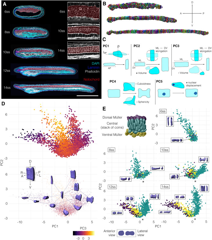
Fig. 1.
A single-cell morphospace captures notochord cell shape diversity. (A-E) Steps 1-4 of the morphospatial embedding pipeline (see Materials and Methods). (A) Amphioxus embryos at successive somite stages stained with phalloidin to mark cortical actin, which was used for manual cell segmentation. Embryos are also immunostained for laminin (extracellular matrix) and acetylated tubulin (cilia). Notochord is false-coloured in red. (B) Images of fully segmented notochords, from 8 ss (top), 10 ss (middle) and 12 ss (bottom). (C) Cell shape metrics correlated with the first five PCs (direction of correlation indicated by plus and minus). (D) Morphospace containing all notochord cells plotted against PC1 and PC2. (Top) Notochord morphospace for all cells and stages, colour code for PC3. (Bottom) Notochord morphospace with representative surfaces from cell segmentation data in anterolateral view. (E) Morphospace filtered by stage, colour-coded for DV position as indicated on a 8 ss notochord fragment (top left). Representative cells are shown in inlays, in anterior (left) and lateral (right) views. Asterisks mark bifurcation resolving central and Müller cells. All embryos and notochords are in lateral view, anterior side facing left. n=3796 cells, five stages, three embryos per stage.

