Abstract
Glioblastoma multiforme (GBM) is the most frequent type of primary astrocytomas. We examined the association between single nucleotide polymorphisms (SNPs) in Aurora kinase A (AURKA), Aurora kinase B (AURKB), Aurora kinase C (AURKC) and Polo-like kinase 1 (PLK1) mitotic checkpoint genes and GBM risk by qPCR genotyping. In silico analysis was performed to evaluate effects of polymorphic biological sequences on protein binding motifs. Chi-square and Fisher statistics revealed a significant difference in genotypes frequencies between GBM patients and controls for AURKB rs2289590 variant (p = 0.038). Association with decreased GBM risk was demonstrated for AURKB rs2289590 AC genotype (OR = 0.54; 95% CI = 0.33–0.88; p = 0.015). Furthermore, AURKC rs11084490 CG genotype was associated with lower GBM risk (OR = 0.57; 95% CI = 0.34–0.95; p = 0.031). Bioinformatic analysis of rs2289590 polymorphic region identified additional binding site for the Yin-Yang 1 (YY1) transcription factor in the presence of C allele. Our results indicated that rs2289590 in AURKB and rs11084490 in AURKC were associated with a reduced GBM risk. The present study was performed on a less numerous but ethnically homogeneous population. Hence, future investigations in larger and multiethnic groups are needed to strengthen these results.
Subject terms: Cancer genetics, CNS cancer, Biomarkers, Molecular medicine, Oncology, Risk factors
Introduction
Glioblastoma multiforme (GBM) represents the most common and lethal form of primary brain tumor with an annual incidence of 5.26 per 100,000 people1,2 and it stands for more than 60% of all brain tumors in adults3. Although a significant number of modern therapies against GBM is available, it is still a deadly disease with a poor prognosis4. Precise chromosomal segregation in dividing cancer cells as well as disturbances during the spindle assembly checkpoint can contribute to malignant transformation5. Genetic modifications in mitotic genes could increase sensitivity to neoplastic transformation through alterations of gene expression profiles6,7. Aurora kinases are members of serine-threonine kinases family which are of great importance for the cell cycle control8. Aurora kinase A (AURKA) is involved in proper functioning of a few oncogenic signaling processes such as mitotic entry, spindle assembly, centrosome functioning, chromosome alignment and/or segregation and cytokinesis9–11. Aurora kinase B (AURKB) is a component of chromosomal passenger complex and mediates in chromatin modification, spindle checkpoint regulation, cytokinesis and correct kinetochore/microtubule attachment9,12. Aurora kinase C (AURKC) is also a member of chromosomal passenger complex which takes part in mitotic events such as accurate centrosome functioning13, and is required to regulate chromosome segregation during meiosis I14. Polo-like kinase 1 (PLK1) is engaged in several cellular processes including centrosome maturation, mitotic checkpoint activation and spindle assembly, kinetochore/microtubule binding, cytokinesis and cellular proliferation15–17. PLK1 overexpression is proved to be associated with poor prognosis in several cancer entities18. Moreover, it has been demonstrated that polymorphisms in PLK1 affect its expression, thus possess the ability to potentially influence the risk of disease onset and progression18.
In our case–control study, we evaluated the impact of single nucleotide polymorphisms rs1047972, rs2273535, rs8173 and rs911160 (AURKA), rs2289590 and rs2241909 (AURKB), rs11084490 and rs758099 (AURKC) and rs42873 (PLK1) in mitotic checkpoint genes on glioblastoma multiforme development in Bosnia and Herzegovina population. Using bioinformatic analysis of genetic variants, we estimated the impact of the polymorphic DNA sequences in introns and untranslated regions (UTRs) within AURKA, AURKB, AURKC and PLK1 genes on transcription factors binding sites.
Methods
Design of the study and study groups
Our study group consisted of 129 patients with diagnosed glioblastoma multiforme (GBM) at the Clinical Pathology and Cytology at the University Clinical Center Sarajevo, Bosnia and Herzegovina. Of that, 68 were men and 61 women, with a mean age of 58 years at the moment of diagnosis (data were missing in 4 cases) (Table 1). The formalin fixed paraffin embedded (FFPE) cancer tissue sections were collected in the course of surgical procedures. Written informed consents, which allow the use of samples in this study, were obtained from all patients prior to the surgery. On the other side, 203 healthy blood donors (ethnicity matched to the cases), upon regular medical examinations, were randomly selected and signed up as a control group for the present study. Control samples had no history of neoplastic formation, were not related to the patients and/or to each other. Three milliliters of blood were taken from each control individual and kept at − 80 °C. An informed written consent was obtained from the participants, with personal and medical information being enciphered in order to ensure maximum anonymity in compliance with the World Medical Association’s Declaration of Helsinki. This study was approved by the University Clinical Centre Sarajevo Ethical Committee (No. 0302-36765).
Table 1.
General information for glioblastoma multiforme patients.
| Variable | GBM patients | |
|---|---|---|
| N | N (%) | |
| Total sample | 129 | |
| Gender | ||
| Female | 61 | (47.3) |
| Male | 68 | (52.7) |
| Age at diagnosis (years)a | ||
| < 58 | 56 | (44.8) |
| ≥ 58 | 69 | (55.2) |
| Mean | 58 | |
| Range | 19–81 | |
GBM glioblastoma multiforme. aData were missing in 4 cases.
DNA extraction
Genomic DNA from glioblastoma multiforme (GBM) formalin fixed paraffin embedded tissues was extracted using the Chemagic FFPE DNA Kit special (PerkinElmer Inc., Waltham, MA, USA). DNA washing and elution was performed on Chemagic Magnetic Separation Module I robot (PerkinElmer Inc., Waltham, MA, USA), following manufacturer’s recommendations. All sample transfers were conducted with the four-eye principle to avoid mixing errors of the samples. DNA from peripheral blood lymphocytes (controls) was isolated using the Promega™ Wizard™ Genomic DNA Purification Kit Protocol (Promega Corp., Fitchburg, WI, USA) in concordance with the manufacturer’s instructions. The qualitative/quantitative analysis of the extracted DNA was performed on the DropSense96 photometer (Trinean, Gentbrugge, Belgium) and Synergy™ 2 Multi Mode Reader (BioTek, Inc., Winooski, VT, USA).
SNP selection
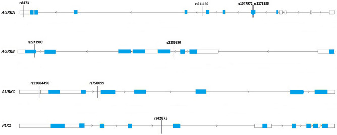
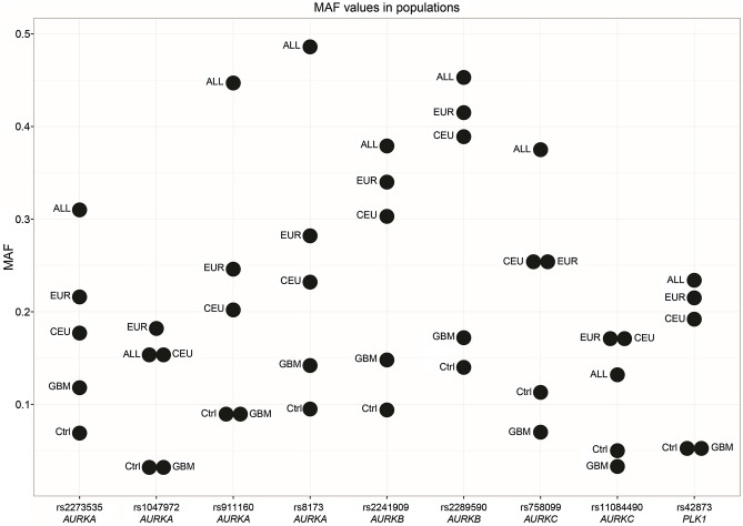
In total, nine single nucleotide polymorphisms (SNPs) in segregation genes, precisely rs1047972, rs2273535, rs8173 and rs911160 (AURKA), rs2289590 and rs2241909 (AURKB), rs11084490 and rs758099 (AURKC) and rs42873 (PLK1) were chosen. The locations of the selected variants in mitotic genes are shown in Fig. 1, whereby gene structures were obtained from the Research Collaboratory for Structural Bioinformatics (RCSB) Protein Data Bank (PDB)19. The parameters described below are used for the selection of the genetic variants: (1) previously established association related to certain tumors; (2) minor allele frequency (MAF) fewer or equal to 10% in the population of Utah residents with Northern and Western European ancestry (CEU) as highlighted by the Phase 3 1000 Genomes; and (3) tagging single nucleotide polymorphisms (tagSNPs) status, which was computed in silico using LD Tag SNP Selection (tagSNP) (https://snpinfo.niehs.nih.gov)20. To predict tagSNPs status, following parameters were used: (a) 1 kb of the upstream–downstream sequences from gene; (b) linkage disequilibrium (LD) lower threshold of 0.8; and (c) minor allele frequency range from 0.05 to 0.5 for the CEU subpopulation (Table 2; Fig. 2).
Figure 1.
The positions of rs1047972, rs2273535, rs8173 and rs911160 (AURKA), rs2289590 and rs2241909 (AURKB), rs11084490 and rs758099 (AURKC) and rs42873 (PLK1) genetic variants within mitotic checkpoint genes. White boxes represent untranslated regions (UTRs). Blue boxes refer to protein coding regions (exons). The black lines connecting the boxes indicate non-coding regions (introns). The gene structures were downloaded from the Research Collaboratory for Structural Bioinformatics (RCSB) Protein Data Bank (PDB), GRCh38 Genome Assembly.
Table 2.
Basic characteristics of the studied genetic variants.
| SNP | Variant type | Gene | Base change | NCBI assembly location (Build GRCh38)a | TaqMan SNP assay ID | MAFb | ||||
|---|---|---|---|---|---|---|---|---|---|---|
| GBM patients | Control group | ALL | EUR | CEU | ||||||
| rs1047972 | Missense | AURKA | C/T | Chr.20:56386407 | AHX1IRW | 0.162 | 0.146 | 0.150 | 0.182 | 0.157 |
| rs2273535 | Missense | AURKA | A/T | Chr.20:56386485 | C_25623289_10 | 0.299 | 0.238 | 0.310 | 0.216 | 0.177 |
| rs8173 | 3′ UTR | AURKA | G/C | Chr.20:56369735 | C_8947675_10 | 0.354 | 0.305 | 0.486 | 0.282 | 0.232 |
| rs911160 | Intron | AURKA | G/C | Chr.20:56382507 | C_8947670_10 | 0.300 | 0.276 | 0.447 | 0.246 | 0.202 |
| rs2289590 | Intron | AURKB | C/A | Chr.17:8207446 | C_15770418_10 | 0.375 | 0.415 | 0.453 | 0.415 | 0.389 |
| rs2241909 | Synonymous | AURKB | A/G | Chr.17:8205021 | C_22272900_10 | 0.332 | 0.332 | 0.379 | 0.340 | 0.303 |
| rs11084490 | 5′ UTR | AURKC | C/G | Chr.19:57231104 | C_27847620_10 | 0.152 | 0.223 | 0.132 | 0.165 | 0.177 |
| rs758099 | Intron | AURKC | C/T | Chr.19:57231966 | C_2581008_1_ | 0.162 | 0.302 | 0.375 | 0.255 | 0.253 |
| rs42873 | Intron | PLK1 | G/C | Chr.16:23683411 | C_2392140_10 | 0.354 | 0.208 | 0.234 | 0.215 | 0.192 |
ALL all phase 3 individuals, CEU Utah residents with Northern and Western European ancestry, EUR European population, GBM glioblastoma multiforme, MAF minor allele frequency, SNP single nucleotide polymorphism, UTR untranslated region. ahttps://www.lifetechnologies.com. bMAFs extracted from 1000 Genomes Project Phase 3.
Figure 2.
Minor allele frequencies (MAFs) for polymorphisms rs1047972, rs2273535, rs8173 and rs911160 (AURKA), rs2289590 and rs2241909 (AURKB), rs11084490 and rs758099 (AURKC) and rs42873 (PLK1) in various populations. ALL all individuals from 1000 Genome Project Phase 3 release, Ctrl studied control population, CEU Utah residents with Northern and Western European ancestry, EUR European population, GBM studied glioblastoma multiforme group, MAF minor allele frequency.
Genotyping of SNPs
Genotyping of the studied variants was performed using TaqMan SNP genotyping assays (Applied Biosystems, Foster City, CA), whose ID numbers are shown in Table 2. The polymerase chain reaction (PCR) mixtures (5 µl for GBM samples and 10 µl for the control samples) consisted of 20X TaqMan® assay along with 2X Master Mix (Applied Biosystems, Foster City, CA), and 20 nanograms of genomic DNA. PCR profile was conducted following manufacturer’s recommendations, hence initial denaturation at 95 °C for 10 min, 45 cycles at 92 °C for 15 s and 60 °C for 90 s, using the ViiA 7 Real Time PCR System (Applied Biosystems, Foster City, CA). At least two negative controls were included in each plate. The results of the PCR reaction were analyzed using TaqMan® Genotyper Software (Applied Biosystems, Foster City, CA, USA).
Statistical analysis
The genotype frequencies of the polymorphisms, for both case and control populations were tested for Hardy–Weinberg equilibrium (HWE) using Michael H. Court’s online HWE calculator (http://www.tufts.edu)21. Significance of the differences in genotype frequencies between GBM patients and controls was determined by use of the Chi-square test or Fisher’s exact test. Multinomial logistic regression was used to test the association between investigated genetic variants and the GBM risk. In this regard, odds ratio (OR) with 95% confidence interval (CI) were calculated to evaluate the relative risk. Statistical analyses were performed using SPSS 20.0 software package (SPSS, Chicago, IL, USA). P ≤ 0.05 was chosen as a threshold significance value. Minor allele frequency (MAF) plot was created in R22 using ggplot2 R package23.
Analysis of haplotypes
To determine the haplotype block structure and perform haplotype analysis, which included corrections for multiple comparisons by 10,000 permutations, Haploview software, version 4.224 and SNP tools V1.80 (MS Windows, Microsoft Excel) were used. In order to create the haplotype block, solid spine of the linkage disequilibrium (LD) algorithm with a minimum Lewontin’s D′ value of 0.8 was chosen.
In silico analysis of polymorphisms
Effects of the polymorphic DNA sequences [polymorphisms in non-coding and untranslated regions (UTRs)] on transcription factors binding sites (TFBSs) were assessed in silico. Bioinformatic functional assessment was conducted using PROMO (ALGGEN) software, which is using data from TRANSFAC database V8.325,26. FASTA sequences for the studied variants were extracted from Ensembl release 98 (http://www.ensembl.org/index.html)27. Identification of TFBSs was determined in concordance with the following criteria: human species, all sites and factors.
Results
Genotypes frequencies for studied SNPs
For all the investigated polymorphisms, rs1047972 (AURKA), rs2273535 (AURKA), rs8173 (AURKA), rs911160 (AURKA), rs2289590 (AURKB), rs2241909 (AURKB), rs11084490 (AURKC), rs758099 (AURKC) and rs42873 (PLK1) was determined to be in Hardy–Weinberg equilibrium (HWE) in both, case and control groups (P > 0.05) (Table 3). After Chi-square test and Fisher exact test were performed to calculate distribution at genotype level (results summarized in Table 3), a significant difference in genotypes frequencies between GBM patients and controls for rs2289590 in AURKB (P = 0.038) was detected.
Table 3.
Genotypes frequencies and Hardy–Weinberg equilibrium for the studied polymorphisms.
| Genotypes | Control group | Glioblastoma multiforme patients | ||||||
|---|---|---|---|---|---|---|---|---|
| N (%) | HWE | N (%) | HWE | GBMa | ||||
| χ2 | P value | χ2 | P value | χ2 | P value | |||
| rs1047972 | 202 | 0.152 | 0.696 | 129 | 1.043 | 0.307 | 0.562 | 0.755 |
| CC | 148 (73.3) | 92 (71.3) | ||||||
| CT | 49 (24.2) | 32 (24.8) | ||||||
| TT | 5 (2.5) | 5 (3.9) | ||||||
| rs2273535 | 203 | 0.867 | 0.351 | 127 | 2.359 | 0.124 | 2.966 | 0.227 |
| AA | 120 (59.1) | 66 (52.0) | ||||||
| AT | 69 (34.0) | 46 (36.2) | ||||||
| TT | 14 (6.9) | 15 (11.8) | ||||||
| rs8173 | 200 | 0.017 | 0.895 | 127 | 0.635 | 0.425 | 1.955 | 0.376 |
| CC | 97 (48.5) | 55 (43.3) | ||||||
| CG | 84 (42.0) | 54 (42.5) | ||||||
| GG | 19 (9.5) | 18 (14.2) | ||||||
| rs911160 | 201 | 0.349 | 0.554 | 128 | 0.031 | 0.859 | 0.509 | 0.755 |
| GG | 107 (53.2) | 63 (49.2) | ||||||
| CG | 77 (38.3) | 53 (41.4) | ||||||
| CC | 17 (8.5) | 12 (9.4) | ||||||
| rs2289590 | 200 | 3.523 | 0.060 | 128 | 2.275 | 0.131 | 6.548b | 0.038 |
| AA | 62 (31.0) | 54 (42.2) | ||||||
| AC | 110 (55.0) | 52 (40.6) | ||||||
| CC | 28 (14.0) | 22 (17.2) | ||||||
| rs2241909 | 203 | 1.186 | 0.276 | 128 | 3.795 | 0.051 | 4.809 | 0.090 |
| AA | 87 (42.9) | 62 (48.4) | ||||||
| AG | 97 (47.8) | 47 (36.8) | ||||||
| GG | 19 (9.3) | 19 (14.8) | ||||||
| rs11084490 | 201 | 0.0009 | 0.975 | 121 | 0.676 | 0.410 | 5.207 | 0.074 |
| CC | 121 (60.2) | 88 (72.7) | ||||||
| CG | 70 (34.8) | 29 (24.0) | ||||||
| GG | 10 (5.0) | 4 (3.3) | ||||||
| rs758099 | 203 | 2.107 | 0.146 | 128 | 0.139 | 0.709 | 1.752 | 0.416 |
| CC | 103 (50.8) | 66 (51.6) | ||||||
| CT | 77 (37.9) | 53 (41.4) | ||||||
| TT | 23 (11.3) | 9 (7.0) | ||||||
| rs42873 | 201 | 0.272 | 0.601 | 128 | 0.111 | 0.738 | 0.174 | 0.917 |
| GG | 127 (63.2) | 78 (61.0) | ||||||
| CG | 64 (31.8) | 43 (33.6) | ||||||
| CC | 10 (5.0) | 7 (5.4) | ||||||
GBM glioblastoma multiforme, HWE Hardy–Weinberg equilibrium, χ2 Chi-square statistics. aχ2 analysis between GBM patients and controls. bFisher statistics. Statistically significant values are highlighted in bold characters (P ≤ 0.05).
Impact of polymorphisms on glioblastoma multiforme risk
Patients with rs2289590 (AURKB) heterozygous AC genotype had a lower risk of glioblastoma multiforme (GBM) development in comparison with the reference AA genotype (OR = 0.54, 95% CI = 0.33–0.88, P = 0.015) (Table 4). Furthermore, the rs11084490 (AURKC) CG genotype was also associated with a decreased GBM risk in comparison with the reference CC genotype (OR = 0.57, 95% CI = 0.34–0.95, P = 0.031).
Table 4.
Risk of glioblastoma multiforme associated with the studied genetic variants.
| Genotypes | Glioblastoma multiforme patients | |
|---|---|---|
| OR (95% CI) | P value | |
| rs1047972 | ||
| CC | 1 (ref) | |
| CT | 1.05 (0.62–1.76) | 0.851 |
| TT | 1.60 (0.45–5.70) | 0.462 |
| rs2273535 | ||
| AA | 1 (ref) | |
| AT | 1.21 (0.75–1.95) | 0.431 |
| TT | 1.94 (0.88–4.28) | 0.097 |
| rs8173 | ||
| CC | 1 (ref) | |
| CG | 1.13 (0.70–1.82) | 0.605 |
| GG | 1.67 (0.81–3.44) | 0.165 |
| rs911160 | ||
| GG | 1 (ref) | |
| CG | 1.16 (0.73–1.86) | 0.513 |
| CC | 1.19 (0.53–2.67) | 0.658 |
| rs2289590 | ||
| AA | 1 (ref) | |
| AC | 0.54 (0.33–0.88) | 0.015 |
| CC | 0.90 (0.46–1.75) | 0.762 |
| rs2241909 | ||
| AA | 1 (ref) | |
| AG | 0.68 (0.42–1.09) | 0.113 |
| GG | 1.40 (0.68–2.86) | 0.353 |
| rs11084490 | ||
| CC | 1 (ref) | |
| CG | 0.57 (0.34–0.95) | 0.031 |
| GG | 0.55 (0.16–1.81) | 0.325 |
| rs758099 | ||
| CC | 1 (ref) | |
| CT | 1.07 (0.67–1.71) | 0.764 |
| TT | 0.61 (0.26–1.40) | 0.244 |
| rs42873 | ||
| GG | 1 (ref) | |
| CG | 1.09 (0.67–1.76) | 0.713 |
| CC | 1.14 (0.41–3.11) | 0.799 |
OR odds ratio, CI confidence interval, Ref reference homozygote. ORs, 95% CIs and P values were obtained by multinomial logistic regression analysis. Statistically significant values are highlighted in bold characters (P ≤ 0.05).
On the other side, no significant effects on GBM susceptibility were revealed for rs1047972, rs2273535, rs8173 and rs911160 in AURKA, rs2241909 in AURKB, rs758099 in AURKC and rs42873 in PLK1 (P > 0.05).
Haplotype analysis
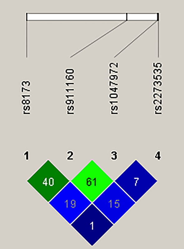
After collecting raw genotyping data for the investigated SNPs in AURKA, namely rs1047972, rs2273535, rs8173 and rs911160, we carried out haplotype analysis using the Haploview software. The outcome of this analysis revealed that no haplotype block was created with an average Lewontin’s D < 0.8 (Fig. 3), therefore no haplotypes were accessible for the examination of their potential association with GBM risk.
Figure 3.

Linkage disequilibrium among single nucleotide polymorphisms in the AURKA gene. The color plot represents Lewontin's D′ values and logarithm of odds (LOD). Blue squares, LOD < 2 and D′ < 1; Green squares, LOD ≥ 2 and D′ < 1. The values within the squares refer to the Lewontin's D′ × 100.
Bioinformatic analysis of the polymorphisms
In silico analysis revealed that polymorphic sequences in transcription factors binding sites (TFBSs), within non-coding and untranslated regions (UTRs) of AURKA, AURKB, AURKC and PLK1 genes, bind various transcription factors (TFs). Our results showed that the region comprising G allele of rs911160 (AURKA) was linked with C/EBPalpha, C/EBPbeta and GR-beta proteins, while for the C allele, extra binding sites for NF-Y, NFI-CTF and NF-1 were recognized (Table 5). As for rs2289590 (AURKB), an additional motif for YY1 binding was identified when C allele was taken into account. In the case of rs11084490 (AURKC), there were no observed differences in transcription factor binding site motif (XBP-1), when different alleles, either C or G, were present. The region including C allele of rs758099 (AURKC) was related with binding sites for NF-1, NF-Y, XBP-1, ENKTF-1, CTF, PEA3 and POU2F1, while for the region surrounding T allele, NF-1, NF-Y, GATA-1 and TFII-I transcription factors were detected. For the polymorphic sequence which include the G allele of rs42873 (PLK1) was demonstrated to be linked with an additional recognition motif for c-Jun DNA-binding factor.
Table 5.
Bioinformatic analysis of the studied genetic variants.
| SNP (gene) | rs911160 (AURKA) | rs2289590 (AURKB) | rs11084490 (AURKC) | rs758099 (AURKC) | rs42873 (PLK1) | |||||
|---|---|---|---|---|---|---|---|---|---|---|
| Alleles | G | C | C | A | C | G | C | T | G | C |
| Transcription factorsa | C/EBPalpha | C/EBPalpha | PEA3 | PEA3 | XBP-1 | XBP-1 | NF-1 | NF-1 | GR-alpha | GR-alpha |
| C/EBPbeta | C/EBPbeta | TFII-I | TFII-I | NF-Y | NF-Y | AP-2alphaA | AP-2alphaA | |||
| GR-beta | GR-beta | YY1 | ENKTF-1 | TFII-I | T3R-beta1 | T3R-beta1 | ||||
| NF-Y | XBP-1 | GATA-1 | c-Jun | |||||||
| NF-1 | CTF | |||||||||
| NFI-CTF | POU2F1 | |||||||||
| PEA3 | ||||||||||
SNP single nucleotide polymorphism.
a Transcription factors binding sites were evaluated using PROMO (ALLGEN) software.
Different transcription factor binding motifs identified for polymorphic alleles of the studied polymorphisms are indicated in bold letters.
Discussion
Our study focused on the assessment of an association between polymorphisms rs1047972, rs2273535, rs8173 and rs911160 (AURKA), rs2289590 and rs2241909 (AURKB), rs11084490 and rs758099 (AURKC) and rs42873 (PLK1), and a risk of glioblastoma multiforme (GBM) development in the population of Bosnia and Herzegovina.
Aurora kinase B (AURKB) is a part of chromosomal passenger complex (CPC), which covers processes such as the segregation of chromatids, cytokinesis and histone modifications28 and for which has been proven to be overexpressed in different types of cancers including brain, prostate and thyroid29. Furthermore, it has been suggested that aurora B overexpression induces abnormalities in chromosome segregation, aneuploidy and tumor development30. We examined the rs2289590 polymorphism in AURKB, and after Chi-square and Fisher exact tests were performed, a significant difference in genotypes frequencies between GBM patients and control group was observed. Additionally, a protective role of the rs2289590 AC genotype against higher GBM risk was found. In silico analysis of rs2289590 polymorphic region detected additional binding site for the Yin-Yang 1 (YY1) transcription factor, in the presence of C allele.
The YY1 transcription factor is implicated in the regulation of basic processes such as development, cell growth and differentiation, cell cycle progression and apoptosis whereby, it has been demonstrated that YY1 overexpression is linked to an uncontrolled cell proliferation, resistance to apoptotic stimuli and metastasis, thus influencing the process of carcinogenesis itself31,32. Transcription factors (TFs) are crucial gene regulators with unique roles during the cell cycle and when their expression is impaired, they fail to provide accurate cellular functioning and stability, which could lead to neoplastic transformation32,33. Single nucleotide polymorphisms (SNPs) in regulatory domains can disturb gene expression profile through potential disruption of sequence specific DNA-binding motifs (removing existing and/or creating new ones), therefore altering the binding of correct TFs34,35. Moreover, it has been suggested that introns, especially long ones, carrying more functional cis-acting elements could accommodate several TFs binding sites, and consequently affect transcription regulation36. Our results for the rs2289590 intron variant in AURKB suggested that binding of an extra YY1 transcription factor when C allele is present, could alter AURKB expression, which might result in lower susceptibility to GBM occurrence. Roles of introns in transcription regulation have been reported in cell cycle and apoptotic genes, emphasizing the significance of intronic genetic variants in carcinogenesis37. In addition to this, SNPs in introns can be used as molecular markers for disease susceptibility and/or as targets in the development of new therapeutics38.
Aurora kinase C (AURKC) is a member of a chromosomal passenger complex, similarly as Aurora kinase B, which plays important role in mitotic events, segregation and centrosome functioning during meiotic events13,14. In cancer cells, the subcellular localization of AURKC is the same as that of AURKB suggesting that they could have similar functions39. AURKC overexpression has been observed in malignant thyroid cell lines and tissues40. Further, it has been demonstrated that AURKC overexpression stimulate centrosome amplification, multinucleation and that its aberrant expression in somatic cells has an oncogenic potential41. In this study, we assessed potential relationship between rs11084490 in AURKC and GBM risk. A link between heterozygous CG genotype and decreased GBM risk was observed. Polymorphism rs11084490 is located within the AURKC 5′ untranslated region. Untranslated regions (UTRs) play role in posttranscriptional regulation of gene expression by a modulation of mRNA stability, nucleo-cytoplasmatic transport, subcellular localization and translation efficiency, thus are involved in fine control of protein product and may affect the quantity and quality of the protein encoded42. Several eukaryotic 5′UTR elements/structures, such as RNA G-quadruplexes (RG4s), hairpins, upstream open reading frames (uORFs) and start codons, Kozak sequences around the initiation codons, iron responsive elements (IREs) and internal ribosome entry sites (IRESs), highly affect mRNA translation43. It has been shown that 5′ uORF-altering SNPs and mutations, by disrupting motifs within 5′UTR alter downstream protein expression, thus are capable of causing modified effects in terms of susceptibility to certain diseases such as esophageal cancer, multiple myeloma and many others44,45. Hence, observed association of the rs11084490 (AURKC) polymorphism with a decreased GBM risk in our study, could be due to altered AURKC translation mediated by heterozygous (CG) genotype affecting some of the above-mentioned functional motifs in AURKC 5′UTR.
Conclusion
The results of the present study demonstrated that AURKB (rs2289590) and AURKC (rs11084490) polymorphisms reduce the risk of glioblastoma multiforme development. These findings undoubtedly indicate the existence of the possible positive roles of genetic variations in AURKB and AURKC genes during brain carcinogenesis. Our data could be beneficial to the future assessments of the functional impact of these polymorphisms. However, our study is based on a reduced number of cases which in a way represents its limitation, and it is therefore necessary that larger prospective studies confirm these allegations.
Acknowledgements
This work was supported by the Slovenian Research Agency (ARRS) (Grant Nos. P1-0390, J3-5504 and BI-BA/14-15-010) and the Federal Ministry of Education and Science of Bosnia and Herzegovina (FMON) (Grant No. 05-39-116-23/14).
Author contributions
P.H. and R.K. designed this study. A.M. and M.R. performed the experiments and analyzed the data. N.B. and I.E. recruited patients and provided the samples. A.M. and P.H. prepared manuscript draft and draft figures and tables. All authors approved the final manuscript.
Competing interests
The authors declare no competing interests.
Footnotes
Publisher's note
Springer Nature remains neutral with regard to jurisdictional claims in published maps and institutional affiliations.
References
- 1.Omuro A, DeAngelis LM. Glioblastoma and other malignant gliomas: A clinical review. JAMA. 2013;310:1842–1850. doi: 10.1001/jama.2013.280319. [DOI] [PubMed] [Google Scholar]
- 2.Veliz I, et al. Advances and challenges in the molecular biology and treatment of glioblastoma—Is there any hope for the future. Ann. Transl. Med. 2015;3:7. doi: 10.3978/j.issn.2305-5839.2014.10.06. [DOI] [PMC free article] [PubMed] [Google Scholar]
- 3.Rock K, et al. A clinical review of treatment outcomes in glioblastoma multiforme the validation in a non-trial population of the results of a randomised Phase III clinical trials: Has a more radical approach improved survival? Br. J. Radiol. 2012;85:e729–e733. doi: 10.1259/bjr/83796755. [DOI] [PMC free article] [PubMed] [Google Scholar]
- 4.Hanif F, Muzaffar K, Perveen K, Malhi SM, Simjee ShU. Glioblastoma multiforme: A review of its epidemiology and pathogenesis through clinical presentation and treatment. Asian Pac. J. Cancer Prev. 2017;18:3–9. doi: 10.22034/APJCP.2017.18.1.3. [DOI] [PMC free article] [PubMed] [Google Scholar]
- 5.Vaclavicek A, et al. Genetic variation in the major mitotic checkpoint genes does not affect familial breast cancer risk. Breast Cancer Res. Treat. 2007;106:205–213. doi: 10.1007/s10549-007-9496-9. [DOI] [PubMed] [Google Scholar]
- 6.Tomonaga T, Nomura F. Chromosome instability and kinetochore dysfunction. Histol. Histopathol. 2007;22:191–197. doi: 10.14670/HH-22.191. [DOI] [PubMed] [Google Scholar]
- 7.McLean MH, El-Omar EM. Genetics of gastric cancer. Nat. Rev. Gastroenterol. Hepatol. 2014;11:664–674. doi: 10.1038/nrgastro.2014.143. [DOI] [PubMed] [Google Scholar]
- 8.Glover DM, Leibowitz MH, McLean DA, Parry H. Mutations in aurora prevent centrosome separation leading to the formation of monopolar spindles. Cell. 1995;81:95–105. doi: 10.1016/0092-8674(95)90374-7. [DOI] [PubMed] [Google Scholar]
- 9.Gavriilidis P, Giakoustidis A, Giakoustidis D. Aurora kinases and potential medical applications of Aurora kinase inhibitors: A review. J. Clin. Med. Res. 2015;7:742–751. doi: 10.14740/jocmr2295w. [DOI] [PMC free article] [PubMed] [Google Scholar]
- 10.Katsha A, Belkhiri A, Goff L, El-Rifai W. Aurora kinase A in gastrointestinal cancers: Time to target. Mol. Cancer. 2015;14:106. doi: 10.1186/s12943-015-0375-4. [DOI] [PMC free article] [PubMed] [Google Scholar]
- 11.Scrofani J, Sardon T, Meunier S, Vernos I. Microtubule nucleation in mitosis by a RanGTP-dependent protein complex. Curr. Biol. 2015;25:131–140. doi: 10.1016/j.cub.2014.11.025. [DOI] [PubMed] [Google Scholar]
- 12.Tang A, et al. Aurora kinases: Novel therapy targets in cancers. Oncotarget. 2017;8:23937–23954. doi: 10.18632/oncotarget.14893. [DOI] [PMC free article] [PubMed] [Google Scholar]
- 13.Sasai K, et al. Aurora-C kinase is a novel chromosomal passenger protein that can complement Aurora-B kinase function in mitotic cells. Cell Motil. Cytoskeleton. 2004;59:249–263. doi: 10.1002/cm.20039. [DOI] [PubMed] [Google Scholar]
- 14.Fellmeth JE, et al. Expression and characterization of three Aurora kinase C splice variants found in human oocytes. Mol. Hum. Reprod. 2015;21:633–644. doi: 10.1093/molehr/gav026. [DOI] [PMC free article] [PubMed] [Google Scholar]
- 15.Strebhardt K. Multifaceted polo-like kinases. Drug targets and antitargets for cancer therapy. Nat. Rev. Drug Discov. 2010;9:643–660. doi: 10.1038/nrd3184. [DOI] [PubMed] [Google Scholar]
- 16.de Carcer G, Manning G, Malumbres M. From Plk1 to Plk5: Functional evolution of polo-like kinases. Cell Cycle. 2011;10:2255–2262. doi: 10.4161/cc.10.14.16494. [DOI] [PMC free article] [PubMed] [Google Scholar]
- 17.Lens SM, Voest EE, Medema RH. Shared and separate functions of polo-like kinases and aurora kinases in cancer. Nat. Rev. Cancer. 2010;10:825–841. doi: 10.1038/nrc2964. [DOI] [PubMed] [Google Scholar]
- 18.Akdeli N, et al. A 3′UTR polymorphism modulates mRNA stability of the oncogene and drug target Polo-like Kinase 1. Mol. Cancer. 2014;13:87. doi: 10.1186/1476-4598-13-87. [DOI] [PMC free article] [PubMed] [Google Scholar]
- 19.Burley SK, et al. RCSB Protein Data Bank: Sustaining a living digital data resource that enables breakthroughs in scientific research and biomedical education. Protein Sci. 2018;27:316–330. doi: 10.1002/pro.3331. [DOI] [PMC free article] [PubMed] [Google Scholar]
- 20.Xu Z, Taylor JA. SNPinfo: Integrating GWAS and candidate gene information into functional SNP selection for genetic association studies. Nucleic Acids Res. 2009;37:W600–W605. doi: 10.1093/nar/gkp290. [DOI] [PMC free article] [PubMed] [Google Scholar]
- 21.Chahil JK, et al. Genetic polymorphisms associated with breast cancer in Malaysian cohort. Indian J. Clin. Biochem. 2015;30:134–139. doi: 10.1007/s12291-013-0414-0. [DOI] [PMC free article] [PubMed] [Google Scholar]
- 22.R Core Team. R: A Language and Environment for Statistical Computing (R Foundation for Statistical Computing, 2020). http://www.r-project.org/index.html.
- 23.Wickham H. ggplot2: Elegant Graphics for Data Analysis. Springer; 2009. [Google Scholar]
- 24.Barrett JC. Haploview: Visualization and analysis of SNP genotype data. Cold Spring Harb. Protoc. 2009;10:pdb. ip 71. doi: 10.1101/pdb.ip71. [DOI] [PubMed] [Google Scholar]
- 25.Messeguer X, et al. PROMO: Detection of known transcription regulatory elements using species-tailored searches. Bioinformatics. 2002;18:333–334. doi: 10.1093/bioinformatics/18.2.333. [DOI] [PubMed] [Google Scholar]
- 26.Farre D, et al. Identification of patterns in biological sequences at the ALGGEN server: PROMO and MALGEN. Nucleic Acids Res. 2003;31:3651–3653. doi: 10.1093/nar/gkg605. [DOI] [PMC free article] [PubMed] [Google Scholar]
- 27.Yates AD, et al. Ensembl 2020. Nucleic Acids Res. 2020;48:D682–D688. doi: 10.1093/nar/gkz1138. [DOI] [PMC free article] [PubMed] [Google Scholar]
- 28.Vader G, Medema RH, Lens SM. The chromosomal passenger complex: Guiding aurora-B through mitosis. J. Cell Biol. 2006;173:833–837. doi: 10.1083/jcb.200604032. [DOI] [PMC free article] [PubMed] [Google Scholar]
- 29.Gautschi O, et al. Aurora kinases as anticancer drug targets. Clin. Cancer Res. 2008;14:1639–1648. doi: 10.1158/1078-0432.CCR-07-2179. [DOI] [PubMed] [Google Scholar]
- 30.González-Loyola A, et al. Aurora B overexpression causes aneuploidy and p21Cip1 repression during tumor development. Mol. Cell Biol. 2015;35:3566–3578. doi: 10.1128/MCB.01286-14. [DOI] [PMC free article] [PubMed] [Google Scholar]
- 31.Rizkallah R, Hurt MM. Regulation of the transcription factor YY1 in mitosis through phosphorylation of its DNA-binding domain. Mol. Biol. Cell. 2009;20:4766–4776. doi: 10.1091/mbc.e09-04-0264. [DOI] [PMC free article] [PubMed] [Google Scholar]
- 32.Gordon S, Akopyan G, Garban H, Bonavida B. Transcription factor YY1: Structure, function, and therapeutic implications in cancer biology. Oncogene. 2006;25:1125–1142. doi: 10.1038/sj.onc.1209080. [DOI] [PubMed] [Google Scholar]
- 33.Broos S, et al. ConTra v2: A tool to identify transcription factors binding sites across species, update 2011. Nucleic Acids Res. 2011;39:W74–W78. doi: 10.1093/nar/gkr355. [DOI] [PMC free article] [PubMed] [Google Scholar]
- 34.Kumar S, Ambrosini G, Bucher P. SNP2TFBS—A database of regulatory SNPs affecting predicted trabscription factor binding site affinity. Nucleic Acids Res. 2017;45:D139–D144. doi: 10.1093/nar/gkw1064. [DOI] [PMC free article] [PubMed] [Google Scholar]
- 35.Wang X, Tomso DJ, Liu X, Bell DA. Single nucleotide polymorphisms in transcriptional regulatory regions and expression of environmentally responsive genes. Toxicol. Appl. Pharmacol. 2005;207:84–90. doi: 10.1016/j.taap.2004.09.024. [DOI] [PubMed] [Google Scholar]
- 36.Li H, Chen D, Zhang J. Analysis of intron sequence features associated with transcriptional regulation in human genes. PLoS One. 2012;7:e46784. doi: 10.1371/journal.pone.0046784. [DOI] [PMC free article] [PubMed] [Google Scholar]
- 37.Jaboin JJ, et al. The Aurora kinase A polymorphisms are not associated with recurrence-free survival in prostate cancer patients. J. Cancer Sci. Ther. 2012;4:016–022. doi: 10.4172/1948-5956.1000105. [DOI] [Google Scholar]
- 38.Xu GZ, Liu Y, Zhang Y, Yu J, Diao B. Correlation between VEGFR2 rs2071559 polymorphism and glioma risk among Chinese population. Int. J. Clin. Exp. Med. 2015;8:16724–16728. [PMC free article] [PubMed] [Google Scholar]
- 39.Tsou JH, et al. Aberrantly expressed AURKC enhances the transformation and tumourigenicity of epithelial cells. J. Pathol. 2011;225:243–254. doi: 10.1002/path.2934. [DOI] [PubMed] [Google Scholar]
- 40.Ulisse S, et al. Expression of Aurora kinases in human thyroid carcinoma cell lines and tissues. Int. J. Cancer. 2006;119:275–282. doi: 10.1002/ijc.21842. [DOI] [PubMed] [Google Scholar]
- 41.Khan J, et al. Overexpression of active aurora-C kinase results in cell transformation and tumour formation. PLoS One. 2011;6:e26512. doi: 10.1371/journal.pone.0026512. [DOI] [PMC free article] [PubMed] [Google Scholar]
- 42.Shamran HA, et al. Impact of single nucleotide polymorphism in IL-4 and IL-4R genes and systemic concentration of IL-4 on the incidence of glioma in Iraqi patients. Int. J. Med. Sci. 2014;11:1147–1153. doi: 10.7150/ijms.9412. [DOI] [PMC free article] [PubMed] [Google Scholar]
- 43.Leppek K, Das R, Barna M. Functional 5′UTR mRNA structures in eukaryotic translation regulation and how to find them. Nat. Rev. Mol. Cell Biol. 2018;19:158–174. doi: 10.1038/nrm.2017.103. [DOI] [PMC free article] [PubMed] [Google Scholar]
- 44.Calvo SE, Pagliarini DJ, Mootha VK. Upstream open reading frames cause widespread reduction of protein expression and are polymorphic among humans. Proc. Natl. Acad. Sci. U.S.A. 2009;106:7507–7512. doi: 10.1073/pnas.0810916106. [DOI] [PMC free article] [PubMed] [Google Scholar]
- 45.Chatterjee, S., Rao, S. J. & Pal, J. K. Pathological mutations in 5′ untranslated regions of human genes in eLS, 1–8 (John Wiley & Sons, Ltd: Chichester, 2017).


