



Abstract
Structural brain lesions, including the broad range of malformations of cortical development (MCD) and glioneuronal tumors, are among the most common causes of drug‐resistant focal epilepsy. Epilepsy surgery can provide a curative treatment option in respective patients. The currently available pre‐surgical multi‐modal diagnostic armamentarium includes high‐ and ultra‐high resolution magnetic resonance imaging (MRI) and intracerebral EEG to identify a focal structural brain lesion as epilepsy underlying etiology. However, specificity and accuracy in diagnosing the type of lesion have proven to be limited. Moreover, the diagnostic process does not stop with the decision for surgery. The neuropathological diagnosis remains the gold standard for disease classification and patient stratification, but is particularly complex with high inter‐observer variability. Here, the identification of lesion‐specific mosaic variants together with epigenetic profiling of lesional brain tissue became new tools to more reliably identify disease entities. In this review, we will discuss how the paradigm shifts from histopathology toward an integrated diagnostic approach in cancer and the more recent development of the DNA methylation‐based brain tumor classifier have started to influence epilepsy diagnostics. Some examples will be highlighted showing how the diagnosis and our mechanistic understanding of difficult to classify structural brain lesions associated with focal epilepsy has improved with molecular genetic data being considered in decision making.
Keywords: DNA methylation, focal epilepsy, malformation of cortical development (MCD), somatic mutation, structural brain lesion
The identification of lesion‐specific somatic variants, gene fusion, and epigenetic profiles has helped to rationalize disease classification in cancer and more recently also in epilepsy. Many structural brain lesions which are commonly associated with epilepsy including low‐grade epilepsy‐associated tumors (LEAT) and the broad spectrum of malformations of cortical development (MCD) have proven difficult to diagnose based on their histopathology alone. Here, molecular diagnostic approaches can be a game‐changer and help provide more accurate diagnoses as well as identify new clinically meaningful disease entities.

1. MOLECULAR DIAGNOSTICS—LESSONS LEARNED FROM BRAIN TUMORS
Histopathological diagnosis of brain tumors is robust for many entities. Examples of clearly recognizable primary intracranial tumors through standard staining and immunohistochemistry are the very most frequent meningioma and glioblastoma. However, recent developments have taught that other entities believed to be well established needed significant adjustments. Upon in‐depth molecular analysis, the so‐called primitive neuroectodermal tumors (PNET) proved a mixed bag (1) so that this entity was entirely purged from the WHO classification. Medulloblastomas (2) and ependymomas (3) received an overhaul and by now are classified by their genetic features in many places.
A major driving force behind the current dynamic was the recognition of epigenetic profiling as a powerful tool for classification, ultimately leading to the development of the brain tumor classifier (4). This tool employs a fraction of CpG sites encoded on a commercially available chip which exhibits best discrimination between predefined reference groups of tumors. The approach is assumed to favor a positive bias for differentiation‐specific over tumor‐specific methylation statuses explaining why the tool's strength is in classification and only to a lesser degree in grading. Major impact has been exerted by the novel options of analyzing large sets of methylation profiles generated in different sites. Therefore, the pool of comparable cases has dramatically increased by now reaching over 50,000 cases, resulting in the detection of many novel tumor entities. In contrast to previous approaches, usually starting with morphological parameters, an increasing number of novel tumor entities are now identified by unique genetic features with the detection of potential pathological hallmarks and clinical characterization to follow. Examples of novel tumor entities or variants detected on grounds of DNA methylation patterns are diffuse glioneuronal tumors with oligodendroglioma‐like features and nuclear clusters (DGONC; (5)), primary mismatch repair‐deficient IDH‐mutant astrocytoma (6), isomorphic diffuse glioma (IDG; (7)), primary intracranial spindle cell sarcoma with rhabdomyosarcoma‐like features DICER1‐mutant (SCS‐DICER1; (8)), and others (Figure 1). Noteworthy is that these novel entities frequently are morphologically quite heterogeneous and, therefore, evaded detection in the pre‐molecular era.
FIGURE 1.

Molecular brain tumor classification. t‐SNE summarizing examples of LEATs and novel tumor entities or variants detected on the grounds of DNA methylation patterns. AG, angiocentric glioma; DNET, dysembryoplastic neuroepithelial tumor; DGONC, diffuse glioneuronal tumor with oligodendroglioma‐like features and nuclear clusters; GG, ganglioglioma; IDG, isomorphic diffuse glioma; HGNET‐BCOR, high‐grade neuroepithelial tumor with BCOR alteration; HGNET‐MN1, high‐grade neuroepithelial tumor with MN1 alteration; PA, pilocytic astrocytoma; PGNT, papillary glioneuronal tumor; PLNTY, Polymorphous low‐grade neuroepithelial tumor of the young; SEGA, Subependymal giant cell astrocytoma; SCS‐DICER1, spindle cell sarcoma DICER1‐mutant; NB‐FOXR2, CNS neuroblastoma FOXR2 activated
The other development in brain tumor classification is emphasizing pathognomonic entity‐specific mutations. This process started with WHO defining astrocytoma IDH‐mutant and oligodendroglioma IDH‐mutant and 1p/19q co‐deleted. More recently added entities include solitary fibrous tumor with the NAB2‐STAT6 translocation (9), supratentorial ependymoma with either RELA‐ or YAP fusions (3), medulloblastomas with mutations in the WNT or SHH pathways and high‐grade gliomas with H3 mutations (10). Interestingly, this list now also includes low grade glial, glioneuronal, and neuronal tumors such as pilocytic astrocytoma exhibiting KIAA1549‐BRAF fusions (11), papillary glioneuronal tumor with PKRCA fusions (PGNT; (12)), polymorphous low‐grade neuroepithelial tumor of the young with FGFR2/3 fusions (PLNTY; (13)), and extraventricular neurocytoma with FGFR1‐TACC fusions (14).
Focal lesions underlying epilepsy have extensively been studied by histology. However, morphological hallmarks for the distinction of malformations or low‐grade glioneuronal tumors proved difficult to establish due to the frequently subtle nature of the changes. Therefore, the recent advances in the molecular characterization of brain tumors and especially lower grade lesions will exert great influence on the research of brain malformations. By now, both developments, methylation‐based analysis, and DNA‐ or RNA sequencing, are actively employed in the research of epilepsy‐associated brain lesions.
2. GENETIC VARIANTS IN EPILEPSY‐ASSOCIATED GLIONEURONAL TUMORS
Low‐grade epilepsy‐associated tumors (LEATs) represent mostly benign brain tumors associated with drug‐resistant epilepsy. They consist primarily of neuronal and mixed neuronal‐glial variants. Characteristic entities comprise the ganglioglioma (GG), the dysembryoplastic neuroepithelial tumor (DNET), the angiocentric glioma (AG), the IDG, the PGNT, and the PLNTY (Figure 1; Table 1), which together account for over 70% of tumors identified in a retrospective surgical epilepsy case series (15). Early‐onset drug‐resistant epilepsy (mean age at onset <15 years) is often the principal or only neurological symptom in these patients (16). Another characteristic feature is their frequent localization in the temporal lobe (17, 18). While for common glial entities, the diagnostic criteria and parameters determining the clinical outcome are well established, and the situation is more difficult for LEAT. For GG and DNET, the neuropathological stratification is particularly complex with high inter‐observer variability (19). This is resembled by inconsistent diagnose frequencies, showing variations from 6% to 49% for GG and 7% to 80% for DNT in large series. Striving for more reliable diagnostic approaches, recent studies started investigating the molecular background of LEAT. The molecular‐genetic landscape of LEAT is different from other tumor types. They lack the IDH1/2 mutations as well as 1p/19q co‐deletions commonly found in high‐grade gliomas. Instead, genetic drivers include BRAF V600E mutations mainly in GG and FGFR1 mutations in DNET (17, 20). Gene expression studies identified alterations in the RAS–RAF–MAPK, and PI3K–AKT–mTOR signaling pathways, which affect cell proliferation and differentiation, autophagy, protein synthesis, and cell survival. BRAF V600E mutations have been negatively associated with recurrence‐free survival in WHO grade I pediatric GG showing the high relevance of molecular‐genetic markers for patient management and disease prognosis (21). However, the pathophysiological mechanisms that give rise to epileptic seizures in brain tumor patients remain to be fully elucidated. Koh et al. recently developed the first mouse model of GG, in which somatic BrafV637E mutations (orthologous to human oncogenic BRAF V600E) in neural progenitors during embryonic brain development recapitulated histopathological features of GG and lead to epileptic seizures 4 weeks postnatally, independent of mTOR signaling (22). Consistent with this finding, Goz et al. showed that BRAFV600E expression in neural progenitor cells resulted in hyperexcitable phenotypes in neocortical pyramidal neurons (Figure 2; (23)). Epilepsy models of DNET are yet missing and need to be developed based on their known germline and somatic FGFR1 mutations.
TABLE 1.
Common and newly identified genetic variants in focal MCD and LEAT
| Lesion | Molecular marker | Variant type | Pathway | Role in cortical development | Refs. |
|---|---|---|---|---|---|
| FCD 1 | EGFR dup, PDGFR dup, SLC35A2 | Somatic | RTK/MAPK signaling, Glycosylation | Unclear (non‐coding) | (24) |
| FCD 2A | TSC1/2, MTOR, PIK3CA, AKT3, DEPDC5, RHEB | Somatic, germline | mTOR signaling | Migration, proliferation, differentiation, autophagy | (25, 26, 27, 28, 29) |
| FCD 2B | MTOR, RPS6, TSC1 NPRL2/3 | Somatic, germline | mTOR signaling | Migration, proliferation, differentiation, autophagy | (30, 31, 32) |
| HME | 1q trisomy, AKT3, PIK3CA, RHEB, MTOR | Somatic | mTOR signaling | Migration, proliferation, differentiation, autophagy | (33, 34) |
| PMG | 1q trisomy, 22q11 del, 1p36 del | Somatic, germline | (35, 36) | ||
| mMCD | SLC35A2 | Somatic | Glycosylation | (37) | |
| MOGHE | SLC35A2 | Somatic | Glycosylation | (38) | |
| NH | 6q27 del, C6orf70, FLNA, ARFGEF | Germline | Migration | (39, 40) | |
| AG | BRAF fusions, MYB fusions | Somatic | (41, 42) | ||
| DNET | FGFR1 dup, FGFR1 fusion | Somatic | RTK/MAPK signaling | (18, 43) | |
| GG | BRAF V600E | Somatic | (22) | ||
| IDG | MYB/MYBL1 fusions | Somatic | (7) | ||
| PGNT | PKRCA fusions | Somatic | (12) | ||
| PLNTY | FGFR1 fusions | Somatic | RTK/MAPK signaling | (13) |
Abbreviations: AG, angiocentric glioma; DNET, dysembryoplastic neuroepithelial tumor; FCD, focal cortical dysplasia; GG, ganglioglioma; HME, hemimegalencephaly; IDG, isomorphic diffuse glioma; LEAT, low‐grade epilepsy‐associated tumor; MAPK, MAP kinase; MCD, malformation of cortical development; mMCD, mild MCD; MOGHE, mMCD with oligodendroglial hyperplasia and epilepsy; MYB, MYB proto‐oncogene, transcription factor; NH, nodular heterotopia; PMG, polymicrogyria; PGNT, papillary glioneuronal tumor; RHEB, Ras homolog enriched in brain; RTK, receptor tyrosine kinase; SLC35A2, solute carrier family 35 member A2.
FIGURE 2.
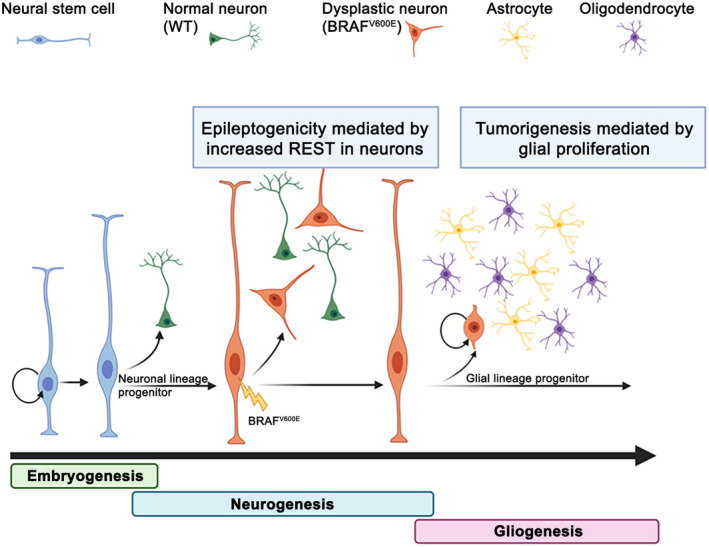
REST‐mediated intrinsic epileptogenicity by BRAF in glioneuronal tumors. Adapted from Dr. Jeong Ho Lee (22) (with permission). When the BRAF V600E variant was introduced into progenitor cells of neuronal lineage, cells were intrinsically epileptogenic. In contrast, tumorigenic properties were attributed to a BRAF V600E variant introduced into glial cells. WT, wild‐type BRAF
3. EPILEPSY GENETICS AND THE BROAD SPECTRUM OF CORTICAL MALFORMATIONS
MCD is the most frequent causes of focal refractory childhood epilepsies, carrying a lifelong disability perspective and reduced quality of life (15, 16). They represent a wide range of cortical lesions resulting from derangements of normal intrauterine developmental processes and involving cells implicated in forming the cortical mantle. The pathological features depend mainly on the defect's timing in the developmental processes and the cause, for example, abnormal proliferation or apoptosis, neuronal migration, or layering, or differentiation. Classification of MCD has proven challenging as sometimes two or more forms of MCD may coexist (e.g., polymicrogyria/PMG may be next to nodular heterotopia/NH (44)). Also, a particular defect in corticogenesis may give rise to more than one morphological subcategory of MCD (e.g., migration defects may cause lissencephaly, focal cortical dysplasia/FCD, PMG, and heterotopia/HET). Conversely, a morphological subtype of MCD may have more than one mechanism for its formation (e.g., PMG may be a consequence of proliferation and migration defects (45)).
Identifying specific gene defects has provided a deeper understanding of the mechanisms involved in brain malformations and epileptogenesis (Table 1). With the ever‐increasing sensitivity of sequencing and new analysis tools available, the epilepsy‐associated genome has been rapidly expanding with the identification of de novo germline variants and, more recently, somatic variants occurring during development (31, 46). In particular, many recent studies using advanced sequencing technologies have revealed that germline and low‐level somatic mutations activating the GATOR1 or mTORC1 pathway account for up to ~60% of hemimegalencephaly (HME) or FCD type 2 characterized by cortical dyslamination plus dysmorphic neurons (FCD type 2A) or balloon cells (FCD type 2B) (29, 47, 48). Hyperactivation of mTOR kinase in a small subset of mouse neocortical excitatory neurons seems sufficient to cause focal seizures as well as cortical dyslamination and dysmorphic neurons that fully recapitulate the phenotypes of FCD type 2 (29, 49, 50, 51).
4. SLC35A2 BRAIN SOMATIC MOSAICISM IN MOGHE
Beyond mTOR signaling, somatic mutations in UDP‐galactose transporter SCL35A2 leading to aberrant N‐glycosylation were reported in FCD1, mMCD, and non‐lesional focal epilepsy (37, 47, 52). Loss of SLC35A2 protein function abolishes the transport of UDP‐galactose from the cytosol into the Golgi apparatus, resulting in the synthesis of truncated glycans in human resected tissues (37). For long only pathogenic de novo germline variants in this X‐linked gene were reported causing a rare type of congenital disorder of glycosylation (CDG) where the majority of affected individuals are primarily characterized by varying degrees of neurological impairments including intellectual disability and epilepsy, and dysmorphisms (53). Recently, somatic pathogenic SLC35A2 mutations were also identified in 9 out of 20 (45%) cases with variable mosaic rates from 7 to 52% with a mild malformation of cortical development with oligodendroglial hyperplasia (MOGHE) phenotype (38). MOGHE is a focal MCD causing childhood‐onset seizures, often refractory to anti‐seizure medication and therefore subjected to epilepsy surgery. It is characterized by the clusters of increased oligodendroglial cell densities, patchy zones of hypomyelination, and heterotopic neurons in the white matter (54). Because of the high frequency of SLC35A2 mutations among the MOGHE population, Bonduelle et al. hypothesized that previously published SLC35A2 cases might also belong to the MOGHE spectrum (Table 1). Histopathological reassessment using NeuN and OLIG2 staining confirmed that 17 previously reported SL35A2 cases, indeed, showed heterotopic neurons and oligodendroglial cell clusters of high density, the two main features characteristic of MOGHE. In addition, analysis by droplet digital PCR of pools of microdissected cells from MOGHE tissue revealed a variant enrichment in clustered oligodendroglial cells and heterotopic neurons, indicating that variants likely occurred in a neuroglial progenitor cell during brain development. The histopathological enrichment of OLIG2‐positive oligodendroglial cells in the white matter and identification of brain somatic SLC35A2 variants are of high specificity to MOGHE and distinguish it from mMCD, FCD1, and non‐lesional focal epilepsy.
5. BRAIN SOMATIC 1q TRISOMY
PMG is a group of highly heterogeneous and difficult to classify MCD (55, 56). Their description is primarily based on their localization (by MRI; e.g., focal, multifocal, or generalized, unilateral or bilateral symmetric/asymmetric) and correlation with clinical aspects including developmental course, growth anomalies, and dysmorphism, seizure history, family history, and genetic testing of blood for PMG‐associated genes (24). Over 40 genes with germline mutations have so far been linked to PMG types and syndromes, but only very recently also brain somatic events were reported. In a series of 26 PMG patients, Kobow et al. identified a group of seven patients exhibiting a unique molecular fingerprint, including the invariable detection of a brain mosaic duplication of the entire long arm of chromosome 1 and a specific genomic DNA methylation signature. This molecular profile was associated with a combination of characteristic clinical features, including a unilateral rather than focal and isolated PMG lesion, early‐onset epilepsy in the first months of life, and severe combinatorial developmental delay, thereby defining a new distinct PMG entity (35). In general, copy number variants are thought to contribute up to 5% of epilepsy cases. Smaller deletion syndromes (e.g., 22q11) had also been identified in PMG, but this was the first report of a brain somatic 1q duplication in PMG. Despite the large size of the chromosomal imbalance, none of the PMG 1q patients displayed HME in contrast to PMG patients without this genetic variant. This may be explained by the gradient distribution of 1q triploid nuclei. About 50% of all cells in the center of the PMG lesion displayed 1q trisomy in FISH analysis, but not in adjacent architecturally normal‐appearing perilesional tissue supporting mosaicism with a clear association to the malformed cortex. Intriguingly, Poduri et al. had previously reported two HME patients without PMG and also brain somatic trisomy of chromosome 1q.(34) The authors argued that the activation of AKT3, either by duplication or by a point mutation (as identified in another HME patient), contributed to hemispheric brain overgrowth. The big structural differences between the described PMG 1q and HME 1q patients in the respective studies (i.e., lesion size, cortical thickness, lamination and gyration, histomorphology) may be related to differences in mutation load and timing of appearance during development together with complex and manifold functions mediated by the vast number of genes linked to chromosome 1q.
6. DNA METHYLATION‐BASED DISEASE CLASSIFICATION IN EPILEPSY
DNA methylation profiling of lesional brain tissue became a promising new option to reliably identify disease entities, particularly for human brain tumors (4). While epigenetic research in developmental biology and cancer has been at the forefront for decades, it was not before early into the millennium that epigenetics became a research focus in epilepsy. In fact, it is only about now that epigenetics is stepping out of a niche with accumulating evidence for epigenetic pathomechanisms in epilepsy and utility of epigenetic marks for disease diagnosis and prediction of disease progression or treatment response. Just about a decade passed since the first report of gene‐specific altered DNA methylation in human TLE associated with distinct histopathology, that is, hippocampal sclerosis and granule cell dispersion (57). Since then, evidence was provided for a targetable key gene regulatory role of DNA methylation in the process of epileptogenesis and chronification of seizures (58) as well as for the etiology‐dependence of such molecular findings in different animal models of epilepsy (59). This finding was validated in structural brain lesions associated with human focal epilepsy, mainly MCDs including major FCD subtypes and PMG (35, 61). These data support the development of a DNA methylation‐based disease classification system also for focal epilepsy, and the high level of standardization of such an approach has great promise to reduce the substantial inter‐observer variability observed in current MCD histopathological diagnostics (61). Significant limitations at this point include conceptual barriers (the field of epigenetics in epilepsy is still in its infancy), the availability of technical infrastructure at epilepsy centers around the globe, and consequently, the restricted number of samples that have yet been analyzed for DNA methylation. Collaborative research studies will be needed to address these issues and to be able to capture rare and extremely rare disease entities (62) as well as to evaluate the predictive value of DNA methylation for, for example, genotype, disease progression, or treatment response.
7. A NEW APPROACH TO MOLECULAR DISEASE DIAGNOSIS AND FUTURE CHALLENGES
In 2016, the World Health Organization provided guidelines for making an integrated diagnosis in a subset of brain tumors. Since then, there have been periodic updates and clarifications reflecting the expanding knowledge on the molecular pathology of brain tumors, but at the same time raising a challenge in rapidly incorporating new molecular findings into diagnostic practice (45, 64). Although in epileptology, we are only about to start accumulating such molecular data, and the precision of identified markers has already been demonstrated, supporting an integrated diagnostic approach in structural brain lesions associated with epilepsy. Our current genetic knowledge of MCD shows some mechanistic overlap with LEAT and other cancers, which is not only limited to the mTOR pathway regulating proliferation, migration, autophagy, metabolism, and cellular functions but also points at a previously unanticipated role for RTK/MAP kinase signaling. This is in line with other findings showing that some candidate genes or genetic variants may be shared between different diseases and phenotypes (64).
Unlike the higher mutational burden often found in brain tumors (VAF thresholds usually >5% (65)), the level of somatic mosaicisms in MCD can be too low to be detected by conventional sequencing technology. Accurate detection of low‐level somatic mosaicism requires high‐depth sequencing and advanced bioinformatic analysis followed by independent validation sequencing (e.g., digital droplet PCR or site‐specific amplicon sequencing, see the review of Khoshkhoo et al. in this mini‐symposium), which might be challenging in most of the current clinical laboratories (66). However, similar to the benefits from the advances in cancer genomics, precise genetic diagnosis of germline and somatic mutations in FCD and MOGHE can provide better genetic consultation of childhood intractable epilepsy and potential therapeutic alternatives based on their molecular genetic profiles (67).
In addition to the sensitivity problem in detecting brain somatic mutations at low allele frequencies, there remains a limited understanding of the consequences of such mutations, particularly their interpretation concerning relevance for seizure development. In the currently accepted pathogenic mechanism for FCD2 and HME, early occurring mutations affecting a progenitor cell in the ventricular zone are associated with HME, whereas late occurring mutations are considered to cause a more focal lesion. Seizures may persist or relapse in up to 40% of patients treated with focal resections for FCD and could originate from the incompletely removed neighboring dysplastic cortex. It has been suggested that MTOR mutations originating before hemispheric cleavage could favor its passing asymmetrically to daughter cells on either side, but only one hemisphere receiving enough mutated cells to develop a macroscopically visible dysplasia (68). Alternatively, a second somatic mutational hit may have occurred, involving a different mTOR pathway gene or any of many other epilepsy genes. This second hit, not present in the resected dysplastic tissue, would be sufficient to activate epileptogenesis but not to determine an MRI‐visible malformation (68). Pelorosso et al. recently showed how a somatic MTOR mutation in the dysplastic hemisphere and a systemic mosaic RPS6 mutation synergistically concurred in determining an HME/epilepsy phenotype in which seizure onset had occurred in the seemingly intact hemisphere after hemispherectomy (69).
To date, a large fraction of FCD2 and MOGHE is caused by somatic mutations that are found exclusively in the brain, restricting the molecular diagnosis to surgical or autopsy specimens. Recently two studies independently provided the proof of principle that somatic mutations may be detected in the cell‐free DNA present in the CSF collected by dural punction during surgery of patients with FCD, GG, or lissencephaly (60, 71). It remains to be proven that even low‐level somatic mutations can be reliably detected from CSF obtained by lumbar punction, but—if reveals to be true—offers tremendous opportunities to obtain a genetic diagnosis before surgery and in patients that are not eligible to surgery (70). This will also allow envisioning targeted therapies acting on the affected neurobiology pathways, that is, galactose supplementation in SLC35A2 cases or mTOR inhibitors in respective FCD cases. Similarly, the identification of epigenetic diagnostic markers, including DNA methylation profiles from peripheral blood, could promote epilepsy diagnosis and prognosis.
ACKNOWLEDGMENTS
Figure 2 was created with BioRender.com. It has been adapted from Koh et al. 2018 (22).
DATA AVAILABILITY STATEMENT
The original contributions referred to in Figure 1 of this review have been provided by AvD and are publicly available. This data can be found in GEO under accession numbers GSE136361 and GSE109381.
REFERENCES
- 1.Sturm D, Orr BA, Toprak UH, Hovestadt V, Jones DTW, Capper D, et al. New brain tumor entities emerge from molecular classification of CNS‐PNETs. Cell. 2016;164:1060–72. [DOI] [PMC free article] [PubMed] [Google Scholar]
- 2.Ramaswamy V, Remke M, Bouffet E, Bailey S, Clifford SC, Doz F, et al. Risk stratification of childhood medulloblastoma in the molecular era: the current consensus. Acta Neuropathol. 2016;131(6):821–31. [DOI] [PMC free article] [PubMed] [Google Scholar]
- 3.Pajtler Kristian W, Witt H, Sill M, Jones David TW, Hovestadt V, Kratochwil F, et al. Molecular classification of ependymal tumors across all CNS compartments, histopathological grades, and age groups. Cancer Cell. 2015;27:728–43. [DOI] [PMC free article] [PubMed] [Google Scholar]
- 4.Capper D, Jones DTW, Sill M, Hovestadt V, Schrimpf D, Sturm D, et al. DNA methylation‐based classification of central nervous system tumours. Nature. 2018;555:469–74. [DOI] [PMC free article] [PubMed] [Google Scholar]
- 5.Deng MY, Sill M, Sturm D, Stichel D, Witt H, Ecker J, et al. Diffuse glioneuronal tumour with oligodendroglioma‐like features and nuclear clusters (DGONC)—a molecularly defined glioneuronal CNS tumour class displaying recurrent monosomy 14. Neuropathol Appl Neurobiol. 2020;46:422–30. [DOI] [PubMed] [Google Scholar]
- 6.Suwala AK, Stichel D, Schrimpf D, Kloor M, Wefers AK, Reinhardt A, et al. Primary mismatch repair deficient IDH‐mutant astrocytoma (PMMRDIA) is a distinct type with a poor prognosis. Acta Neuropathol. 2021;141:85–100. [DOI] [PMC free article] [PubMed] [Google Scholar]
- 7.Wefers AK, Stichel D, Schrimpf D, Coras R, Pages M, Tauziède‐Espariat A, et al. Isomorphic diffuse glioma is a morphologically and molecularly distinct tumour entity with recurrent gene fusions of MYBL1 or MYB and a benign disease course. Acta Neuropathol. 2020;139:193–209. [DOI] [PMC free article] [PubMed] [Google Scholar]
- 8.Koelsche C, Mynarek M, Schrimpf D, Bertero L, Serrano J, Sahm F, et al. Primary intracranial spindle cell sarcoma with rhabdomyosarcoma‐like features share a highly distinct methylation profile and DICER1 mutations. Acta Neuropathol. 2018;136:327–37. [DOI] [PubMed] [Google Scholar]
- 9.Schweizer L, Koelsche C, Sahm F, Piro RM, Capper D, Reuss DE, et al. Meningeal hemangiopericytoma and solitary fibrous tumors carry the NAB2‐STAT6 fusion and can be diagnosed by nuclear expression of STAT6 protein. Acta Neuropathol. 2013;125:651–8. [DOI] [PubMed] [Google Scholar]
- 10.Schwartzentruber J, Korshunov A, Liu XY, Jones DT, Pfaff E, Jacob K, et al. Driver mutations in histone H3.3 and chromatin remodelling genes in paediatric glioblastoma. Nature. 2012;482:226–31. [DOI] [PubMed] [Google Scholar]
- 11.Jones DT, Kocialkowski S, Liu L, Pearson DM, Bäcklund LM, Ichimura K, et al. Tandem duplication producing a novel oncogenic BRAF fusion gene defines the majority of pilocytic astrocytomas. Cancer Res. 2008;68:8673–7. [DOI] [PMC free article] [PubMed] [Google Scholar]
- 12.Hou Y, Pinheiro J, Sahm F, Reuss DE, Schrimpf D, Stichel D, et al. Papillary glioneuronal tumor (PGNT) exhibits a characteristic methylation profile and fusions involving PRKCA. Acta Neuropathol. 2019;137:837–46. [DOI] [PubMed] [Google Scholar]
- 13.Huse JT, Snuderl M, Jones DTW, Brathwaite CD, Altman N, Lavi E, et al. Polymorphous low‐grade neuroepithelial tumor of the young (PLNTY): an epileptogenic neoplasm with oligodendroglioma‐like components, aberrant CD34 expression, and genetic alterations involving the MAP kinase pathway. Acta Neuropathol. 2017;133(3):417–29. [DOI] [PMC free article] [PubMed] [Google Scholar]
- 14.Sievers P, Stichel D, Schrimpf D, Sahm F, Koelsche C, Reuss DE, et al. FGFR1:TACC1 fusion is a frequent event in molecularly defined extraventricular neurocytoma. Acta Neuropathol. 2018;136:293–302. [DOI] [PubMed] [Google Scholar]
- 15.Blumcke I, Spreafico R, Haaker G, Coras R, Kobow K, Bien CG, et al. Histopathological findings in brain tissue obtained during epilepsy surgery. N Engl J Med. 2017;377:1648–56. [DOI] [PubMed] [Google Scholar]
- 16.Lamberink HJ, Otte WM, Blumcke I, Braun KPJ, Writi EEBB, Grp S, et al. Seizure outcome and use of antiepileptic drugs after epilepsy surgery according to histopathological diagnosis: a retrospective multicentre cohort study. Lancet Neurol. 2020;19:748–57. [DOI] [PubMed] [Google Scholar]
- 17.Blumcke I, Aronica E, Becker A, Capper D, Coras R, Honavar M, et al. Low‐grade epilepsy‐associated neuroepithelial tumours—the 2016 WHO classification. Nat Rev Neurol. 2016;12:732–40. [DOI] [PubMed] [Google Scholar]
- 18.Slegers RJ, Blumcke I. Low‐grade developmental and epilepsy associated brain tumors: a critical update 2020. Acta Neuropathol Commun. 2020;8:27. [DOI] [PMC free article] [PubMed] [Google Scholar]
- 19.Blümcke I, Coras R, Wefers AK, Capper D, Aronica E, Becker A, et al. Review: challenges in the histopathological classification of ganglioglioma and DNT: microscopic agreement studies and a preliminary genotype‐phenotype analysis. Neuropathol Appl Neurobiol. 2019;45:95–107. [DOI] [PubMed] [Google Scholar]
- 20.Stone TJ, Keeley A, Virasami A, Harkness W, Tisdall M, Izquierdo Delgado E, et al. Comprehensive molecular characterisation of epilepsy‐associated glioneuronal tumours. Acta Neuropathol. 2018;135:115–29. [DOI] [PMC free article] [PubMed] [Google Scholar]
- 21.Dahiya S, Haydon DH, Alvarado D, Gurnett CA, Gutmann DH, Leonard JR. BRAFV600E mutation is a negative prognosticator in pediatric ganglioglioma. Acta Neuropathol. 2013;125:901–10. [DOI] [PubMed] [Google Scholar]
- 22.Koh HY, Kim SH, Jang J, Kim H, Han S, Lim JS, et al. BRAF somatic mutation contributes to intrinsic epileptogenicity in pediatric brain tumors. Nat Med. 2018;24:1662–8. [DOI] [PubMed] [Google Scholar]
- 23.Goz RU, Akgül G, LoTurco JJ. BRAFV600E expression in neural progenitors results in a hyperexcitable phenotype in neocortical pyramidal neurons. J Neurophysiol. 2020;123:2449–64. [DOI] [PMC free article] [PubMed] [Google Scholar]
- 24.Vasudevaraja V, Rodriguez JH, Pelorosso C, Zhu K, Buccoliero AM, Onozato M, et al. Somatic focal copy number gains of noncoding regions of receptor tyrosine kinase genes in treatment‐resistant epilepsy. J Neuropathol Exp Neurol. 2021;80:160–8. [DOI] [PMC free article] [PubMed] [Google Scholar]
- 25.Jansen LA, Mirzaa GM, Ishak GE, O'Roak BJ, Hiatt JB, Roden WH, et al. PI3K/AKT pathway mutations cause a spectrum of brain malformations from megalencephaly to focal cortical dysplasia. Brain. 2015;138:1613–28. [DOI] [PMC free article] [PubMed] [Google Scholar]
- 26.Lee WS, Baldassari S, Chipaux M, Adle‐Biassette H, Stephenson SEM, Maixner W, et al. Gradient of brain mosaic RHEB variants causes a continuum of cortical dysplasia. Ann Clin Transl Neurol. 2021;8:485–90. [DOI] [PMC free article] [PubMed] [Google Scholar]
- 27.Lim JS, Gopalappa R, Kim SH, Ramakrishna S, Lee M, Kim WI, et al. Somatic mutations in TSC1 and TSC2 cause focal cortical dysplasia. Am J Hum Genet. 2017;100:454–72. [DOI] [PMC free article] [PubMed] [Google Scholar]
- 28.Møller RS, Weckhuysen S, Chipaux M, Marsan E, Taly V, Bebin EM, et al. Germline and somatic mutations in the MTOR gene in focal cortical dysplasia and epilepsy. Neurol Genet. 2016;2:e118. [DOI] [PMC free article] [PubMed] [Google Scholar]
- 29.Ribierre T, Deleuze C, Bacq A, Baldassari S, Marsan E, Chipaux M, et al. Second‐hit mosaic mutation in mTORC1 repressor DEPDC5 causes focal cortical dysplasia‐associated epilepsy. J Clin Invest. 2018;128:2452–8. [DOI] [PMC free article] [PubMed] [Google Scholar]
- 30.Baldassari S, Picard F, Verbeek NE, van Kempen M, Brilstra EH, Lesca G, et al. The landscape of epilepsy‐related GATOR1 variants. Genet Med. 2019;21:398–408. [DOI] [PMC free article] [PubMed] [Google Scholar]
- 31.Marsan E, Baulac S. Review: mechanistic target of rapamycin (mTOR) pathway, focal cortical dysplasia and epilepsy. Neuropathol Appl Neurobiol. 2018;44:6–17. [DOI] [PubMed] [Google Scholar]
- 32.Weckhuysen S, Marsan E, Lambrecq V, Marchal C, Morin‐Brureau M, An‐Gourfinkel I, et al. Involvement of GATOR complex genes in familial focal epilepsies and focal cortical dysplasia. Epilepsia. 2016;57:994–1003. [DOI] [PubMed] [Google Scholar]
- 33.Mirzaa G, Conway R, Graham JM Jr, Dobyns WB, et al. PIK3CA‐related segmental overgrowth. In: Adam MP, Ardinger HH, Pagon RA, Wallace SE, Bean LJH, Stephens K, eds. GeneReviews(®). Seattle: University of Washington; 1993. [Google Scholar]
- 34.Poduri A, Evrony Gilad D, Cai X, Elhosary Princess C, Beroukhim R, Lehtinen Maria K, et al. Somatic activation of AKT3 causes hemispheric developmental brain malformations. Neuron. 2012;74:41–8. [DOI] [PMC free article] [PubMed] [Google Scholar]
- 35.Kobow K, Jabari S, Pieper T, Kudernatsch M, Polster T, Woermann FG, et al. Mosaic trisomy of chromosome 1q in human brain tissue associates with unilateral polymicrogyria, very early‐onset focal epilepsy, and severe developmental delay. Acta Neuropathol. 2020;140:881–91. [DOI] [PMC free article] [PubMed] [Google Scholar]
- 36.Robin NH, Taylor CJ, McDonald‐McGinn DM, Zackai EH, Bingham P, Collins KJ, et al. Polymicrogyria and deletion 22q11.2 syndrome: Window to the etiology of a common cortical malformation. Am J Med Genet Part A. 2006;140A:2416–25. [DOI] [PubMed] [Google Scholar]
- 37.Sim NS, Seo Y, Lim JS, Kim WK, Son H, Kim HD, et al. Brain somatic mutations in SLC35A2 cause intractable epilepsy with aberrant N‐glycosylation. Neurol Genetics. 2018;4(6):e294. [DOI] [PMC free article] [PubMed] [Google Scholar]
- 38.Bonduelle T, Hartlieb T, Baldassari S, Sim NS, Kim SH, Kang HC, et al. Frequent SLC35A2 brain mosaicism in mild malformation of cortical development with oligodendroglial hyperplasia in epilepsy (MOGHE). Acta Neuropathol Commun. 2021;9:3. [DOI] [PMC free article] [PubMed] [Google Scholar]
- 39.Conti V, Carabalona A, Pallesi‐Pocachard E, Parrini E, Leventer RJ, Buhler E, et al. Periventricular heterotopia in 6q terminal deletion syndrome: role of the C6orf70 gene. Brain. 2013;136(Pt 11):3378–94. [DOI] [PubMed] [Google Scholar]
- 40.Guerrini R, Dobyns WB. Malformations of cortical development: clinical features and genetic causes. Lancet Neurol. 2014;13:710–26. [DOI] [PMC free article] [PubMed] [Google Scholar]
- 41.Bandopadhayay P, Ramkissoon LA, Jain P, Bergthold G, Wala J, Zeid R, et al. MYB—QKI rearrangements in angiocentric glioma drive tumorigenicity through a tripartite mechanism. Nat Genet. 2016;48:273–82. [DOI] [PMC free article] [PubMed] [Google Scholar]
- 42.Johnson A, Severson E, Gay L, Vergilio J‐A, Elvin J, Suh J, et al. Comprehensive genomic profiling of 282 pediatric low‐ and high‐grade gliomas reveals genomic drivers, tumor mutational burden, and hypermutation signatures. Oncologist. 2017;22:1478–90. [DOI] [PMC free article] [PubMed] [Google Scholar]
- 43.Rivera B, Gayden T, Carrot‐Zhang J, Nadaf J, Boshari T, Faury D, et al. Germline and somatic FGFR1 abnormalities in dysembryoplastic neuroepithelial tumors. Acta Neuropathol. 2016;131(6):847–63. [DOI] [PMC free article] [PubMed] [Google Scholar]
- 44.Wieck G, Leventer RJ, Squier WM, Jansen A, Andermann E, Dubeau F, et al. Periventricular nodular heterotopia with overlying polymicrogyria. Brain. 2005;128(Pt 12):2811–21. [DOI] [PubMed] [Google Scholar]
- 45.Klingler E, Francis F, Jabaudon D, Cappello S. Mapping the molecular and cellular complexity of cortical malformations. Science. 2021;371(6527):eaba4517. [DOI] [PubMed] [Google Scholar]
- 46.Jamuar SS, Lam A‐TN, Kircher M, D’Gama AM, Wang J, Barry BJ, et al. Somatic mutations in cerebral cortical malformations. N Engl J Med. 2014;371:733–43. [DOI] [PMC free article] [PubMed] [Google Scholar]
- 47.Baldassari S, Ribierre T, Marsan E, Adle‐Biassette H, Ferrand‐Sorbets S, Bulteau C, et al. Dissecting the genetic basis of focal cortical dysplasia: a large cohort study. Acta Neuropathol. 2019;138(6):885–900. [DOI] [PMC free article] [PubMed] [Google Scholar]
- 48.Sim NS, Ko A, Kim WK, Kim SH, Kim JS, Shim KW, et al. Precise detection of low‐level somatic mutation in resected epilepsy brain tissue. Acta Neuropathol. 2019;138:901–12. [DOI] [PubMed] [Google Scholar]
- 49.Hsieh LS, Wen JH, Claycomb K, Huang Y, Harrsch FA, Naegele JR, et al. Convulsive seizures from experimental focal cortical dysplasia occur independently of cell misplacement. Nat Commun. 2016;7:11753. [DOI] [PMC free article] [PubMed] [Google Scholar]
- 50.Kim JK, Cho J, Kim SH, Kang H‐C, Kim D‐S, Kim VN, et al. Brain somatic mutations in MTOR reveal translational dysregulations underlying intractable focal epilepsy. J Clin Investig. 2019;129:4207–23. [DOI] [PMC free article] [PubMed] [Google Scholar]
- 51.Park SM, Lim JS, Ramakrishina S, Kim SH, Kim WK, Lee J, et al. Brain somatic mutations in MTOR disrupt neuronal ciliogenesis, leading to focal cortical dyslamination. Neuron. 2018;99:83–97.e7. [DOI] [PubMed] [Google Scholar]
- 52.Winawer MR, Griffin NG, Samanamud J, Baugh EH, Rathakrishnan D, Ramalingam S, et al. Somatic SLC35A2 variants in the brain are associated with intractable neocortical epilepsy. Ann Neurol. 2018;83:1133–46. [DOI] [PMC free article] [PubMed] [Google Scholar]
- 53.Ng BG, Sosicka P, Agadi S, Almannai M, Bacino CA, Barone R, et al. SLC35A2‐CDG: Functional characterization, expanded molecular, clinical, and biochemical phenotypes of 30 unreported Individuals. Hum Mutat. 2019;40:908–25. [DOI] [PMC free article] [PubMed] [Google Scholar]
- 54.Schurr J, Coras R, Rossler K, Pieper T, Kudernatsch M, Holthausen H, et al. Mild malformation of cortical development with oligodendroglial hyperplasia in frontal lobe epilepsy: a new clinico‐pathological entity. Brain Pathol. 2017;27:26–35. [DOI] [PMC free article] [PubMed] [Google Scholar]
- 55.Oegema R, Barakat TS, Wilke M, Stouffs K, Amrom D, Aronica E, et al. International consensus recommendations on the diagnostic work‐up for malformations of cortical development. Nat Rev Neurol. 2020;16:618–35. [DOI] [PMC free article] [PubMed] [Google Scholar]
- 56.Severino M, Geraldo AF, Utz N, Tortora D, Pogledic I, Klonowski W, et al. Definitions and classification of malformations of cortical development: practical guidelines. Brain. 2020;143(10):2874–94. [DOI] [PMC free article] [PubMed] [Google Scholar]
- 57.Kobow K, Jeske I, Hildebrandt M, Hauke J, Hahnen E, Buslei R, et al. Increased reelin promoter methylation is associated with granule cell dispersion in human temporal lobe epilepsy. J Neuropathol Exp Neurol. 2009;68:356–64. [DOI] [PubMed] [Google Scholar]
- 58.Kobow K, Kaspi A, Harikrishnan KN, Kiese K, Ziemann M, Khurana I, et al. Deep sequencing reveals increased DNA methylation in chronic rat epilepsy. Acta Neuropathol. 2013;126:741–56. [DOI] [PMC free article] [PubMed] [Google Scholar]
- 59.Debski KJ, Pitkanen A, Puhakka N, Bot AM, Khurana I, Harikrishnan KN, et al. Etiology matters—Genomic DNA methylation patterns in three rat models of acquired epilepsy. Sci Rep. 2016;6:25668. [DOI] [PMC free article] [PubMed] [Google Scholar]
- 60.Kim S, Baldassari S, Sim NS, Chipaux M, Dorfmüller G, Kim DS, et al. (2021) Detection of brain somatic mutations in CSF from refractory epilepsy patients. Ann Neurol. 10.1002/ana.26080. [DOI] [PubMed] [Google Scholar]
- 61.Kobow K, Ziemann M, Kaipananickal H, Khurana I, Muhlebner A, Feucht M, et al. Genomic DNA methylation distinguishes subtypes of human focal cortical dysplasia. Epilepsia. 2019;60:1091–103. [DOI] [PMC free article] [PubMed] [Google Scholar]
- 62.Coras R, de Boer OJ, Armstrong D, Becker A, Jacques TS, Miyata H, et al. Good interobserver and intraobserver agreement in the evaluation of the new ILAE classification of focal cortical dysplasias. Epilepsia. 2012;53:1341–8. [DOI] [PubMed] [Google Scholar]
- 63.Perez E, Capper D. Invited Review: DNA methylation‐based classification of paediatric brain tumours. Neuropathol Appl Neurobiol. 2020;46:28–47. [DOI] [PubMed] [Google Scholar]
- 64.Brock S, Cools F, Jansen AC. Neuropathology of genetically defined malformations of cortical development—a systematic literature review. Neuropathol Appl Neurobiol. 2021. 10.1111/nan.12696. [DOI] [PMC free article] [PubMed] [Google Scholar]
- 65.Keller R, Basta R, Salerno L, Elia M. Autism, epilepsy, and synaptopathies: a not rare association. Neurol Sci. 2017;38:1353–61. [DOI] [PubMed] [Google Scholar]
- 66.Sukhai MA, Misyura M, Thomas M, Garg S, Zhang T, Stickle N, et al. Somatic tumor variant filtration strategies to optimize tumor‐only molecular profiling using targeted next‐generation sequencing panels. J Mol Diagn. 2019;21:261–73. [DOI] [PubMed] [Google Scholar]
- 67.Song P, Chen SX, Yan YH, Pinto A, Cheng LY, Dai P, Patel AA, Zhang DY. Detecting and quantitating low fraction DNA variants with low‐depth sequencing. bioRxiv. 2020. 10.1101/2020.04.26.061747. [DOI] [PMC free article] [PubMed] [Google Scholar]
- 68.Witters P, Tahata S, Barone R, Õunap K, Salvarinova R, Grønborg S, et al. Clinical and biochemical improvement with galactose supplementation in SLC35A2‐CDG. Genet Med. 2020;22:1102–7. [DOI] [PMC free article] [PubMed] [Google Scholar]
- 69.Guerrini R, Cavallin M, Pippucci T, Rosati A, Bisulli F, Dimartino P, et al. Is focal cortical dysplasia/epilepsy caused by somatic MTOR mutations always a unilateral disorder? Neurol Genet. 2021;7:e540. [DOI] [PMC free article] [PubMed] [Google Scholar]
- 70.Pelorosso C, Watrin F, Conti V, Buhler E, Gelot A, Yang X, et al. Somatic double‐hit in MTOR and RPS6 in hemimegalencephaly with intractable epilepsy. Human Mol Genet. 2019;28(22):3755–65. [DOI] [PMC free article] [PubMed] [Google Scholar]
- 71.Ye Z, Chatterton Z, Pflueger J, Damiano JA, McQuillan L, Harvey AS, et al. Cerebrospinal fluid liquid biopsy for detecting somatic mosaicism in brain. Brain Commun. 2021;3(1):fcaa235. 10.1093/braincomms/fcaa235. [DOI] [PMC free article] [PubMed] [Google Scholar]
Associated Data
This section collects any data citations, data availability statements, or supplementary materials included in this article.
Data Availability Statement
The original contributions referred to in Figure 1 of this review have been provided by AvD and are publicly available. This data can be found in GEO under accession numbers GSE136361 and GSE109381.