Abstract
目的
采用前瞻性随机对照研究,比较经股内侧肌下微创入路与传统手术入路行人工全膝关节置换术(total knee arthroplasty,TKA)的疗效。
方法
将 2011 年 1 月—2012 年 4 月收治并符合选择标准的 94 例(104 膝)拟行 TKA 的骨关节炎患者纳入研究,随机分为传统入路组(46 例、52 膝)和微创入路组(48 例、52 膝);其中传统入路组 45 例(51 膝)、微创入路组 45 例(49 膝)获得完整随访,纳入最终分析。两组患者性别、年龄、体质量指数、侧别、骨关节炎分级以及术前髋膝踝角(hip-knee-ankle angle,HKA)、美国特种外科医院评分(HSS)、美国膝关节协会评分(KSS)、疼痛视觉模拟评分(VAS)、膝关节活动度等一般资料比较,差异均无统计学意义(P>0.05)。比较两组手术时间、切口长度、总失血量、术后输血例数、住院时间、术后开始行直腿抬高锻炼时间及切口愈合情况;术后膝关节主动活动时 VAS 评分以及膝关节活动度,膝关节功能 HSS 及 KSS 评分;X 线片测量的 HKA、股骨解剖轴线与股骨假体关节面夹角(股骨角)、胫骨解剖轴线与胫骨假体关节面夹角(胫骨角)、股骨假体屈曲角、胫骨平台后倾角。
结果
两组患者均获随访,传统入路组随访时间 60.0~72.5 个月,平均 66.4 个月;微创入路组为 60.0~71.2 个月,平均 65.6 个月。与传统入路组相比,微创入路组切口明显缩短、手术时间延长、患者开始直腿抬高锻炼时间提前,比较差异有统计学意义(P<0.05)。两组总失血量以及术后输血例数、住院时间比较,差异均无统计学意义(P>0.05)。微创入路组并发症发生率为 8.2%(4/49),传统入路组为 0,差异无统计学意义(P=0.054)。术后 1、3 d,微创入路组 VAS 评分及膝关节活动度明显优于传统入路组(P<0.05);14 d 时两组以上指标比较,差异无统计学意义(P>0.05)。术后 1、3、6、12 个月以及 3、5 年,两组 HSS 评分及 KSS 评分比较,差异均无统计学意义(P>0.05)。X 线片复查提示,随访期间假体无松动表现。末次随访时,两组 HKA、股骨角、胫骨角、股骨假体屈曲角以及胫骨平台后倾角测量值及其异常发生率比较,差异均无统计学意义(P>0.05)。
结论
经股内侧肌下微创入路行 TKA 有利于膝关节功能早期恢复,但远期疗效与传统手术入路 TKA 一致;同时因显露范围有限增加了手术并发症发生风险。
Keywords: 人工全膝关节置换术, 手术入路, 微创手术, 随机对照研究
Abstract
Objective
To compare the clinical and radiographic results between primary total knee arthroplasty (TKA) via mini-subvastus or conventional approach through a prospective randomized controlled study.
Methods
Ninety-four patients (104 knees) undergoing primary TKA between January 2011 and April 2012 were evaluated and randomly divided into 2 groups. Forty-six patients (52 knees) underwent TKA via conventional approach (conventional approach group), and 48 patients (52 knees) underwent TKA via mini-subvastus approach (mini-subvastus approach group). In these patients, 45 cases (51 knees) in conventional approach group and 45 cases (49 knees) in mini-subvastus approach group were followed up and recruited in the final analysis. There was no significant difference in age, gender, body mass index, sides, osteoarthritis grading, American Knee Society Score (KSS), Hospital for Special Surgery (HSS) score, visual analogue scale (VAS), range of motion (ROM) of knee between 2 groups (P>0.05). The clinical indexes were recorded and analyzed, including the operation time, length of incision, total blood loss, blood transfusion after operation, hospital stay time, the time of performing straight leg raise, incision condition, VAS score, ROM of knee, HSS score, and KSS score, hip-knee-ankle angle (HKA), femoral anatomic axis and the femoral prosthesis joint angle (femoral angle), tibia anatomic axis and tibial prosthesis joint angle (tibial angle), femoral prosthesis flexion angle (FPFA), and posterior slope angle (PSA) of the tibial plateau.
Results
All patients were followed up. The average follow-up time was 66.4 months (range, 60.0-72.5 months) in conventional approach group and 65.6 months (range, 60.0-71.2 months) in mini-subvastus approach group. Compared with the conventional approach group, the incision of mini-subvastus approach group shortened, the operation time prolonged, and the time when patients started straight leg raise exercises was earlier; showing significant differences between 2 groups (P<0.05). There was no significant difference in hospital stay time, total blood loss, and numbers of blood transfusion between 2 groups (P>0.05). The complication rate was 8.2% (4/49) in mini-subvastus approach group and 0 in conventional approach group, showing no significant difference between 2 groups (P=0.054). At 1 and 3 days, the VAS and the ROM of knee in mini-subvastus approach group were significantly better than those in conventional approach group (P<0.05); but there was no significant difference in above indexes between 2 groups at 14 day (P>0.05). There was no significant difference in HSS and KSS scores between 2 groups at the 1, 3, 6, 12 months and 3, 5 years (P>0.05). X-ray film showed no aseptic loosening in all cases. There was no significant difference in the measured values and incidence of abnormal patient of HKA, femoral angle, tibial angle, FPFA, and PSA at last follow-up between 2 groups (P>0.05).
Conclusion
TKA via mini-subvastus approach is helpful to the early function recovery of knee, but the long-term effectiveness is consistent with TKA via conventional approach. As the limited exposure, TKA via mini-subvastus spproach may has high risk of operative complications.
Keywords: Total knee arthroplasty, surgical approach, minimally invasive operation, randomized controlled study
人工全膝关节置换术(total knee arthroplasty,TKA)是目前治疗晚期骨关节炎的有效方法。TKA 传统手术入路采用膝前正中切口,需要切开股四头肌,翻转髌骨,扰乱和破坏伸膝装置,具有创伤大、患者疼痛明显等不足[1]。为解决以上问题,Hofmann 等[2]首先提出经股内侧肌下微创入路,切口长度仅 8~13 cm,旨在保留伸膝装置完整性并减少对髌骨血供的破坏。由于该入路显露范围有限,术中可能会影响假体安装质量,影响远期疗效,所以对于采用股内侧肌下微创入路 TKA 的临床意义尚存在争议。目前相关报道多为近期疗效,中远期疗效报道较少。为此,安徽医科大学第二附属医院骨科开展了一项为期 5 年的前瞻性随机对照研究,旨在通过与传统手术入路相比,分析经股内侧肌下微创入路能否改善术后早期膝关节功能,有利于实现患者早期功能锻炼,是否会影响假体安装位置和假体的生存质量,以及对中远期临床疗效的影响。报告如下。
1. 临床资料
1.1. 患者选择标准
纳入标准:① 临床结合 X 线片检查诊断为骨关节炎,患者膝关节疼痛且严重影响生活质量,经正规口服药物保守治疗无效;② 初次 TKA 患者;③ 膝关节屈曲畸形<15°,或内翻畸形<15°,或外翻畸形<15°;④ 膝关节活动范围>90°。排除标准:① 合并严重内科疾病,存在手术禁忌证者;② 体质量指数(body mass index,BMI)>30 kg/m2;③ 患膝有手术史;④ 存在潜在感染风险者。
2011 年 1 月—2012 年 4 月收治因骨关节炎拟行初次 TKA 患者共 130 例(148 膝),其中 94 例(104 膝)符合选择标准纳入研究。应用抽签法将患者随机分为传统入路组(46 例、52 膝)和微创入路组(48 例、52 膝)。其中,传统入路组 1 例(1 膝)失访;微创入路组 1 例(1 膝)失访,1 例(1 膝)因关节外疾病(术后 17 个月出现脑血管意外)死亡,1 例(1 膝)术后 1 年因外伤导致假体周围骨折行内固定术。排除以上患者,最终纳入传统入路组 45 例(51 膝)、微创入路组 45 例(49 膝)进行分析。本研究经安徽医科大学第二附属医院伦理委员会批准,患者均知情同意。
1.2. 一般资料
传统入路组:男 21 例(23 膝),女 24 例(28 膝);年龄 58~73 岁,平均 66.5 岁。BMI(26.2±3.5)kg/m2,左膝 19 例,右膝 20 例;双膝 6 例。骨关节炎 Kellgren-Lawrence 分级:Ⅲ级 35 膝,Ⅳ级 16 膝。术前髋膝踝角(hip-knee-ankle angle,HKA)为(0.8±1.0)°。
微创入路组:男 19 例(20 膝),女 26 例(29 膝);年龄 58~70 岁,平均 65.7 岁。BMI(25.3±3.1)kg/m2。左膝 20 例,右膝 21 例;双膝 4 例。骨关节炎 Kellgren-Lawrence 分级:Ⅲ级 32 膝,Ⅳ级 17 膝。术前 HKA 为(1.0±1.1)°。
两组患者性别、年龄、BMI、侧别、骨关节炎分级以及术前 HKA、美国特种外科医院评分(HSS)、美国膝关节协会评分(KSS)、疼痛视觉模拟评分(VAS)、膝关节活动度等一般资料比较,差异均无统计学意义(P>0.05)。
1.3. 手术方法
两组手术均由同一组医生完成。全麻下,患者取仰卧位,患肢上充气止血带后手术,压力为 34.7 kPa;均采用 GEMINII®MKⅡ后稳定型假体(Link 公司,德国)。其中双膝置换者均分为两期手术,手术间隔 1~24 周,平均 7.6 周。
微创入路组:切口起于髌骨上缘内侧,沿髌骨内侧、髌韧带内侧向下,止于胫骨结节内侧;术中根据需要可以向上适当延长。依次切开皮肤、皮下、深筋膜,暴露后沿股内侧肌内下缘作 L 形切口,由后向前切开关节,再向下沿髌旁内侧入路显露关节腔,将股四头肌及髌骨推向外侧,不翻转髌骨,外侧安放拉弧形 Hoffman 拉钩显露关节面。然后,依次完成股骨远端、胫骨近端侧方截骨、股骨远端前后侧及斜面截骨,恢复下肢力线。安装试模后测试,平衡内外侧软组织,“no-thumb”试验评价髌骨运动轨迹满意后,安装假体。放置引流管后关闭切口,弹力绷带加压包扎。
传统入路组:作膝前正中皮肤切口,起于髌上 5~10 cm,向下至胫骨结节内侧,切口长度达 15~20 cm。切口深层常规髌旁内侧入路显露,切口上部沿着股内侧肌外红白交界处切开,下部沿髌骨内侧缘及髌韧带内侧切开,完全向外翻转髌骨。其余手术操作与微创入路组一致。
1.4. 术后处理
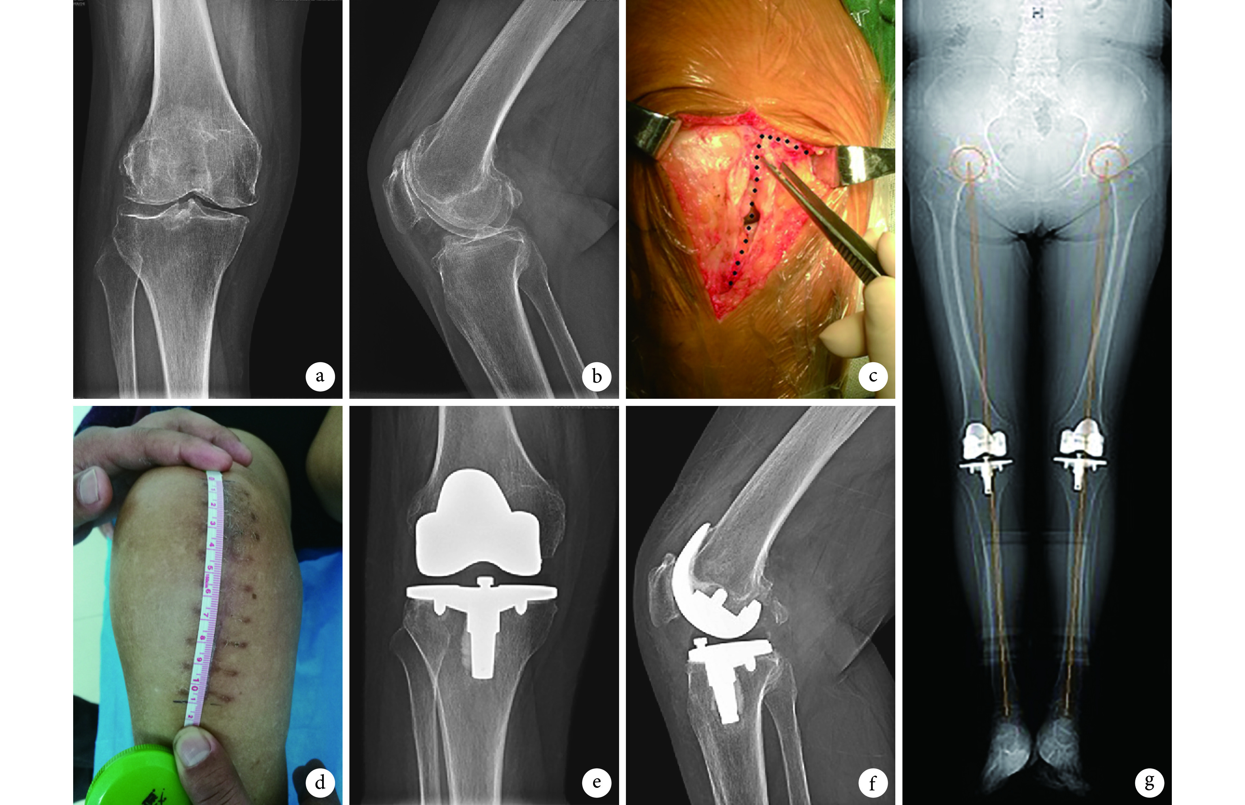
两组患者术后处理方法一致。术后常规预防感染、下肢深静脉血栓形成以及镇痛治疗。术后第 1 天指导患者行膝关节伸屈及伸直抬腿锻炼。术后第 2 天拔除引流管后,指导患者在助步器辅助下行走,之后逐渐负重,1~2 个月后完全负重行走。
1.5. 疗效评价指标
术后 1、3、6、12 个月以及之后每年随访 1 次。记录手术时间、切口长度、总失血量(术中失血量+术后引流量)、术后输血例数、住院时间,术后开始行直腿抬高锻炼时间及切口愈合情况;术后 1、3、14 d 膝关节主动活动时 VAS 评分以及膝关节活动度。膝关节功能采用 HSS 及 KSS 评分评价。
随访时摄双下肢全长及患侧膝关节正侧位 X 线片,测量下肢机械轴线(HKA)、股骨解剖轴线与股骨假体关节面夹角(股骨角)、胫骨解剖轴线与胫骨假体关节面夹角(胫骨角)、股骨假体屈曲角和胫骨平台后倾角,以上指标与术中设定角度差值大于3° 评定为异常病例[3-4]。观察假体有无松动移位,周围有无透亮带。
1.6. 统计学方法
采用 SPSS13.0 统计软件进行分析。计量资料以均数±标准差表示,组间比较采用独立样本 t 检验;计数资料组间比较采用 χ2 检验或 Fisher’s 确切概率法;检验水准 α=0.05。
2. 结果
传统入路组术中 2 例(2 膝)出现髌股关节匹配不理想,给予外侧髌旁软组织松解;微创入路组术中髌股关节均达到良好匹配,无需髌旁软组织松解。与传统入路组相比,微创手术组切口明显缩短、手术时间延长,患者开始直腿抬高锻炼时间提前;比较差异均有统计学意义(P<0.05)。两组总失血量以及术后输血例数、住院时间比较,差异均无统计学意义(P>0.05)。见表 1。传统入路组随访时间 60.0~72.5 个月,平均 66.4 个月;微创入路组为 60.0~71.2 个月,平均 65.6 个月。微创入路组术中 1 例(1 膝)发生髌韧带止点部分撕脱,给予锚钉缝合固定;术后 2 例(2 膝)切口出现水疱渗出,1 例(1 膝)切口出现脂肪液化渗出,经换药后愈合。两组其余患者切口均Ⅰ期愈合。两组均无关节感染、下肢深静脉血栓形成、关节强直、膝关节不稳等并发症发生。微创组并发症发生率为 8.2%(4/49)、传统入路组为 0;两组并发症发生率比较,差异无统计学意义(P=0.054)。
表 1.
Comparison of the clinical indexes between 2 groups
两组患者手术一般资料比较
| 组别
Group |
膝数
n |
切口长度(cm)
Length of incision (cm) |
手术时间(min)
Operation time (min) |
总失血量(mL)
Total blood loss (mL) |
术后输血例数
Number of blood transfusion |
住院时间(d)
Hospital stay time (days) |
开始直腿抬高锻炼时间(d)
Time of performing straight leg raise (days) |
| 传统入路组
Conventional approach group |
51 | 17.1±1.1 | 65.5±6.8 | 374.5±27.6 | 8 | 8.0±1.2 | 3.3±0.4 |
| 微创入路组
Mini-subvastus approach group |
49 | 12.4±0.6 | 75.2±4.0 | 381.8±23.6 | 7 | 7.7±1.5 | 1.9±07 |
| 统计值
Statistic |
t=12 226
P= 0.000 |
t=–7.434
P= 0.000 |
t=–0.763
P= 0.449 |
χ2=0.038
P=0.845 |
t=0.334
P=0.740 |
t=6.014
P=0.015 |
术后 1、3 d,微创入路组 VAS 评分及膝关节活动度明显优于传统入路组,比较差异有统计学意义(P<0.05);14 d 时两组以上指标比较,差异无统计学意义(P>0.05);见表 2。术后 1、3、6、12 个月以及 3、5 年,两组 HSS 评分及 KSS 评分比较,差异均无统计学意义(P>0.05)。见表 3、4。
表 2.
Comparison of the pre- and post-operative VAS score and ROM of knee between 2 groups (
 )
)
两组手术前后 VAS 评分及膝关节活动度比较(
 )
)
| 组别
Group |
膝数
n |
VAS 评分
VAS score |
膝关节活动度(°)
ROM of knee (°) |
|||||||
| 术前
Preoperative |
术后 1 d
One day after operation |
术后 3 d
Three days after operation |
术后 14 d
Fourteen days after operation |
术前
Preoperative |
术后 1 d
One day after operation |
术后 3 d
Three days after operation |
术后 14 d
Fourteen days after operation |
|||
| 传统入路组
Conventional approach group |
51 | 6.3±0.9 | 5.4±1.1 | 6.1±0.9 | 2.9±1.2 | 99.5±4.9 | 40.1±6.8 | 51.8±8.1 | 91.7±4.8 | |
| 微创入路组
Mini-subvastus approach group |
49 | 6.4±1.1 | 3.0±0.7 | 4.9±1.2 | 2.6±0.6 | 98.6±5.9 | 47.2±8.2 | 61.4±10.5 | 93.3±3.3 | |
| 统计值
Statistic |
t=–0.560
P= 0.579 |
t=2.538
P=0.014 |
t=3.698
P=0.001 |
t=0.678
P=0.502 |
t=1.448
P=0.655 |
t=–2.511
P= 0.018 |
t=–3.341
P= 0.022 |
t=–0.338
P= 0.756 |
||
表 3.
Comparison of pre- and post-operative HSS scores between 2 groups (
 )
)
两组手术前后 HSS 评分比较(
 )
)
| 组别
Group |
膝数
n |
术前
Preoperative |
术后 1 个月
One month after operation |
术后 3 个月
Three months after operation |
术后 6 个月
Six months after operation |
术后 12 个月
Twelve months after operation |
术后 3 年
Three years after operation |
术后 5 年
Five years after operation |
| 传统入路组
Conventional approach group |
51 | 65.6±5.9 | 73.1±5.4 | 76.5±5.1 | 80.5±5.6 | 84.3±4.2 | 84.2±3.4 | 83.9±4.1 |
| 微创入路组
Mini-subvastus approach group |
49 | 62.5±5.4 | 75.4±5.1 | 76.2±6.3 | 80.6±4.6 | 83.5±4.5 | 84.9±3.9 | 83.8±3.7 |
| 统计值
Statistic |
t=–1.532
P= 0.136 |
t=–1.412
P= 0.166 |
t=1.259
P=0.216 |
t=–1.006
P= 0.321 |
t=1.142
P=0.261 |
t=–1.105
P= 0.289 |
t=0.846
P=0.403 |
表 4.
Comparison of pre- and post-operative KSS scores between 2 groups (
 )
)
两组手术前后 KSS 评分比较(
 )
)
| 组别
Group |
膝数
n |
术前
Preoperative |
术后 1 个月
One month after operation |
术后 3 个月
Three months after operation |
术后 6 个月
Six months after operation |
术后 12 个月
Twelve months after operation |
术后 3 年
Three years after operation |
术后 5 年
Five years after operation |
| 传统入路组
Conventional approach group |
51 | 117.5±8.4 | 133.5±7.6 | 143.3±7.6 | 162.2±7.3 | 168.5±6.6 | 171.9±8.9 | 168.1±8.2 |
| 微创入路组
Mini-subvastus approach group |
49 | 113.2±8.5 | 136.4±8.2 | 145.8±5.8 | 164.2±6.7 | 170.2±7.5 | 170.4±8.5 | 168.8±9.1 |
| 统计值
Statistic |
t=1.433
P=0.158 |
t=–0.717
P= 0.478 |
t=–0.478
P= 0.816 |
t=–0.525
P= 0.602 |
t=–0.654
P= 0.559 |
t=0.505
P=0.314 |
t=–0.367
P= 0.992 |
X 线片复查示,随访期间假体无松动、移位,假体周围无连续透亮线、骨溶解表现。见图 1。末次随访时,两组 HKA、股骨角、胫骨角、股骨假体屈曲角以及胫骨平台后倾角比较,差异均无统计学意义(P>0.05);其异常发生率比较差异亦无统计学意义(P>0.05)。见表 5。
图 1.
A 63-year-old female patient with bilateral knee osteoarthritis underwent TKA via conventional surgical approach for left knee and via mini-subvastus approach for right knee
患者,女,63 岁,双膝骨关节炎行 TKA,左膝行传统入路、右膝行股内侧肌下微创入路
a、b. 术前右膝正侧位 X 线片;c. 右膝术中 L 形(蓝色虚线)切开髌旁内侧腱膜至股内侧肌下缘;d.术后 1 个月右膝切口外观;e、f. 术后 1 个月右膝正侧位 X 线片;g. 术后 5 年双下肢全长 X 线片
a, b. Anteroposterior and lateral X-ray films of the right knee before operation; c. Dissected from medial margin of patella to border of the vastus medialis for deep tissue, like L-shape (blue dotted line) during operation; d. Appearance of the right knee incision at 1 month after operation; e, f. Anteroposterior and lateral X-ray films of right knee at 1 month after operation; g. Full-length X-ray film of bilateral lower extremities at 5 years after operation

表 5.
Comparison of measured indexes of X-ray film at last follow-up between 2 groups
末次随访时两组 X 线片测量指标比较
| 组别
Group |
膝数
n |
HKA | 股骨角
Femoral angle |
胫骨角
Tibial angle |
胫骨平台后倾角
PSA of tibial plateau |
股骨假体屈曲角
FPFA |
|||||||||
| 测量值
(°) Measured value (°) |
异常发生率
Incidence of abnormal patient |
测量值
(°) Measured value (°) |
异常发生率
Incidence of abnormal patient |
测量值
(°) Measured value (°) |
异常发生率
Incidence of abnormal patient |
测量值
(°) Measured value (°) |
异常发生率
Incidence of abnormal patient |
测量值
(°) Measured value (°) |
异常发生率
Incidence of abnormal patient |
||||||
| 传统入路组
Conventional approach group |
51 | 0.8±0.6 | 3.9% (2) | 96.3±1.8 | 1.9% (1) | 87.7±2.2 | 3.9% (2) | 5.8±1.0 | 1.9% (1) | 2.8±0.9 | 0 (0) | ||||
| 微创入路组
Mini-subvastus approach group |
49 | 1.2±0.9 | 8.2% (4) | 95.8±2.2 | 4.1% (2) | 87.5±1.7 | 8.2% (2) | 5.4±0.8 | 6.1% (3) | 3.3±1.3 | 2.4% (1) | ||||
| 统计值 Statistic |
t=–1.594
P= 0.121 |
χ2=0.223
P=0.372 |
t=1.554
P=0.129 |
χ2=0.001
P=0.972 |
t=0.236
P=0.814 |
χ2=0.000
P=1.000 |
t=0.643
P=0.524 |
χ2=0.304
P=0.581 |
t=–1.417
P= 0.165 |
P=0.490 | |||||
3. 讨论
3.1. 微创 TKA 技术
微创 TKA 具有减少软组织损伤,减轻患者术后疼痛和利于功能恢复等优势,临床应用越来越多。目前,膝关节微创手术主要采用微创内侧髌旁入路、股内侧肌下入路、经股内侧肌入路、股四头肌保留入路等[5-6]。以上入路均遵循以下原则:① 保护伸膝装置;② 术中屈膝显露时不翻转髌骨;③ 术中选择相对较短的切口(一般认为小于 15 cm);④ 术中避免关节脱位,实现原位截骨。但微创手术术中视野相对较小,不便于操作,术中可能会出现韧带撕裂和股骨髁骨折,本研究中微创组 1 例患者术中出现髌韧带止点部分撕脱。我们结合临床实践总结了以下注意事项:① 充分松解关节囊,使推移牵开髌骨更容易;② 先股骨远端截骨或股骨端截骨,才能充分显露胫骨侧;③ 术中采用伸屈膝关节方法,以达到尽可能显露关节面的目的;④ 术中胫骨平台后外侧难以显露,可以挑选或自行制备双叉式 Hoffman 拉钩,充分阻挡外侧软组织和髌骨,利于术中安全截骨;⑤ 假体安装完成后,选择合适工具彻底清除周围残留骨水泥。
3.2. 微创手术适应证
微创 TKA 手术适应证包括既往无膝关节外伤及手术史、BMI <30~35 kg/m 2、无严重内外翻畸形(<15°))、膝关节活动度>90°[7-8]。此外,患者如合并以下情况则不宜选择微创手术:① 髌骨畸形及低位髌骨者,此类患者术中髌骨推开困难;② 股骨髁及胫骨平台宽大者,可能发生假体安装困难;③ 类风湿性骨关节炎患者,尤其合并骨质疏松者,术中容易发生髌韧带及侧副韧带撕脱。
3.3. 微创手术疗效分析
股内侧肌下微创入路可以充分保护伸膝装置,促进股四头肌功能恢复[9],同时保护了股内侧肌,而股内侧肌是膝关节伸直过程中唯一限制髌骨外移的肌肉,因此采用该入路术后可获较理想的髌骨活动轨迹[10-11]。早期文献报道,微创 TKA 与传统 TKA 相比不仅无明显优势,反而会增加手术时间、出血和感染风险[12-13]。但随着微创技术的发展,大量研究发现微创手术有利于术后患者早期康复。van Hemert 等[14]报道与传统髌旁内侧入路相比,股内侧肌下微创入路术后 3 d 切口 VAS 评分较低,但术后 2 周膝关节功能评分无明显差异。Boerger 等[15]研究发现,股内侧肌下微创入路术后第 1 天 VAS 评分较传统入路明显降低,有利于术后患者早期进行直腿抬高锻炼,但术后 1 个月两组关节活动度无明显差异。Koh 等[16]进行了自身对照研究,对一组行双膝关节置换患者,一侧应用股四头肌下入路,另一侧行传统髌旁内侧入路,微创组术后 1 周股四头肌等长收缩力量明显优于传统组,但其他评价指标,如疼痛评分和功能评分,即使在术后早期两组也无明显差异。
TKA 术后疼痛是影响术后早期功能锻炼的重要因素,本研究中微创入路组术后 1、3 d 时 VAS 评分明显低于传统入路组,术后疼痛减轻主要与微创入路保护了深层软组织有关。微创入路组患者在术后 3 d 内即可完成直腿抬高功能锻炼,较传统入路组显著缩短,与文献报道结果类似[17]。但两组术后 1 个月 HSS 评分及 KSS 评分差异均无统计学意义,提示微创手术早期疼痛减轻有利于患者功能锻炼,但远期功能恢复并无明显差异。
另外微创入路组手术时间明显延长,分析原因可能与术者早期微创技术欠熟练有关。微创入路组切口虽明显缩短,但术后 2 例切口出现水疱渗出,1 例出现脂肪液化渗出,考虑为术中过度牵拉软组织导致,并发症发生率高于传统入路组,虽然差异无统计学意义,但也值得临床医师注意。两组总出血量及输血例数比较差异无统计学意义。
3.4. 微创手术假体安放位置
微创手术视野不佳可能对术中截骨和假体放置造成一定影响[18],但多数学者认为术中正规操作可以避免上述问题。Flören 等[19]对 74 例微创 TKA 患者随访 1 年,结果显示假体位置及下肢力线良好,下肢力线角度与传统手术患者无显著差异,作者认为微创手术可以达到满意的假体位置。Bonutti 等[20]对 51 例患者(102 膝)分别行股内侧肌下和经股内侧肌入路人工全膝关节表面置换术,术后2年随访发现两组假体安装角度及下肢力线满意。本研究中末次随访时两组患者下肢力线、假体位置均无明显差异。虽然两组均存在假体位置异常病例,但发生率比较差异无统计学意义。
综上述,经股内侧肌下微创入路 TKA 保护了伸膝装置,能减轻术后疼痛,有利于患者术后早期功能锻炼,但由于显露范围有限,增加了手术并发症发生风险,远期膝关节功能与传统入路手术相比并无明显差异。但本研究患者数量有限,且微创入路组患者均为早期收治,存在术者操作技术相对不熟练等问题,可能影响结果;其次患者术中常规给予镇痛药物,对术后疼痛评分有一定影响。因此,本研究结论尚有待进一步完善明确。
Funding Statement
安徽省自然科学基金面上项目(1608085MH167)
Natural Science Foundation of Anhui Province (1608085MH167)
References
- 1.Vaishya R, Vijay V, Demesugh DM, et al Surgical approaches for total knee arthroplasty. J Clin Orthop Trauma. 2016;7(2):71–79. doi: 10.1016/j.jcot.2015.11.003. [DOI] [PMC free article] [PubMed] [Google Scholar] [Retracted]
- 2.Hofmann AA, PIaster RL, Murdock LE Subvastus (Southern) approach for primary total knee arthroplasty. Clin Orthop Relat Res. 1991;(269):70–77. [PubMed] [Google Scholar]
- 3.Hasegawa M, Miyazaki S, Yamaguchi T, et al Comparison of midterm outcomes of minimally invasive computer-assisted vs minimally invasive jig-based total knee arthroplasty. J Arthroplasty. 2017;32(1):43–46. doi: 10.1016/j.arth.2016.07.005. [DOI] [PubMed] [Google Scholar]
- 4.Verburg H, Mathijssen NM, Niesten DD, et al Comparison of mini-midvastus and conventional total knee arthroplasty with clinical and radiographic evaluation: A prospective randomized clinical trial with 5-year follow-up. J Bone Joint Surg (Am) 2016;98(12):1014–1022. doi: 10.2106/JBJS.15.00654. [DOI] [PubMed] [Google Scholar]
- 5.Yoo JH, Park SH, Han CD, et al Radiologic outcomes according to varus deformity in minimally invasive surgery total knee arthroplasty. Yonsei Med J. 2016;57(1):225–231. doi: 10.3349/ymj.2016.57.1.225. [DOI] [PMC free article] [PubMed] [Google Scholar]
- 6.Layher F, Zipfel M, Sander K, et al Functional comparison of the outcome after midvastus and medial parapatellar surgical approach in total knee arthroplasty. Z Orthop Unfall. 2016;154(1):50–57. doi: 10.1055/s-0035-1558074. [DOI] [PubMed] [Google Scholar]
- 7.Huang AB, Wang HJ, Yu JK, et al Are there any clinical and radiographic differences between quadriceps-sparing and mini-medial parapatellar approaches in total knee arthroplasty after a minimum 5 years of follow-up? Chin Med J (Engl) 2015;128(14):1898–1904. doi: 10.4103/0366-6999.160521. [DOI] [PMC free article] [PubMed] [Google Scholar]
- 8.曾建伟, 沈彬, 杨静, 等 微创切口与传统切口对全膝关节置换术后切口外侧皮肤感觉障碍影响的对比研究. 中华解剖与临床杂志. 2015;20(1):1–6. [Google Scholar]
- 9.Unwin O, Hassaballa M, Murray J, et al Minimally invasive surgery (MIS) for total knee replacement; medium term results with minimum five year follow-up. Knee. 2017;24(2):454–459. doi: 10.1016/j.knee.2017.01.010. [DOI] [PubMed] [Google Scholar]
- 10.Huang AB, Wang HJ, Yu JK, et al Optimal patellar alignment with minimally invasive approaches in total knee arthroplasty after a minimum five year follow-up. Int Orthop. 2016;40(3):487–492. doi: 10.1007/s00264-015-2896-2. [DOI] [PubMed] [Google Scholar]
- 11.Teng Y, Du W, Jiang J, et al Subvastus versus medial parapatellar approach in total knee arthroplasty: meta-analysis. Orthopedics. 2012;35(12):e1722–1731. doi: 10.3928/01477447-20121120-16. [DOI] [PubMed] [Google Scholar]
- 12.Whiteside LA Mini incision: occasionally desirable, rarely necessary: in the affirmative. J Arthroplasty. 2006;21(4 Suppl 1):16–18. doi: 10.1016/j.arth.2006.02.098. [DOI] [PubMed] [Google Scholar]
- 13.Cuckler JM The ugly underbelly of the MIS movement: in the affirmative. J Arthroplasty. 2007;22(4 Suppl 1):99–101. doi: 10.1016/j.arth.2006.12.055. [DOI] [PubMed] [Google Scholar]
- 14.van Hemert WL, Senden R, Grimm B, et al Early functional outcome after subvastus or parapatellar approach in knee arthroplasty is comparable. Knee Surg Sports Traumatol Arthrosc. 2011;19(6):943–951. doi: 10.1007/s00167-010-1292-0. [DOI] [PMC free article] [PubMed] [Google Scholar]
- 15.Boerger TO, Aglietti P, Mondanelli N, et al Mini-subvastus versus medial parapatellar approach in total knee arthroplasty. Clin Orthop Relat Res. 2005;(440):82–87. doi: 10.1097/01.blo.0000185755.09777.2d. [DOI] [PubMed] [Google Scholar]
- 16.Koh IJ, Kim MW, Kim MS, et al The Patient's perception does not differ following subvastus and medial parapatellar approaches in total knee arthroplasty: A simultaneous bilateral randomized study. J Arthroplasty. 2016;31(1):112–117. doi: 10.1016/j.arth.2015.08.004. [DOI] [PubMed] [Google Scholar]
- 17.Shah NA, Jain NP Total knee arthroplasty in valgus knees using minimally invasive medial-subvastus approach. Indian J Orthop. 2016;50(1):25–33. doi: 10.4103/0019-5413.173517. [DOI] [PMC free article] [PubMed] [Google Scholar]
- 18.Yuan FZ, Wang SJ, Zhou ZX, et al Malalignment and malposition of quadriceps-sparing approach in primary total knee arthroplasty: a systematic review and meta-analysis. J Orthop Surg Res. 2017;12(1):129. doi: 10.1186/s13018-017-0627-7. [DOI] [PMC free article] [PubMed] [Google Scholar]
- 19.Flören M, Reichel H, Davis J, et al The mini-incision mid-vastus approach for total knee arthroplasty. Oper Orthop Traumatol. 2008;20(6):534–543. doi: 10.1007/s00064-008-1509-2. [DOI] [PubMed] [Google Scholar]
- 20.Bonutti PM, Zywiel MG, Ulrich SD, et al A comparison of subvastus and midvastus approaches in minimally invasive total knee arthroplasty. J Bone Joint Surg (Am) 2010;92(3):575–585. doi: 10.2106/JBJS.I.00268. [DOI] [PubMed] [Google Scholar]


