Abstract
Aims
The main objective of this study is to analyze the penetration of bone cement in four different full cementation techniques of the tibial tray.
Methods
In order to determine the best tibial tray cementation technique, we applied cement to 40 cryopreserved donor tibiae by four different techniques: 1) double-layer cementation of the tibial component and tibial bone with bone restrictor; 2) metallic cementation of the tibial component without bone restrictor; 3) bone cementation of the tibia with bone restrictor; and 4) superficial bone cementation of the tibia and metallic keel cementation of the tibial component without bone restrictor. We performed CT exams of all 40 subjects, and measured cement layer thickness at both levels of the resected surface of the epiphysis and the endomedular metaphyseal level.
Results
At the epiphyseal level, Technique 2 gave the greatest depth compared to the other investigated techniques. At the endomedular metaphyseal level, Technique 1 showed greater cement penetration than the other techniques.
Conclusion
The best metaphyseal cementation technique of the tibial component is bone cementation with cement restrictor. Additionally, if full tibial component cementation is to be done, the cement volume used should be about 40 g of cement, and not the usual 20 g.
Cite this article: Bone Joint Res 2021;10(8):467–473.
Keywords: Total knee arthroplasty, Cementation techniques
Keywords: tibial components, bone cement, tibial tray’s, tibial bone, knee arthroplasty, epiphysis, metaphysis, t-test, bone plugs, tourniquets
Article focus
This study investigated different cementation techniques to discover which represented a better cement penetration level.
Key messages
The usual quantity of 20 g of cement used for tibial tray fixation in knee prostheses may not suffice for complete cementation.
The use of tibial intramedullary bone restrictor in primary knee prostheses improves metaphyseal cementation.
The interdigitation of cement at the tibial metaphysis level is greater with the double bone and metal cementation of tibial keel.
Strengths and limitations
One possible limitation of this study is that the bone densities of the study specimens are homogeneous.
As noteworthy data, we indicate that our study is pioneering in exploring cement penetration at the metaphyseal level, and in introducing the evaluation of bone plugs in prosthetic knee surgery.
Introduction
The aseptic loosening of tibial components remains the first cause of failure of total knee arthroplasty (TKA). Hence, one of the main aspects to bear in mind during this surgery is the correct fixation of the tibial tray to bone.1-4 Some authors prefer tibial component complete cementation to reduce micromovements at the cement-bone interface, which reduces patient pain, and to increase prosthesis longevity.1-3 Others prefer only cementing the inferior surface of tibial component, and avoiding not only increasing bone loss during possible revision surgery, but also reducing stress shielding on the tibial tray surface.5-11
Surgical bone cement is not only a glue used to adhere the prosthesis to bone, but also a complex entity that produces mechanical binding through interdigitation with the bone trabeculae.12 If this process is not properly followed, micromovements between interfaces could cause the prosthesis to mechanically loosen.13,14 The micromovement of tibial component depends on cement penetration into bone, and even a specific technicality such as the rapid change in viscosity can affect bone penetration. One study has shown that ideal cement penetration in spongy bone tissue under the tibial tray is between 3 mm and 5 mm.5 Cement interdigitation under 3 mm has been deemed vulnerable because it predisposes the interface to micromovements, whereas penetration less than 1.5 mm could lead to the generation of radiolucent lines. Excessive cement penetration over 5 mm could correlate with higher bone necrosis rates due to the exothermic reaction occurring while the cement’s compounds harden, which can then reach 110°C.5 As every surgeon deals with this problem differently, it might be necessary to design new studies to prove which technique offers the best results and thereby homogenate clinical practice in this domain.15,16 Therefore, the purpose of the present study was to analyze differences in cement penetration by comparing four distinct full cementation techniques of the tibial tray in cadaveric specimens.
Methods
Participants
The study required 40 cryogenized cadaver tibiae. In order to carry out this study, all the experiments were followed in accordance with the Ethics Committee of the University Clinical Hospital of Valencia and with the approval of the Ethics Committee of the University of Valencia for experimentation with human biological samples.
Materials
The implant selected for this study was the GENUTECH tibial tray of the primary endoprosthesis of the knee trademarked Surgival (Spain), fabricated of a titanium (Ti6Al4V) alloy. This prosthesis, in addition to having high availability thanks to its manufacturer’s proximity, has shown very good results.16 The tibial tray has a metallic ridge of 0.75 mm on the inferior side, designed to facilitate cement penetration into the bone structure. Its stem (3.75 cm long) is modular in design, which facilitates the insertion of a shaft into its inferior end, which may vary in length. In this case, only the shortest shaft (15 mm) was used with all the subjects. This stem is designed to sit in a significant cement mantle. Both the tray and stem were completely cemented in all cases. The distribution of the anatomical pieces for all the experiments was completely random. Four groups were established per cementation technique and each group had ten individuals/implants.
The chemical compound employed as surgical bone cement in the experiment was polymethylmethacrylate (PMMT), trademarked Type 1 high-viscosity by Surgival. It was stored beforehand and prepared under controlled conditions at an average temperature of 20°C and 70% relative humidity. High viscosity cement was used, instead of another type of viscosity, on the recommendation of the prosthesis manufacturer. This is a cement that sets at 12 minutes.
Cementation techniques
A graphic representation of the four techniques is seen in Figure 1.
Fig. 1.
a) Technique 1 – Double cementation with bone restrictor. b) Technique 2 – Metallic cementation without bone restrictor. c) Technique 3 – Bone cementation with bone restrictor. d) Mixed cementation, superficial bone, and metaphyseal metallic without bone restrictor.
Technique 1. Double cementation (bone and metallic surfaces) with bone restrictor (Figure 1a): 20 g of bone cement was manually applied to the bone bed carved for the keel and on the resected epiphyseal surface of the tibial bone; 20 g was also applied to the inferior surface of the tibial tray and to the tibial shaft. Before applying cement, the intramedullary cavity was sealed by a bone plug obtained from the previous bone resection of the articular tibial surface.
Technique 2. Metallic cementation (metallic surfaces) without bone restrictor (Figure 1b): 20 g of bone cement was directly applied to the inferior metallic surface of the tibial tray and to the tibial shaft. The intramedullary cavity was not sealed with a bone plug.
Technique 3. Bone cementation (bone surfaces) with bone restrictor (Figure 1c): 20 g of bone cement was manually applied to both the interior of the medullary cavity carved for the tibial stem and the resected epiphyseal surface of the tibial bone. Before introducing cement, the intramedullary cavity was sealed by a bone plug obtained from the previous resected articular surface.
Technique 4. Mixed cementation (epiphysis bone and metallic keel and stem surfaces) without bone restrictor (Figure 1d): 20 g of bone cement was manually applied to both the resected epiphyseal surface of the tibial bone and the metallic stem of the tibial component of the prosthesis. The intramedullary cavity was not sealed by a bone plug.
There was no indication for a bone restrictor in techniques 2 and 4, because they entailed no risk of the cement material migrating inside the medullary channel.
It should be mentioned that, since this was a cadaver study, no tourniquet was used. However, in a usual clinical practice, tourniquets are used because it is thought that cementation (key point of implant survival) improves if the operated limb is kept bloodless, as supported by most authors. However, recent research advises against the use of a tourniquet in primary TKA and using intravenous tranexamic acid instead, thus reducing total blood loss and hidden blood loss.17 In our study, negative intramedullary pressure was not used to aspirate the cement.
Experimental procedures
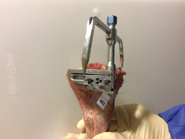
After defrosting the study pieces for 48 hours at a constant temperature of 22°C, they were measured from the articular bone surface of the medial tibial plateau and then cut at the proximal metaphysis at a depth of 6 mm. In order to standardize osteotomy, an intramedullary guide was used, as was the resection guide that came with the instrument set prepared for the endoprosthesis (Figure 2). As the intramedullary guide was used, we considered it necessary to introduce a bone restrictor to prevent cement from leaking into the medullary canal.
Fig. 2.
Tibial cutting block set with pins at + 4 mm to achieve a tibial proximal metaphysis cut at a depth of 6 mm, measured from the articular bone surface of the medial tibial plateau.
The bone bed for the stem tibial component was carved into the tibial metaphysis using the impactor of the surgical kit for the Genutech prosthesis. Cement was manually prepared without using a vacuum, and always following manufacturer’s instructions. It was applied in a paste-like state to the surgical bed after four minutes. Neither jet lavage or pressure cementation was employed during the procedure.
Tibial trays were covered by a layer of transparent film to ensure that cement would not adhere to metallic parts, and to also facilitate extraction after the compound hardened (14 minutes). Each tibial component was impacted 12 times to guarantee correct and complete bone setting.
In order to achieve a cement-bone interface free of bone debris and blood, bone lavage with a pulsating gun, a pressurized tourniquet for selective limb ischaemia, or a negative pressure intrusion was used.
Radiological study
Each bone piece was studied by CT, performed with a 'Prime Aquilion' CT scanner model (Canon Medical Systems, UK). Central coronal cuts were taken of each sample. All the images were analyzed with the Vitrea software (Canon Group) at wavelengths 900/50.
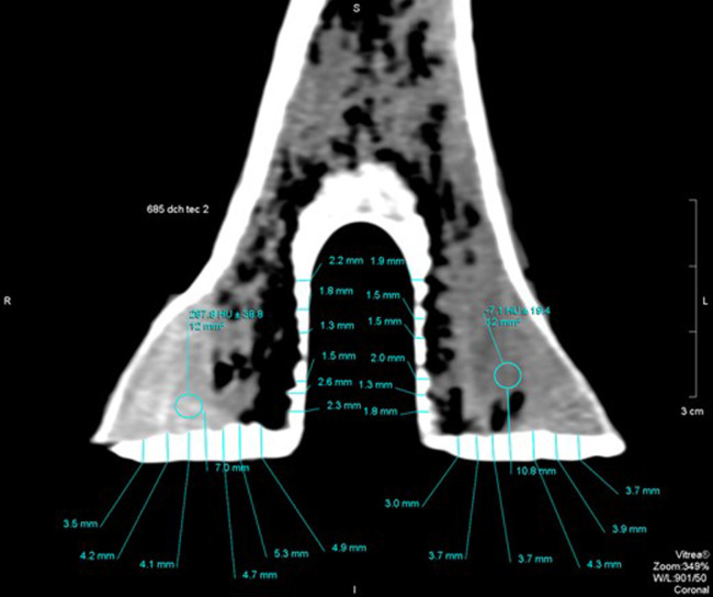
Two specific areas were defined in every image: the superficial epiphysial area (SEA) and the endomedular metaphyseal area (EMA). In the SEA, 12 measurements were taken by screening medially to laterally, six on the medial tibial plateau and six on the lateral tibial plateau, and at similar intervals (S1 to S12). In the EMA, measurements were similarly planned: six from the surface towards the medial interface depth (M1 to M6) and six more in the lateral zone from the surface towards the depth in similar intervals (M7 to M12) (Figure 3).
Fig. 3.
Central coronal CT slide of one of the studied samples. Measurement of the thickness of both the superficial and the metaphyseal cement-bone interfaces.
Statistical analysis
Data were analyzed and represented in graphs using the R software, version 3.4.2 (R Core Team 2017; R Foundation for Statistical Computing, Austria). A pairwise t-test was run for each cementation technique. The data obtained with the CT scanner were processed separately by splitting the data of Coronal SEA (cementation on epiphysial surface) and Coronal EMA (cementation on tibial metaphysis).
Descriptive statistics were given as mean and standard deviation (SD). The four cementation techniques were compared by Welch’s t-test as variances were not considered equal, and the p-values were adjusted in the multiple comparisons by Holm’s method. In each study group, 120 values were compared, and testing normality was not necessary. Data were analyzed for variances homogeneity (Levène’s test). A value of p < 0.05 was considered statistically significant for all the comparisons.
Results
Superficial epiphyseal area
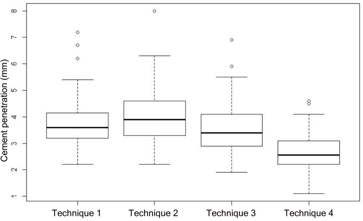
After analyzing the obtained data, the technique with the deepest penetration at the epiphysis cut was Technique 1, with a thickness of 4.056 mm (SD 0.954). Technique 2 and Technique 4 scored second and third with 3.758 mm (SD 0.841) and 3.59 mm (SD 0.887), respectively. Lastly, Technique 3 had the lowest results with 2.683 mm (SD 0.683) (Figure 4).
Fig. 4.
Box plot of the results obtained from the superficial epiphyseal cementation. The means and standard deviations of the cement penetrations in Techniques 1, 2, 3, and 4 are illustrated.
All four techniques were compared by Welch’s t-test and a value of less than 2.2e-16 (p < 0.05) was obtained. This allowed the assumption that the presented techniques induced different outcomes. Of the multiple regressions, the only non-significant one was that which compared techniques 1 and 4 (p > 0.05).
Endomedular metaphyseal area
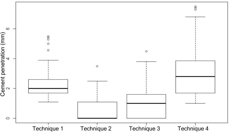
After reviewing the data obtained on cement penetration at the prosthesis stem height, the technique with the deepest penetration was Technique 1, with a thickness of 3.019 mm (SD 1.483), followed by Technique 3 with 2.274 mm (SD 0.880). Techniques 4 and 2 obtained the lowest scores with 0.921 mm (SD 0.962) and 0.553 mm (SD 0.752), respectively (Figure 5).
Fig. 5.
Box plot of the results obtained from the metaphyseal cementation. The means and standard deviations of the cement penetrations in Techniques 1, 2, 3, and 4 are illustrated.
Welch’s t-test was once again conducted to compare the four techniques, and statistically significant values of 2.2e-16 were obtained. This allowed the assumption that the compared techniques induced different outcomes. In this case, all the performed multiple regressions were statistically significant (p < 0.05).
Discussion
In this study, we explored four cementation techniques to analyze if there were any differences in cement penetration. The analyses revealed that at the SEA level, Technique 1, which is based on double cementation (bone and metallic surfaces) with bone restrictor, showed the deepest penetration, followed by Techniques 3 (bone cementation with restrictor), 4 (mixed cementation without restrictor), and 2 (metallic cementation without restrictor) in this order. The cement penetration differences were statistically significant among all the techniques (p < 0.05), except between Techniques 1 and 4 (p > 0.05). The analyses also revealed that at EMA level, Technique 1 showed the deepest penetration, followed by Techniques 2, 4, and 3 in this order. These cement penetration differences were statistically significant among all the techniques (p < 0.05). All methods achieved superficial cement penetration.
Firstly, the need for total cementation is discussed given the controversy among research groups. Two main groups postulate the following theses: the first defends total tibial tray cementation, including the stem (complete cementation),1-3 whereas the second favours only the cementation of the inferior surface of the tibial tray (superficial cementation).5-11 Some authors refer to following prosthesis stem cementation (complete cementation), which would diminish bone density under the tibial tray’s surface as a result of Wolff’s Law, which could lead to premature tibial tray loosening.6,7 Furthermore, bone loss would be greater for a full cemented prosthesis.9,10 Moreover, full cemented prosthesis increases the cement’s interdigitation area, which would reduce the micromovements of the interface by providing the implant with additional stability, which could increase prosthesis longevity and reduce postinterventional pain.1-3
Some studies have reviewed cement penetration in bone tissue and showed that, in order to achieve biomechanical stability, the interface thickness should lie between 3 mm and 5 mm. Interdigitation thinner than 3 mm could lead to a vulnerable bone-cement interface and predispose to micromovements, which would favour the appearance of radiolucent lines. However, a cement mantle thicker than 5 mm could render bone tissue prone to develop necrosis due to the exothermic reaction taking place while the cement hardens, which can reach up to 110°C and thus reduce prosthesis longevity.5
Differences also appear among the research groups that work with bone phantoms: the group of Vanlommel et al5 concluded that double superficial cementing (cementation of the inferior surface of the metallic tibial tray and the epiphyseal bone surface of the tibia) was the ideal technique to allow an interface thickness between 3 mm and 5 mm. Pérez et al15 concluded that the digital pressurization technique on the tibial bone surface achieved greater cement penetration around the stem than the technique in which cement was applied directly to the inferior surface of the tibial tray.
Our results confirm the work of Grupp et al8 and Peters et al11 on cadaveric bones, as they show that the technique with the best horizontal cementation outcome was double cementation (bone and metallic surfaces) with bone restrictor (Technique 1).
Secondly, based on the results of this study, it is noteworthy that the results in the horizontal cementation of Technique 3 (bone cementation with restrictor) differed vastly from those of Technique 4 (mixed cementation without restrictor), but their execution was relatively similar. The reason for such marked disparity could be due to more cement being used inside the carved space for the metallic keel in Technique 3, which would leave less to fill the tibial epiphyseal surface. Bearing this in mind, the complete metaphyseal cementation with restrictor (Technique 3) should be complemented with larger quantities of cement (30 g to 40 g) because metaphysis cementation entails employing a considerable amount of cement.
Thirdly, in the techniques in which cement is directly applied to the metallic surface of the stem (Techniques 2 and 4), penetration into the metaphyseal bone was fairly poor, especially in Technique 2, where cement thickness barely reached 0.76 mm. One possible explanation could lie in the tibial tray being impacted in bone, and the cement is pulled towards the surface instead of into the metaphysis as a result of bone friction. In the techniques in which surgical bone cement is firstly applied to the carved bone cavity (Techniques 1 and 3), cement penetration into metaphyseal bone was good. One possible explanation for this could be that when the tibial tray is impacted into bone, cement has no escape route due to both the bone restrictor and the metallic keel being pushed into the metaphyseal bone trabeculae.
Furthermore, as Technique 1 (double cementation with bone restrictor) uses twice the amount of cement than the other techniques, it ensures deeper cement penetration into the bone structure, with acceptable widths at both the epiphyseal and metaphyseal levels. This makes it an admissible technique for adequate cementation, although it requires larger surgical bone cement volumes.
Finally, we conclude that the full cementation technique on the tibial tray and metallic stem can obtain good bone cement interdigitation at the epiphyseal surface, but rather poor interdigitation at the metaphyseal level. This outcome could be explained by the escaping effect during tibial implant impaction. For this reason, if the intention is to cement the stem, direct endomedular metaphysis cementation should take place instead of cementing the tibial keel and stem. Additionally, endomedular channel cementation should always include a restrictor fabricated with resected bone, especially if an intramedullary guide is used. This would block the channel and prevent cement from leaking while forcing it towards bone, as seen with Techniques 1 and 3. Ultimately, if the intention is to perform complete tibial tray cementation of both superficial and metaphyseal, the amount of cement should be increased to an average of 40 g instead of the usual 20 g. Although it was not one of the objectives, the results show that a bone plug with an adequate volume of cement should be used if the cementation of the keel is considered necessary. For those who only cement the tray, no plug is necessary and the cement on the implant or bone reaches the appropriate thickness according to modern definitions.
Author contributions
J. R. Rodríguez-Collell: Investigation, Data curation, Visualization, Writing - review and editing.
D. Mifsut: Investigation, Writing - original draft, Writing - review and editing.
A. Ruiz-Sauri: Data curation, Visualization, Writing - original draft, Writing - review and editing.
L. Rodríguez-Pino: Formal analysis, Writing - review and editing.
E. M. González-Soler: Investigation, Writing - review and editing.
A. A. Valverde-Navarro: Investigation, Writing - original draft, Writing - review and editing.
Funding statement
This research received no specific grant from any funding agency in the public, commercial or not-for-profit sectors. No benefits in any form have been received or will be received from a commercial party related directly or indirectly to the subject of this article. Open access was self-funded.
Acknowledgements
The authors thank the body donors to science and teaching, and their families, for their collaboration.
Ethical review statement
This study followed the instructions and supervision of the Ethics Committee of the University Clinical Hospital of Valencia and the Ethics Committee of the University of Valencia for experimentation with human biological samples.
References
- 1.Luring C, Perlick L, Trepte C, et al. Micromotion in cemented rotating platform total knee arthroplasty: cemented tibial stem versus hybrid fixation. Arch Orthop Trauma Surg. 2006;126(1):45–48. [DOI] [PubMed] [Google Scholar]
- 2.Bert JM, McShane M. Is it necessary to cement the tibial stem in cemented total knee arthroplasty? Clin Orthop Relat Res. 1998;356:73–78. [DOI] [PubMed] [Google Scholar]
- 3.Billi F, Kavanaugh A, Schmalzried H, Schmalzried TP. Techniques for improving the initial strength of the tibial tray-cement interface bond. Bone Joint J. 2019;101-B(1_Supple_A):53–58. [DOI] [PubMed] [Google Scholar]
- 4.Cerquiglini A, Henckel J, Hothi H, et al. Analysis of the Attune tibial TraY backside: a comparative retrieval study. Bone Joint Res. 2019;8(3):136–145. [DOI] [PMC free article] [PubMed] [Google Scholar]
- 5.Vanlommel J, Luyckx JP, Labey L, Innocenti B, De Corte R, Bellemans J. Cementing the tibial component in total knee arthroplasty. J Arthroplasty. 2011;26(3):492–496. [DOI] [PubMed] [Google Scholar]
- 6.Skwara A, Figiel J, Knott T, Paletta JRJ, Fuchs-Winkelmann S, Tibesku CO. Primary stability of tibial components in TKA: in vitro comparison of two cementing techniques. Knee Surg Sports Traumatol Arthrosc. 2009;17(10):1199–1205. [DOI] [PubMed] [Google Scholar]
- 7.Chong DYR, Hansen UN, van der Venne R, Verdonschot N, Amis AA. The influence of tibial component fixation techniques on resorption of supporting bone stock after total knee replacement. J Biomech. 2011;44(5):948–954. [DOI] [PubMed] [Google Scholar]
- 8.Grupp TM, Saleh KJ, Holderied M, et al. Primary stability of tibial plateaus under dynamic compression-shear loading in human tibiae - Influence of keel length, cementation area and tibial stem. J Biomech. 2017;59:9–22. [DOI] [PubMed] [Google Scholar]
- 9.Galasso O, Jenny JY, Saragaglia D, Miehlke RK. Full versus surface tibial baseplate cementation in total knee arthroplasty. Orthopedics. 2013;36(2):e151–e158. [DOI] [PubMed] [Google Scholar]
- 10.Schlegel UJ, Bishop NE, Püschel K, Morlock MM, Nagel K. Comparison of different cement application techniques for tibial component fixation in TKA. Int Orthop. 2015;39(1):47–54. [DOI] [PubMed] [Google Scholar]
- 11.Peters CL, Craig MA, Mohr RA, Bachus KN. Tibial component fixation with cement: full- versus surface-cementation techniques. Clin Orthop Relat Res. 2003;409:158–168. [DOI] [PubMed] [Google Scholar]
- 12.Lutz MJ, Halliday BR. Survey of current cementing techniques in total knee replacement. ANZ J Surg. 2002;72(6):437–439. [DOI] [PubMed] [Google Scholar]
- 13.Schlegel UJ, Püschel K, Morlock MM, Nagel K. An in vitro comparison of tibial tray cementation using gun pressurization or pulsed lavage. Int Orthop. 2014;38(5):967–971. [DOI] [PMC free article] [PubMed] [Google Scholar]
- 14.Kopec M, Milbrandt JC, Duellman T, Mangan D, Allan DG. Effect of hand packing versus cement gun pressurization on cement mantle in total knee arthroplasty. Can J Surg. 2009;52(6):490–494. [PMC free article] [PubMed] [Google Scholar]
- 15.Pérez Mañanes R, Martín JV, Martínez MV. Influencia de la técnica de cementación sobre La calidad del manto de cemento en La artroplastia de rodilla. Estudio experimental sobre un modelo sintético. Rev Esp Cir Ortop Traumatol. 2011;55(1):39–49. [Google Scholar]
- 16.Arias-de la Torre J, Martínez O, Espallargues M, Departament de Salut. Generalitat de Catalunya . Resultados de las prótesis de rodilla fabricadas POR Surgival (informe 2005-2016). Barcelona: Agència de Qualitat I Avaluació Sanit ries de Catalunya. 2018. https://scientiasalut.gencat.cat/handle/11351/334/discover
- 17.Zhao HY, Yeersheng R, Kang XW, Xia YY, Kang PD, Wang WJ. The effect of tourniquet uses on total blood loss, early function, and pain after primary total knee arthroplasty: a prospective, randomized controlled trial. Bone Joint Res. 2020;9(6):322–332. [DOI] [PMC free article] [PubMed] [Google Scholar]


