. 2021 Sep 3;10:e68466. doi: 10.7554/eLife.68466

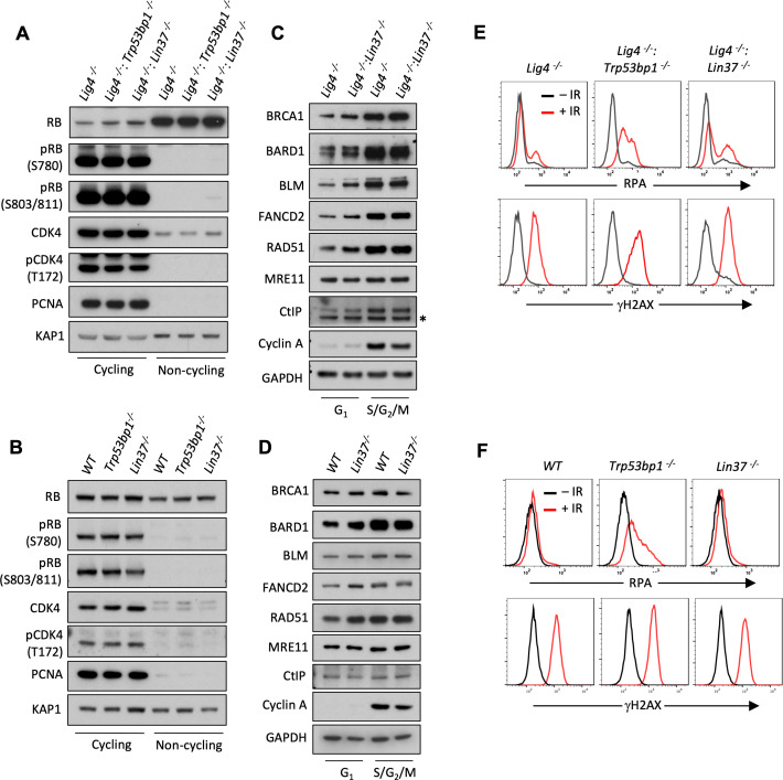
Figure 7. LIN37 function in DNA end protection is restricted to G0.

(A, B) Western blot analysis of indicated proteins in cycling and non-cycling abl pre-B cells (A) or MCF10A cells (B). (C, D) Western blot analysis of indicated proteins in cycling G1 or S/G2/M abl pre-B cells (C) or MCF10A cells (D), isolated by flow cytometric cell sorting based on the PIP-FUCCI reporter. Representative of two independent experiments. Asterisk indicates non-specific recognizing bands. (E, F) Flow cytometric analysis of chromatin-bound RPA and γH2AX before and after IR treatment of G1-phase Lig4−/−, Lig4−/−:Trp53bp1/− and Lig4−/−:Lin37/− abl pre-B cells (E) or WT, Trp53bp1/−, and Lin37/− MCF10A cells (F). Representative of three experiments. IR, ionizing radiation; WT, wild type.

Figure 7—source data 1. RNA-Seq result and GO analysis in cycling G1 Lig4/− and Lig4/−:Lin37−/− abl pre-B cells.
GO, gene ontology; RNA-Seq, RNA sequencing.
Figure 7—source data 2. GO analysis of genes upregulated in non-cycling G0 and/or cycling G1 Lig4/−:Lin37−/− abl pre-B cells.
GO, gene ontology.

Figure 7.

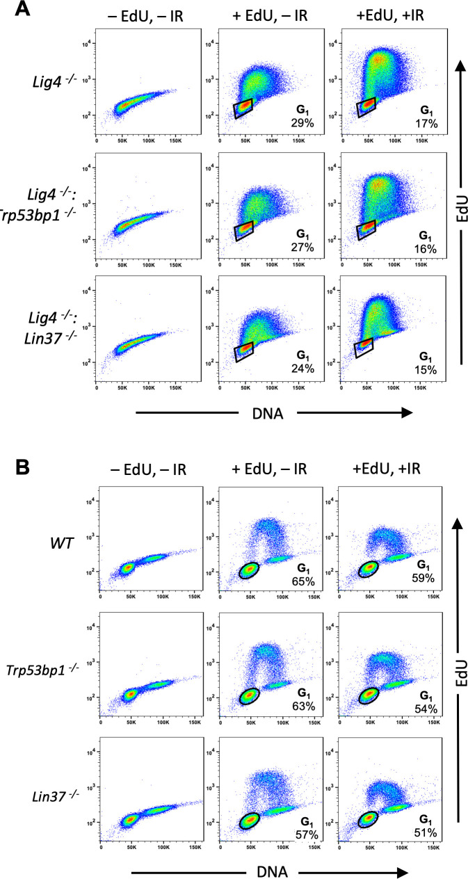
Figure 7—figure supplement 1. Identification of G1-phase cells in proliferating cells.

Figure 7—figure supplement 1.

(A, B) Flow cytometric analysis of EdU incorporation and DNA content (7-AAD) of cycling Lig4−/−, Lig4−/−:Trp53bp1/−, and Lig4−/−:Lin37/− abl pre-B cells (A) or cycling WT, Trp53bp1/−, and Lin37/− MCF10A cells (B). Cells were treated (+EdU) or not treated (−EdU) with EdU and analyses were carried out before or after IR. The percentage of G1-phase cells is shown. IR, ionizing radiation; WT, wild type.