Abstract
Bacteroidetes are efficient degraders of complex carbohydrates, much thanks to their use of polysaccharide utilization loci (PULs). An integral part of PULs are highly specialized carbohydrate-active enzymes, sometimes composed of multiple linked domains with discrete functions—multicatalytic enzymes. We present the biochemical characterization of a multicatalytic enzyme from a large PUL encoded by the gut bacterium Bacteroides eggerthii. The enzyme, BeCE15A-Rex8A, has a rare and novel architecture, with an N-terminal carbohydrate esterase family 15 (CE15) domain and a C-terminal glycoside hydrolase family 8 (GH8) domain. The CE15 domain was identified as a glucuronoyl esterase (GE), though with relatively poor activity on GE model substrates, attributed to key amino acid substitutions in the active site compared to previously studied GEs. The GH8 domain was shown to be a reducing-end xylose-releasing exo-oligoxylanase (Rex), based on having activity on xylooligosaccharides but not on longer xylan chains. The full-length BeCE15A-Rex8A enzyme and the Rex domain were capable of boosting the activity of a commercially available GH11 xylanase on corn cob biomass. Our research adds to the understanding of multicatalytic enzyme architectures and showcases the potential of discovering novel and atypical carbohydrate-active enzymes from mining PULs.
Subject terms: Carbohydrates, Biochemistry, Enzymes, Hydrolases
Introduction
The human gut microbiota (HGM) is characterized by very high cell densities of diverse microbial communities. One of its major roles is the degradation of recalcitrant dietary fiber and simultaneous secretion of short-chain fatty acids, which have been associated with numerous health benefits1. Understanding the HGM-host relationship is a major research field2,3 and the composition of the HGM is highly variable and influenced by factors such as diet, genetics, C-section vs. natural delivery, breastfeeding, gender, age, and medication4. Whether or not there is an “ideal” HGM composition is not fully known5, but in healthy adults the dominant bacterial phyla are typically Bacteroidetes and Firmicutes6. Both of these phyla include numerous species capable of efficiently degrading complex polysaccharides, which are a major component of dietary fiber.
To facilitate the complete degradation of recalcitrant dietary fiber, many Bacteroidetes species utilize polysaccharide utilization loci (PULs)7, which are discrete gene clusters encoding all proteins necessary to metabolize a specific polysaccharide8,9. The starch utilization system (Sus) from the anaerobic human gut symbiont Bacteroides thetaiotaomicron was the first PUL described and serves as a template for the identification and description of new PULs10,11. In addition to enzymes, carbohydrate-binding proteins, and a sugar sensor/regulatory protein, the Sus also encodes the proteins SusC and SusD; SusC is an integral outer membrane maltooligosaccharide transporter and SusD is a surface-tethered maltooligosaccharide-binding protein12. Homologs of SusC/D are found in all PULs and thereby enable the prediction of PULs from genomic sequences. Both characterized and putative PULs are collected in the database PULDB, which is part of the carbohydrate-active enzymes database CAZy (www.cazy.org;13,14). PULs encode sets of carbohydrate-active enzymes (CAZymes) with activities corresponding to their glycan target and consequently the number of CAZymes may vary greatly between different PULs. Several PULs have been characterized to date and have been found to target a wide range of homo- and heteroglycans such as plant hemicelluloses and pectins, crystalline chitin, fungal mannans, and algal polysaccharides15–19. Based on the co-localization of CAZyme-encoding genes targeting specific glycans, PULs can be used to assess the diversity of glycans in the natural environment as well as for the discovery of novel enzymes or enzyme architectures in Bacteroidetes species20–22.
Bacteroides eggerthii is a Bacteroidetes member that has been isolated from both human and fish feces23,24, indicating a successful adaptation to different host diets. While this bacterium has not been studied extensively to date, it has shown to be abundant in patients with type 2 diabetes25. B. eggerthii 1_2_48FAA is predicted to encode 39 PULs, and consequently a plethora of corresponding putative CAZymes14. Only a handful of enzymes from B. eggerthii have been characterized to date, including the heparinase Hep I26, the endo-xylanase BeXyn5A27, and the “multicatalytic” arabinofuranosidase-feruloyl esterase BeGH43/FAE28. Multicatalytic enzymes contain multiple connected catalytic domains with discrete functions, and while only few have been characterized so far, multicatalytic enzymes often display synergistic activities between these domains. For example, the CAZymes CelA (N-terminal glycoside hydrolase family 9 (GH9) endo-cellulase and C-terminal GH48 exo-cellulase) from Caldicellulosiruptor bescii29, ChiA (N-terminal exo- and C-terminal endo-chitinase domain) from Flavobacterium johnsoniae16,30, and BoCE6-CE1 (acetyl-feruloyl esterase) from Bacteroides ovatus20, have significantly enhanced substrate turnover capabilities as full-length enzymes compared to their individual catalytic domains. In some cases, intramolecular synergy has however not been observed for multicatalytic enzymes20,31,32. This might be caused by a lack of appropriate substrates, analytics, or appropriate reaction conditions, or simply that there is no synergy between the catalytic domains.
Of the predicted PULs in the genome of B. eggerthii, PUL 27 is one of the largest and it is anticipated to confer xylan degradation abilities to the bacterium based on its encoded CAZymes13,14. Two of the aforementioned characterized B. eggerthii enzymes, BeXyn5A and BeGH43/FAE, are also encoded by this PUL, and both enzymes have been shown to act on complex xylan27,28. In addition to BeGH43/FAE, the PUL encodes one more putative multicatalytic enzyme comprising an N-terminal carbohydrate esterase family 15 (CE15) and a C-terminal GH8 domain. Characterized CE15 members to date are glucuronoyl esterases (GEs), which have the proposed role of cleaving ester linkages between lignin and (4-O-methyl)-d-glucuronate decorations of glucuronoxylan and glucuronoarabinoxylan (GAX) in lignin carbohydrate complexes (LCCs)33–39. LCCs confer strength and rigidity to the plant cell wall and represent major obstacles in industrial enzymatic biomass hydrolysis processes40–43. In contrast to CE15, various activities have been demonstrated in GH8 including chitosanase, cellulase, licheninase, endo-β-1,4-xylanase and reducing-end xylose-releasing exo-oligoxylanase (Rex) enzymes13. These enzyme activities are found in multiple CAZy families, apart from Rex activity which is unique to GH8. The first Rex was isolated from Bacillus halodurans C-125 and, while it showed no activity on xylan, it released xylose moieties from the reducing end of xylooligosaccharides (XOs) longer than xylobiose, with xylotriose being the preferred substrate44. The combination of a CE15 domain and a GH8 domain into one single enzyme suggests a common substrate for the two catalytic domains, similar to the recently studied GE-xylanase CkXyn10C-GE15A from the hyperthermophilic bacterium Caldicellulosiruptor kristjanssonii, which additionally incorporates five carbohydrate-binding modules (CBMs)32. The polyspecificity of GH8 however precludes conclusive functional prediction of the B. eggerthii enzyme as a GE-xylanase fusion.
Here, our aim was to characterize the atypical multicatalytic enzyme from B. eggerthii comprising a CE15 and a GH8 domain to gain insight into its biological role. This enzyme architecture was found to be extremely rare, with the few identifiable homologs existing in the Bacteroidetes phylum. Biochemical characterization of the CE15 domain showed that it was active on standard GE substrates, though only with minor activity. This low activity was attributed to an amino acid substitution close to the catalytic serine, though changing the residue to the most conserved amino acid within the broader family did not increase activity. Assays on a wide range of substrates revealed the C-terminal GH8 domain to be a Rex, and the full-length protein was named BeCE15A-Rex8A. Direct synergy between the two catalytic domains could not be observed on GAX-rich corn cob biomass, possibly attributable to the minimal GE activity, though the Rex domain was able to boost the activity of a GH11 xylanase.
Results and discussion
Sequence based analysis
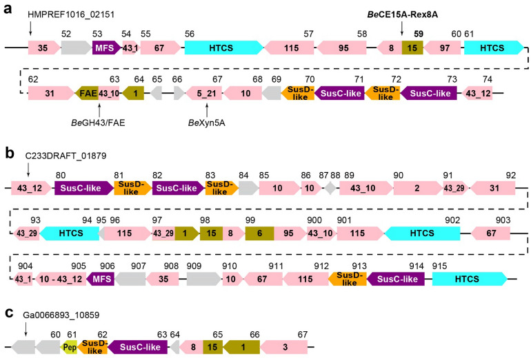
The 39 predicted PULs of B. eggerthii 1_2_48FAA range from solitary SusC/D pairs to loci spanning more than 30 genes. PUL 27 spans 24 genes (locus tags HMPREF1016_02151—HMPREF1016_02174; Fig. 1a)14. Curiously, no gene corresponding to HMPREF1016_02158 was listed in the PULDB. Translation of the intergenic sequence between HMPREF1016_02157 and HMPREF1016_02159 revealed a putative GH95 domain (785 amino acids) which is in agreement with enzymes found in previously studied PULs targeting GAX15. The collective enzyme repertoire of PUL 27, in addition to the previously characterized BeGH43/FAE (HMPREF1016_02163) and BeXyn5A (HMPREF1016_02167), strongly supports the hypothesis of the PUL targeting complex glycans, with putative xylanase (GH10), β-xylosidase or α-l-arabinofuranosidase (GH43), α-glucuronidase (GH67, GH115), α-xylosidase (GH31), α-galactosidase (GH95, GH97) and feruloyl or acetyl esterase (CE1) activities (Table S1)45–47. No PUL with a similar architecture was found in the PULDB14.
Figure 1.
Overview of PULs containing CE15-GH8 enzymes as predicted by the PULDB14. (a) PUL 27 of B. eggerthii, (b) PUL 20 of B. gallinarum, and (c) PUL 6 of Prevotella sp. BP1-145 (identical to PUL 1 of Prevotella sp. BP1-148). Locus tags are shown above each corresponding gene, drawn to scale. Enzymes are shown with enzyme family numbers indicated: glycoside hydrolases in pink, carbohydrate esterases in brown, sugar transporters in purple (MFS—major facilitator superfamily), putative regulators in blue (HTCS—hybrid two-component system), peptidase in yellow-green (Pep), and proteins of unknown function in gray. Intergenic regions are shown with dashed lines and are not drawn to scale (283 bp between HMPREF1016_01261 and 01262, 30 bp between C233DRAFT_01892 and 01893, and 0 bp between C233DRAFT_01903 and 01904).
The product of HMPREF1016_02159 in PUL 27 has a very unusual enzyme architecture, encoding a predicted multicatalytic enzyme comprising an N-terminal CE15 domain and a C-terminal GH8 domain, and a very short potential linker region. When compared to sequences in the NCBI protein database, a similar architecture was only found in three other species from the Bacteroides genus (Bacteroides sp. NSJ-48, B. stercoris, and B. gallinarum), each encoding one uncharacterized protein with 89–94% sequence identity (100% seq. coverage) to the B. eggerthii enzyme46,48. Furthermore, two homologs with lower similarity were found encoded by the more distantly related Prevotella sp. BP1-148 and Prevotella sp. BP1-145 (55% seq. id., 97% seq. coverage). Of these, only the B. gallinarum enzyme is found in a very large PUL likely targeting xylan (encoding enzymes from e.g. GH10, GH43, GH67, GH115, CE1; Fig. 1b), and the Prevotella enzymes are encoded by two identical small PULs that in addition to the CE15-GH8 enzyme only encode CAZymes from GH3 and CE1 (Fig. 1c)14. The CE15-GH8 architecture thus appears confined to the Bacteroidetes phylum and is strongly suggested to be involved in xylan turnover.
The individual domains of the B. eggerthii enzyme were compared to characterized enzymes from CE15 and GH8, respectively. The CE15 domain was most similar to OtCE15B from the soil bacterium Opitutus terrae (seq. id. 44%, coverage 97%)37. OtCE15B and the here investigated CE15 domain were phylogenetically more closely related to characterized fungal GEs than to other characterized GEs of bacterial origin and both contained a key disulfide bridge locking the catalytic serine and histidine in place as it is common in fungal GEs (Fig. S1). The catalytic triad was found to be conserved in BeCE15A (Ser230, Glu253, His357).
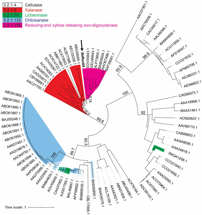
In contrast to CE15, many more members belonging to GH8 have been biochemically characterized13. Previous work has shown phylogeny to be a useful tool to predict enzyme specificities in GH8 using a limited number of sequences49. As the number of characterized GH8 members have since grown significantly, we constructed a new phylogenetic tree using the catalytic domains of all characterized members of GH8 as well as the B. eggerthii GH8 domain (Fig. 2). The tree was largely in agreement with the previous one, with different specificities mostly clustering into separate clades, including a clade encompassing all xylanases characterized to date. Rex enzymes formed a separate branch within the xylanase clade. The B. eggerthii GH8 domain was found to be most similar to BiRex8A from Bacteroides intestinalis50; the enzymes share 84% sequence identity which strongly indicates a similar function. BiRex8A was characterized simultaneously with BiXyn8A from the same organism, where the latter was shown to be an endo-xylanase, as BiXyn8A hydrolyzed both wheat arabinoxylan and oat spelt xylan into XOs50. BiRex8A on the other hand showed no xylanase activity but was instead able to release xylose moieties from the reducing end of XOs. The same study demonstrated that both BiRex8A and BiXyn8A shared the same conserved catalytic residues50, which are also conserved in the BeRex8A domain (Glu483, Asp541 and Asp679; Fig. S2).
Figure 2.
Phylogenetic tree of biochemically characterized GH8 domains. Proteins are labelled with Genbank accession numbers. The C-terminal Rex domain of BeCE15A-Rex8A is indicated in bold and marked with an arrow. Branches are colored by activity, with xylanase in red, Rex in magenta, licheninase in green, chitosanase in blue, and cellulase uncolored. Branches representing enzymes with dual specificity are striped with the corresponding colors.
Biochemical characterization of the BeCE15A-Rex8A CE15 domain
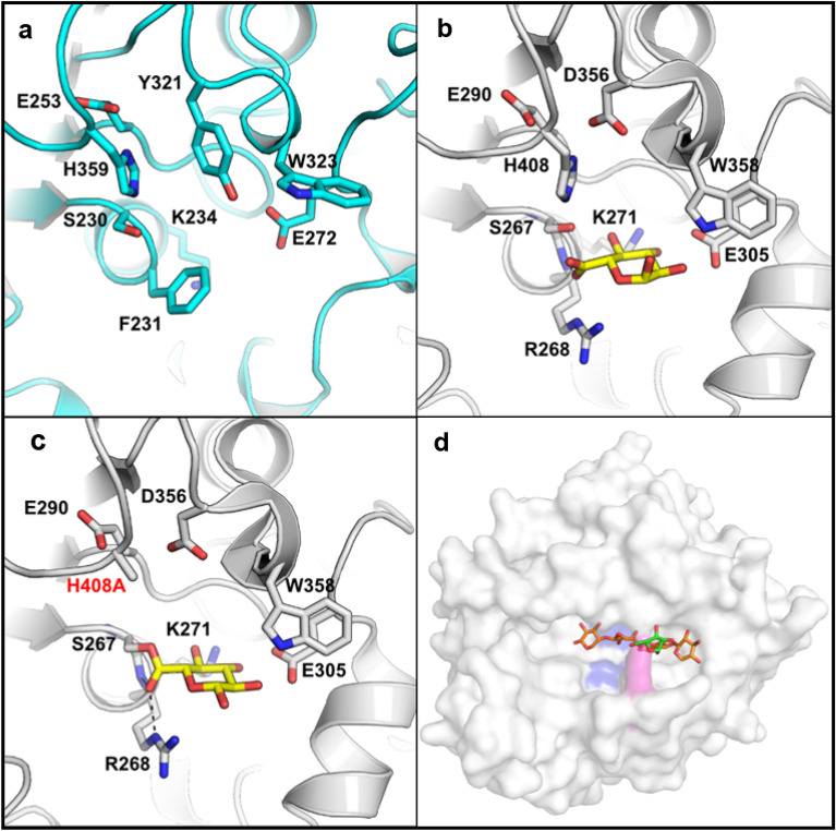
To confirm the putative functions of BeCE15A-Rex8A, the enzyme was heterologously produced in E. coli both as a full-length enzyme (91.5 kDa) and as the individual domains BeCE15A (46.8 kDa; amino acid residues 32-413) and BeRex8A (50.0 kDa; amino acid residues 414—812). BeCE15A-Rex8A and BeCE15A were assayed on the standard GE substrates benzyl glucuronoate (BnzGlcA), allyl glucuronoate (AllylGlcA), methyl glucuronoate (MeGlcA) and methyl galacturonoate (MeGalA) (Fig. 3). In contrast to previously studied GEs, none of the reactions were saturable up to concentrations of 40 mM substrate, precluding determination of either kcat or KM parameters. However, the catalytic efficiency (kcat/KM) could be determined using linear regression and showed that BeCE15A-Rex8A and BeCE15A were most active on BnzGlcA, with the activity decreasing successively on AllylGlcA, MeGlcA, and MeGalA (Table 1). This is in accordance with other characterized bacterial GEs, and consistent with the hypothesis that GEs prefer bulky substrates that are ester-linked to the O-6 position of a uronic acid moiety37,51,52, mimicking lignin or a lignin fragment in LCCs. In GEs, the rate-limiting step has been proposed to be the deacylation of the acyl-enzyme intermediate, given the similar kcat values determined for various enzymes to date52. The low kcat/KM values of BeCE15A may thus be a result of high KM values, indicating a poor fit of the model substrates in the active site. The isolated BeCE15A was approximately as active on the model substrates as the full-length enzyme, with roughly equal catalytic efficiencies on BnzGlcA, and 1.5-fold higher catalytic efficiency on AllylGlcA. This indicates that the truncation of BeCE15A-Rex8A into BeCE15A did not negatively affect the GE. The observed catalytic efficiencies were minimal compared to the majority of previously studied GEs reported in literature and the activity on BnzGlcA was approximately 500-fold lower than that of TtCE15A from Teredinibacter turnerae, which to date has the highest reported kcat/KM value for this substrate51. However, enzymes with even lower kcat/KM values on BnzGlcA than BeCE15A have previously been studied, including the most closely related characterized enzyme OtCE15B from O. terrae (kcat/KM value of 18.6 s−1 M−1), which is approximately fourfold lower than that of BeCE15A37. OtCE15B is an exception among studied GEs, as it has a tyrosine residue in the equivalent position of the conserved active site arginine residue believed to partake in forming the oxyanion hole and stabilizing the transition state during catalysis37,52,53. BeCE15A has an unexpected non-polar phenylalanine residue (Phe231) in equivalent position, which would not be able to electrostatically stabilize the transition state with its side chain (Fig. 4). To investigate whether replacement of this residue with the expected arginine residue would improve the activity on GE model substrates, we constructed an F231R variant of BeCE15A. Instead of increasing the activity, the result was however a complete loss of GE activity. Similarly, a substitution with a tyrosine (F231Y), as present in OtCE15B, also led to a complete loss of GE activity (data not shown).
Figure 3.
Substrates used to assay glucuronoyl esterase activity of BeCE15A: (a) benzyl glucuronoate, (b) allyl glucuronoate, (c) methyl glucuronoate, and (d) methyl galacturonoate. (e) The suggested target of the full-length BeCE15A-Rex8A enzyme, consists of a xylooligosaccharide (or longer xylan chain as indicated by the small arrow) decorated with GlcA, which is further ester-linked to lignin or lignin fragments.
Table 1.
Activity of BeCE15-Rex8A and BeCE15A on GE model substrates.
| Enzyme | Substrate | kcat/KM (s−1 mM−1) |
|---|---|---|
| BeCE15A-Rex8A | BnzGlcA | 0.0693 ± 0.0009 |
| AllylGlcA | 0.0159 ± 0.0005 | |
| MeGlcA | Trace | |
| MeGalA | Trace | |
| BeCE15A | BnzGlcA | 0.0753 ± 0.0008 |
| AllylGlcA | 0.0235 ± 0.0002 | |
| MeGlcA | 0.0075 ± 0.0003 | |
| MeGalA | 0.0008 ± 0.0002 |
“Trace” stands for trace activity detected for 0.02 µM min−1 mg−1 protein. The reactions were not saturable up to 40 mM and linear regression was used to calculate the kcat/KM values using GraphPad Prism 8. The results are based on triplicate measurements and presented with standard errors of the mean. The BeCE15A variants F231R and F231Y were also assayed, but no activity could be detected.
Figure 4.
Active site of a homology model of BeCE15A and model structure of BeRex8A. (a) A homology model of the BeCE15A was generated with Phyre256 using the CE15 from Hypocrea jecorina (PDB ID: 3PIC) as a template and compared to structures of (b) the wild type OtCE15A (PDB ID: 6SYR) in complex with glucuronate (yellow sticks) and (c) a H408A variant of OtCE15A (PDB ID: 6SZ4) which was trapped with glucuronate covalent adduct and shows the interaction with the usually conserved active site arginine. The equivalent position in BeCE15A is predicted to be a phenylalanine (Phe231). (d) The model structure of BeRex8A was generated by Phyre256 and based on PbRex8A (PDB ID: 6TRH). The catalytic residues are shown in blue. The Arg670 residue is shown in purple. The arabinoxylooligosaccharide from PbRex8A is modelled into the active site. The figure was made using PyMOL 2.3.
Biochemical characterization of the BeCE15A-Rex8A GH8 domain
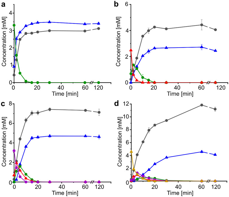
As GH8 is a polyspecific family, the GH8 domain of BeCE15A-Rex8A was assayed on a range of polysaccharides: cellulose, birchwood and beechwood xylan, wheat arabinoxylan, linear ivory nut mannan, mixed linkage β-glucan from barley, as well as starch. No activity could be detected on any of these substrates even after prolonged incubations. Previously studied Rex enzymes have been shown to either be inactive or have minimal activity on polymeric xylan and instead are active on XOs44,49,50,54,55. Similarly, BeRex8A was able to hydrolyze XOs ranging from xylotriose (X3) to xylohexaose (X6), and only trace activity (above 0.02 µM min−1 mg−1 protein) was observed on X2 when incubated for prolonged periods of time (Fig. 5). X1 and X2 were the end products of all reactions. Our time-course analysis shows highly similar hydrolysis progress curves to those of BiRex8A from Bacteroides intestinalis50, with the substrates being sequentially shortened into intermediate products, themselves acting as new substrates, and with a concomitant accumulation of X1 and X2 as end products (Fig. 5). BeRex8A was not active on pNP-xylobiose or borohydrate-reduced xylotriose, further supporting the Rex activity.
Figure 5.
Hydrolysis of xylooligosaccharides by BeRex8A. Substrates used were (a) xylotriose, (b) xylotetraose, (c) xylopentaose, and (d) xylohexaose. Concentrations are shown for xylose (gray circle), xylobiose (blue triangle), xylotriose (green circle), xylotetraose (red triangle), xylopentaose (purple circle), and xylohexaose (golden triangle). Data are presented as averages of triplicate experiments with standard errors of the mean.
Attempts to crystallize and determine the structure of BeCE15A-Rex8A or its parts were unfortunately not successful. However, modeling of BeRex8A using Phyre256 yielded a predicted protein structure with 95% coverage and 100% confidence based on the structurally determined E70A variant of PbRex8A from Paenibacillus barcinonensis (PDB ID: 6TRH; 42% seq. id. to BeRex8A; Fig. 4d)57. PbRex8A was previously shown to have minimal activity on xylan and a loop comprised of Leu320-His321-Pro322 blocking the active site after the + 1 subsite was attributed to the inability of the enzyme to efficiently bind polysaccharides and generate products longer than xylose57. Attempts to reduce the size of the loop to open up the active site did however not improve the activity on xylan57. An equivalent to the Leu320-His321-Pro322 loop in PbRex8A is not found in BeRex8A, but the modeled structure shows a similar active site groove, blocked by a single arginine residue (Arg670; Fig. 4d). This arginine residue appeared to form a tunnel allowing access of unsubstituted oligo- or polysaccharides. We hypothesized that this residue may play a role in the specificity for shorter oligos, and the lack of activity on polysaccharide chains, and constructed an R670A variant. However, this variant was inactive on polymeric xylan, similar to the wild type enzyme (data not shown). Deeper structural investigation would be needed to shed light on possible interchangeability between Rex vs. endo-xylanase activity.
Boosting of xylanase hydrolysis of corn cob
Enzymes present in PULs are expected to act in concert in the degradation of a specific polysaccharide. Based on the GE and Rex activities of the individual catalytic domains of BeCE15A-Rex8A, the enzyme was expected to aid in the degradation of complex xylans of the plant cell wall. The reason for combining activities presumed to target complex LCCs and shorter XOs into one single enzyme is however not clear. To gain further insight into the function of BeCE15A-Rex8A, as well as its truncated single domain versions (BeCE15A and BeRex8A), the enzymes were assayed for their ability to boost the action of a commercially available GH11 xylanase (Xyn11A), which has previously been used successfully in similar experiments20,31. Ball-milled corn cob biomass, which has a high content of GAX15, was used as substrate. No release of sugars was observed when no enzyme was added (data not shown), and similarly no released sugars were detected if BeCE15A-Rex8A, BeCE15A or BeRex8A were added without Xyn11A (data not shown). Addition of Xyn11A (control reaction) lead to the release of small amounts of XOs ranging from X1 to X6 (Fig. 6). The main products were X1 and X2 with concentrations reaching 1.6 mM each after 30 h, and substantially more X4 and X6 were released than X3 and X5. Supplementation of Xyn11A with BeCE15A did not alter XO release substantially compared to the control reaction. Supplementation of Xyn11A with BeRex8A, BeCE15A-Rex8A or an equimolar mix of BeCE15A and BeRex8A increased X1 (twofold), X2 (1.3-fold), X3 (5.6-fold) and X5 concentrations (twofold), while X4 and X6 concentrations were reduced to roughly a third compared to the control reaction. The total xylose equivalents from X1-X6 that were released by Xyn11A when supplemented with BeRex8A increased 20–30% compared to the reaction of Xyn11A alone, and do not appear to stem simply from conversion of longer XOs to short ones by the Rex enzyme. Possibly, the apparent improvement of Xyn11A could be a result of reduced product inhibition.
Figure 6.
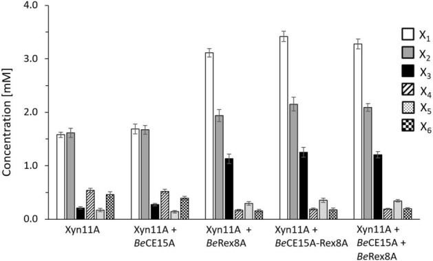
Xylooligosaccharide production profiles from corn cob biomass hydrolysis. The endo-β-1,4-xylanase Xyn11A from Neocallimastix patriciarum was either incubated alone or supplemented with BeCE15A, BeRex8A, BeCE15A-Rex8A or an equimolar mix of BeCE15A and BeRex8A. Reactions in which BeCE15A, BeRex8A, and BeCE15A-Rex8A were incubated without Xyn11A yielded no detected sugars (data not shown), as expected of a Rex enzyme and a GE. Presented are xylose (X1, white), xylobiose (X2, gray), xylotriose (X3, black), xylotetraose (X4, striped), xylopentaose (X5, dotted), and xylohexaose (X6, checkered) concentrations after 30 h of incubation that were determined using high-performance anion-exchange chromatography with pulsed amperometric detection (HPAEC-PAD). Data are shown as average of triplicate experiments with standard errors of the mean.
The reason for the inability of BeCE15A to boost xylanase activity on corn cob biomass is not clear but echoes the results of the CE15 domain from the Caldicellulosiruptor kristjanssonii encoded CkXyn10C-GE15A, which similarly did not appear to boost xylanase activity directly either with commercial enzymes or the linked CkXyn10C xylanase domain32. Possibly, the effect of GEs on xylanases cannot be monitored by sugar release measurements due to the overall complexity of the (un-pretreated) material and reduced access to LCC esters that the enzymes can target. Alternatively, BeCE15A, with its atypical active site residues, may be more specialized to target structures that were not present or accessible in the here utilized corn cob biomass. The low activity of BeCE15A on GE model substrates, similar to that of OtCE15B37, suggests that these enzymes might have a different role in biomass turnover than other so far characterized CE15 enzymes. The main activity of characterized CE15 members to this date has been (4-O-methyl)-glucuronoyl esterase activity13, but the incorporation of a CE15 enzyme into a PUL suggests that the activity of BeCE15A supports xylan degradation. Deeper investigation of atypical enzymes such as BeCE15A and OtCE15B holds the potential of adding to our knowledge on enzymatic biomass degradation and might be an interesting target for the improvement of industrial enzyme cocktails.
Comparing supplementation of Xyn11A with the full-length BeCE15A-Rex8A and an equimolar mix of its single domains BeCE15A + BeRex8A did not reveal significant differences in the XO production profiles over the whole course of the experiment (Fig. S3). Deducing the preferred substrate of a multicatalytic enzyme can be challenging due to the highly specialized nature of these proteins and the vast diversity among polysaccharides, especially in the context of the complex cell wall polymer network. A lack of intramolecular enzyme synergy has also been observed for other multicatalytic enzymes, such as FjCE6-CE1 from F. johnsoniae20, CkXyn10C-GE15A from C. kristjansonii32, and DmCE1B from Dysgonomonas mossii31. Given the complexity of the substrates targeted by these enzymes, which are presumed to be part of LCCs, it is currently unclear whether the lack of observed intramolecular enzyme synergy is the result of missing intramolecular synergy, a lack of the right substrate, or another unknown reason. Typically, multicatalytic enzymes are joined by flexible linkers of varying length or small domains29,32. In BeCE15A-Rex8A, a short potential linker is present between Trp402 and Ala423, although exactly how flexible the linker is remains unclear. While no experimental structural data is available, multiple models constructed using the Phyre256 and I-TASSER58 structural modelling servers suggest that the catalytic domains may be in close contact with each other (Fig. 7). Additionally, the domains appear to be oriented with their active sites facing in opposite directions. Whether the active sites are able to act in close proximity or not, depending on the length and flexibility of the putative linker, is currently unclear and would need support with structural data.
Figure 7.
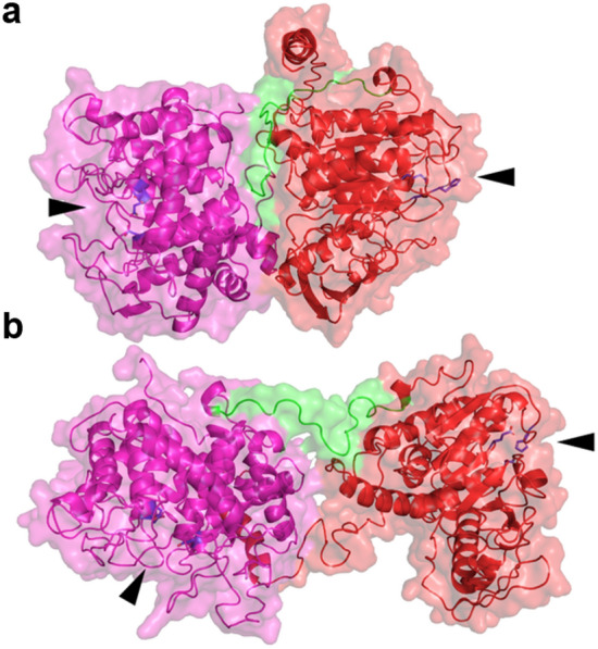
Full length model of BeCE15A-Rex8A using I-TASSER58 (a), and Phyre256 (b). In both models, the GE domain is colored red, the Rex domain is purple, the potential linker region is green, and the active-site residues are blue. In both models the active sites of the two domains are positioned facing away from each other and marked by black arrows.
Analysis using Signal P 5.059 identified a 23 amino acid long signal peptide with 95% likelihood as Sec/SPI, indicating that the protein is likely secreted into the periplasm, but whether BeCE15A-Rex8A is further transported outside the cell is not known. The presumed biological role of GEs would indicate that the target substrate(s) of BeCE15A is found in large LCCs that are unlikely to be imported into the periplasm. Conversely, the Rex activity of BeRex8A would be more in keeping with how final degradation of poly- and oligosaccharides in PULs is believed to mainly occur in the periplasm to prevent “leakage” of metabolizable sugars to surrounding cells8,9,18,19. The low GE activity of the enzyme and atypical active site setup might indicate that GlcA in xylan can be esterified with as of yet unidentified moieties that are hydrolyzed in the periplasm by B. eggerthii. Identification of such motifs would likely require significant efforts, though enzymes such as BeCE15A and OtCE15B could be highly useful tools in such an endeavor.
Conclusion
In this study we biochemically characterized the multicatalytic enzyme BeCE15A-Rex8A. The N-terminal domain was identified as a GE having minimal activity on model substrates and harboring a highly unusual amino acid substitution close to the catalytic serine that might play an important role in substrate turnover or substrate preferences that are yet unidentified in CE15. The C-terminal domain was identified as a Rex, an activity that has been demonstrated in very few enzymes to date. The here described enzyme architecture of BeCE15A-Rex8A was shown to be very rare and confined to a few PULs within the bacterial phylum of Bacteroidetes. This work further highlights the usefulness of mining PULs for the discovery of novel enzyme types and architectures.
Material and methods
Phylogenetic tree
The amino acid sequences of GH8 enzymes listed as characterized were downloaded from CAZy (Nov 2020), trimmed to only contain the catalytic domains, and subsequently aligned using MUSCLE60. The phylogenetic tree was built based on the alignment using IQ-TREE61, with automatic finding of the best substitution model (LG + F + I + G4) and 1000 ultrafast bootstraps. The maximum-likelihood tree was visualized using iTOL62.
Cloning of BeCE15A-Rex8A and protein variants
The putative BeCE15-GH8 was amplified from genomic DNA of B. eggerthii 1_2_48FAA by PCR (primers listed in Table S2) and the products cloned into a modified pET-28a vector, by ligation independent cloning (In-Fusion HD kit; Clontech Laboratories), containing an N-terminal His6 tag and a tobacco etch virus protease cleavage site. A signal peptide predicted at the N-terminal end of the gene encoding BeCE15A-Rex8A (residues 1–31) was not included for protein production. Enzyme variants were created by site-specific mutagenesis by the QuikChange method using the primers listed in Table S263.
Protein production and purification
Cell cultures harboring expression vectors were grown in lysogeny broth at 37 °C and 180 rpm until cells reached mid-log phase (OD600 0.4–0.6), at which point protein production was induced by addition of 0.2 mM isopropyl-β-d-1-thiogalactopyranoside, and cells cultured overnight (16 °C and 180 rpm). The cells were harvested by centrifugation and lysed by sonication. The resulting protein containing crude lysate was purified using immobilized metal ion affinity chromatography as previously described37. Purified protein was concentrated and buffer exchanged (BeCE15A in 50 mM Tris pH 8.0 + 100 mM NaCl; BeRex8A in sodium phosphate pH 6.5 + 100 mM NaCl; and BeCE15A-Rex8A in 50 mM Tris pH 8.0 + 250 mM NaCl + 5% w/v glycerol) using 10 kDa cut-off centrifugal filter units (Amicon Ultra-15, Merck-Millipore) and imidazole concentrations were reduced to < 1 mM. Sodium dodecyl sulfate polyacrylamide gel electrophoresis using Mini-PROTEAN TGX Stain-Free Gels (BIO-RAD) was used to verify molecular weight and protein purity. Protein concentrations were determined using a Nanodrop 2000 Spectrophometer (Thermo Fisher Scientific) using extinction coefficients and molecular weights predicted by Benchling.
Biochemical characterization of the CE15 domain
pH dependency was established for BeCE15A by comparing the activity on BnzGlcA in a range of buffers and pH values (Fig. S4). A pH dependency profile for BeRex8A could not be established as the enzyme fell out of solution at pH values different than 6.5 ± 0.5. Assays on model GE substrates (BnzGlcA, AllylGlcA, MeGlcA, MeGalA) were performed at pH 7.5 for comparison to other GEs as previously described32,37 using the d-Glucuronic/d-Galacturonic Acid Assay Kit (Megazyme). Briefly, concentrations of substrate up to 40 mM were incubated with BeCE15A at room temperature in a coupled enzyme assay with uronate dehydrogenase and the formation of nicotinamide adenine dinucleotide hydride was monitored at 340 nm. Data were analyzed using GraphPad Prism 8.4.2, and kcat/KM values were determined by linear regression.
Biochemical characterization on XOs and complex substrates
All here described substrates were purchased from Megazyme if unless stated otherwise. Reactions were incubated at 37 °C with mixing at 500 rpm and contained BeRex8A (2 µM; in 50 mM sodium phosphate buffer pH 6.0 + 100 mM NaCl) and the different substrates. Screening of possible polysaccharide hydrolyzing ability of the Rex8A domain was done using 1.25% w/v cellulose, birchwood xylan, beechwood xylan (Apollo scientific), wheat arabinoxylan, linear ivory nut mannan, mixed linkage β-glucan from barley, or starch, with sugar release monitored using the dinitrosalicylic acid assay. Xylooligosaccharides tested were xylobiose (X2; 3.2 mM), xylotriose (X3; 3.25 mM), xylotetraose (X4; 3.3 mM), xylopentaose (X5; 2.65 mM) and xylohexaose (X6; 3.33 mM). Samples were flash-frozen in liquid nitrogen, diluted with HCl (0.1 M final concentration) to stop the enzymatic reaction and analyzed using HPAEC-PAD (see below).
Corn cob biomass for xylanase hydrolysis studies was produced by processing corn cob (excluding corn grains) in a kitchen blender followed by ball-milling into a fine powder, washing with water, and then freeze-drying. The corn cob was used as substrate (0.45% w/v) with BeCE15A-Rex8A, BeCE15A or BeRex8A, incubated at 37 °C and 1000 rpm in 100 mM sodium phosphate pH 6.5 including 0.5 µM of each enzyme, in various combinations with and without addition of the commercially available endo-β-1,4-xylanase Xyn11A from N. patriciarum (E-XYLNP; Megazyme; concentration in assay 11 µM). The samples were flash-frozen in liquid nitrogen and stopped by addition HCl (0.1 M final concentration) before being analyzed using HPAEC-PAD.
High-performance anion-exchange chromatography with pulsed amperometric detection
HPAEC-PAD was performed on a Dionex ICS-5000 + (Thermo Fisher Scientific) equipped with a Dionex CarboPac™ PA200 column (Thermo Fisher Scientific). To achieve sufficient separation of the XOs a constant flow of 0.5 mL/min and a multistep gradient (Table S3) were applied using deionized water, 300 mM NaOH, and 1 M NaAc. Prior to use dissolved oxygen was removed from all solutions by sparging with helium gas.
Structural models of BeCE15A-Rex8A
The model for BeRex8A was generated with Phyre256 and based on the structurally determined E70A variant of PbRex8A from P. barcinonensis. Models of full-length BeCE15A-RexA domains combined were generated both with the Phyre2 server56 and with the I-TASSER server58. When selecting a model from I-TASSER, manual inspection of the predicted folding of the individual domains was used (in comparison to crystal structures of other Rex and GE domains) in order to select the most likely model.
Supplementary Information
Acknowledgements
We thank Dr. Eric Martens (University of Michigan) for generously providing us with the strain Bacteroides eggerthii 1_2_48FAA for extraction of genomic DNA for cloning.
Abbreviations
- AllylGlcA
Allyl glucuronic acid ester
- BnzGlcA
Benzyl glucuronoate
- CAZy
Carbohydrate-active enzymes database
- CAZyme
Carbohydrate-active enzyme
- CE
Carbohydrate esterase
- CE15
Carbohydrate esterase family 15
- GAX
Glucuronoarabinoxylan
- GE
Glucuronoyl esterase
- GH
Glycoside hydrolase
- HGM
Human gut microbiota
- HPAEC-PAD
High-performance anion-exchange chromatography with pulsed amperometric detection
- LCC
Lignin-carbohydrate complex
- MeGalA
Methyl galacturonoate
- MeGlcA
Methyl glucuronoate
- PDB
Protein data bank
- PUL
Polysaccharide utilization locus
- PULDB
Polysaccharide-Utilization Loci DataBase
- Rex
Reducing-end xylose-releasing exo-oligoxylanase
- Sus
Starch utilization system
- XO
Xylooligosaccharide
- X1-6
Xylose, xylobiose, xylotriose, xylotetraose, xylopentaose and xylohexaose, respectively
Author contributions
The study was conceived and supervised by J.L. J.L. constructed the phylogenetic tree. D.K. and S.M. cloned the enzymes. S.M. produced the homology model of BeCE15A. D.K. biochemically characterized the enzymes on GE model substrates, polysaccharides, and XOs and generated the model structures of BeCE15A-Rex8A and BeRex8A. C.K. performed the assays on corn cob biomass, carried out HPAEC-PAD and produced the comparative sequence analysis. C.K. and D.K. produced the enzymes and wrote the manuscript. S.M. and J.L. critically appraised and revised the manuscript.
Funding
Open access funding provided by Chalmers University of Technology. This work was supported by the Swedish Research Council (Dnr 2016-03931), the Swedish Research Council Formas (Dnr 2016-01065), the Novo Nordisk Foundation (Grant number NNF17OC0027648), and the Knut and Alice Wallenberg Foundation through the Wallenberg Wood Science Center.
Competing interests
The authors declare no competing interests.
Footnotes
Publisher's note
Springer Nature remains neutral with regard to jurisdictional claims in published maps and institutional affiliations.
These authors contributed equally: Cathleen Kmezik and Daniel Krska.
Supplementary Information
The online version contains supplementary material available at 10.1038/s41598-021-96659-z.
References
- 1.den Besten G, et al. The role of short-chain fatty acids in the interplay between diet, gut microbiota, and host energy metabolism. J. Lipid Res. 2013;54:2325–2340. doi: 10.1194/jlr.R036012. [DOI] [PMC free article] [PubMed] [Google Scholar]
- 2.Martens EC, Chiang HC, Gordon JI. Mucosal glycan foraging enhances fitness and transmission of a saccharolytic human gut bacterial symbiont. Cell Host Microbe. 2008;4:447–457. doi: 10.1016/j.chom.2008.09.007. [DOI] [PMC free article] [PubMed] [Google Scholar]
- 3.Flint HJ, Bayer EA, Rincon MT, Lamed R, White BA. Polysaccharide utilization by gut bacteria: Potential for new insights from genomic analysis. Nat. Rev. Microbiol. 2008;6:121–131. doi: 10.1038/nrmicro1817. [DOI] [PubMed] [Google Scholar]
- 4.Wen L, Duffy A. Factors influencing the gut microbiota, inflammation, and type 2 diabetes. J. Nutr. 2017;147:1468S–1475S. doi: 10.3945/jn.116.240754. [DOI] [PMC free article] [PubMed] [Google Scholar]
- 5.Lozupone CA, Stombaugh JI, Gordon JI, Jansson JK, Knight R. Diversity, stability and resilience of the human gut microbiota. Nature. 2012;489:220–230. doi: 10.1038/nature11550. [DOI] [PMC free article] [PubMed] [Google Scholar]
- 6.Eckburg PB, et al. Diversity of the human intestinal microbial flora. Science. 2005;308:1635–1638. doi: 10.1126/science.1110591. [DOI] [PMC free article] [PubMed] [Google Scholar]
- 7.Bjursell MK, Martens EC, Gordon JI. Functional genomic and metabolic studies of the adaptations of a prominent adult human gut symbiont, Bacteroides thetaiotaomicron, to the suckling period. J. Biol. Chem. 2006;281:36269–36279. doi: 10.1074/jbc.M606509200. [DOI] [PubMed] [Google Scholar]
- 8.Grondin JM, Tamura K, Dejean G, Abbott DW, Brumer H. Polysaccharide utilization loci: Fueling microbial communities. J. Bacteriol. 2017;199:e00860-00816. doi: 10.1128/JB.00860-16. [DOI] [PMC free article] [PubMed] [Google Scholar]
- 9.Larsbrink J, McKee LS. Bacteroidetes bacteria in the soil: glycan acquisition, enzyme secretion, and gliding motility. Adv. Appl. Microbiol. 2020;110:63–98. doi: 10.1016/bs.aambs.2019.11.001. [DOI] [PubMed] [Google Scholar]
- 10.D'Elia JN, Salyers AA. Effect of regulatory protein levels on utilization of starch by Bacteroides thetaiotaomicron. J. Bacteriol. 1996;178:7180–7186. doi: 10.1128/jb.178.24.7180-7186.1996. [DOI] [PMC free article] [PubMed] [Google Scholar]
- 11.Shipman JA, Berleman JE, Salyers AA. Characterization of four outer membrane proteins involved in binding starch to the cell surface of Bacteroides thetaiotaomicron. J. Bacteriol. 2000;182:5365–5372. doi: 10.1128/JB.182.19.5365-5372.2000. [DOI] [PMC free article] [PubMed] [Google Scholar]
- 12.Foley MH, Cockburn DW, Koropatkin NM. The Sus operon: a model system for starch uptake by the human gut Bacteroidetes. Cell. Mol. Life Sci. 2016;73:2603–2617. doi: 10.1007/s00018-016-2242-x. [DOI] [PMC free article] [PubMed] [Google Scholar]
- 13.Lombard V, Golaconda Ramulu H, Drula E, Coutinho PM, Henrissat B. The carbohydrate-active enzymes database (CAZy) in 2013. Nucleic Acids Res. 2014;42:D490–D495. doi: 10.1093/nar/gkt1178. [DOI] [PMC free article] [PubMed] [Google Scholar]
- 14.Terrapon N, et al. PULDB: The expanded database of polysaccharide utilization loci. Nucleic Acids Res. 2018;46:D677–D683. doi: 10.1093/nar/gkx1022. [DOI] [PMC free article] [PubMed] [Google Scholar]
- 15.Rogowski A, et al. Glycan complexity dictates microbial resource allocation in the large intestine. Nat. Commun. 2015;6:1–16. doi: 10.1038/ncomms8481. [DOI] [PMC free article] [PubMed] [Google Scholar]
- 16.Larsbrink J, et al. A polysaccharide utilization locus from Flavobacterium johnsoniae enables conversion of recalcitrant chitin. Biotechnol. Biofuels. 2016;9:1–16. doi: 10.1186/s13068-016-0674-z. [DOI] [PMC free article] [PubMed] [Google Scholar]
- 17.Ficko-Blean E, et al. Carrageenan catabolism is encoded by a complex regulon in marine heterotrophic bacteria. Nat. Commun. 2017;8:1–17. doi: 10.1038/s41467-017-01832-6. [DOI] [PMC free article] [PubMed] [Google Scholar]
- 18.Larsbrink J, et al. A discrete genetic locus confers xyloglucan metabolism in select human gut Bacteroidetes. Nature. 2014;506:498–502. doi: 10.1038/nature12907. [DOI] [PMC free article] [PubMed] [Google Scholar]
- 19.Cuskin F, et al. Human gut Bacteroidetes can utilize yeast mannan through a selfish mechanism. Nature. 2015;517:165–169. doi: 10.1038/nature13995. [DOI] [PMC free article] [PubMed] [Google Scholar]
- 20.Kmezik C, Bonzom C, Olsson L, Mazurkewich S, Larsbrink J. Multimodular fused acetyl-feruloyl esterases from soil and gut Bacteroidetes improve xylanase depolymerization of recalcitrant biomass. Biotechnol. Biofuels. 2020;13:1–14. doi: 10.1186/s13068-020-01698-9. [DOI] [PMC free article] [PubMed] [Google Scholar]
- 21.Razeq FM, et al. A novel acetyl xylan esterase enabling complete deacetylation of substituted xylans. Biotechnol. Biofuels. 2018;11:1–12. doi: 10.1186/s13068-018-1074-3. [DOI] [PMC free article] [PubMed] [Google Scholar]
- 22.Lapébie P, Lombard V, Drula E, Terrapon N, Henrissat B. Bacteroidetes use thousands of enzyme combinations to break down glycans. Nat. Commun. 2019;10:1–7. doi: 10.1038/s41467-019-10068-5. [DOI] [PMC free article] [PubMed] [Google Scholar]
- 23.Kabiri L, Alum A, Rock C, McLain JE, Abbaszadegan M. Isolation of Bacteroides from fish and human fecal samples for identification of unique molecular markers. Can. J. Microbiol. 2013;59:771–777. doi: 10.1139/cjm-2013-0518%M24313449. [DOI] [PubMed] [Google Scholar]
- 24.Holdeman LV, Moore W. New genus, Coprococcus, twelve new species, and emended descriptions of four previously described species of bacteria from human feces. Int. J. Syst. Evol. Microbiol. 1974;24:260–277. [Google Scholar]
- 25.Medina-Vera I, et al. A dietary intervention with functional foods reduces metabolic endotoxaemia and attenuates biochemical abnormalities by modifying faecal microbiota in people with type 2 diabetes. Diabetes Metab. 2019;45:122–131. doi: 10.1016/j.diabet.2018.09.004. [DOI] [PubMed] [Google Scholar]
- 26.Liu C-Y, Su W-B, Guo L-B, Zhang Y-W. Cloning, expression, and characterization of a novel heparinase I from Bacteroides eggerthii. Prep. Biochem. Biotechnol. 2020;50:477–485. doi: 10.1080/10826068.2019.1709977. [DOI] [PubMed] [Google Scholar]
- 27.Dodd D, Moon Y-H, Swaminathan K, Mackie RI, Cann IKO. Transcriptomic analyses of xylan degradation by Prevotella bryantii and insights into energy acquisition by xylanolytic bacteroidetes. J. Biol. Chem. 2010;285:30261–30273. doi: 10.1074/jbc.M110.141788. [DOI] [PMC free article] [PubMed] [Google Scholar]
- 28.Pereira GV, et al. Degradation of complex arabinoxylans by human colonic Bacteroidetes. Nat. Commun. 2021;12:1–21. doi: 10.1038/s41467-020-20314-w. [DOI] [PMC free article] [PubMed] [Google Scholar]
- 29.Brunecky R, et al. Revealing nature's cellulase diversity: the digestion mechanism of Caldicellulosiruptor bescii CelA. Science. 2013;342:1513–1516. doi: 10.1126/science.1244273. [DOI] [PubMed] [Google Scholar]
- 30.Mazurkewich S, et al. Structural insights of the enzymes from the chitin utilization locus of Flavobacterium johnsoniae. Sci. Rep. 2020;10:1–11. doi: 10.1038/s41598-020-70749-w. [DOI] [PMC free article] [PubMed] [Google Scholar]
- 31.Kmezik C, et al. A polysaccharide utilization locus from the gut bacterium Dysgonomonas mossii encodes functionally distinct carbohydrate esterases. J. Biol. Chem. 2021;296:100500. doi: 10.1016/j.jbc.2021.100500. [DOI] [PMC free article] [PubMed] [Google Scholar]
- 32.Krska D, Larsbrink J. Investigation of a thermostable multi-domain xylanase-glucuronoyl esterase enzyme from Caldicellulosiruptor kristjanssonii incorporating multiple carbohydrate-binding modules. Biotechnol. Biofuels. 2020;13:1–13. doi: 10.1186/s13068-020-01709-9. [DOI] [PMC free article] [PubMed] [Google Scholar]
- 33.Špániková S, Biely P. Glucuronoyl esterase: Novel carbohydrate esterase produced by Schizophyllum commune. FEBS Lett. 2006;580:4597–4601. doi: 10.1016/j.febslet.2006.07.033. [DOI] [PubMed] [Google Scholar]
- 34.d'Errico C, et al. Improved biomass degradation using fungal glucuronoyl-esterases-hydrolysis of natural corn fiber substrate. J. Biotechnol. 2016;219:117–123. doi: 10.1016/j.jbiotec.2015.12.024. [DOI] [PubMed] [Google Scholar]
- 35.Arnling Bååth J, Giummarella N, Klaubauf S, Lawoko M, Olsson L. A glucuronoyl esterase from Acremonium alcalophilum cleaves native lignin-carbohydrate ester bonds. FEBS Lett. 2016;590:2611–2618. doi: 10.1002/1873-3468.12290. [DOI] [PubMed] [Google Scholar]
- 36.Mosbech C, Holck J, Meyer AS, Agger JW. The natural catalytic function of CuGE glucuronoyl esterase in hydrolysis of genuine lignin-carbohydrate complexes from birch. Biotechnol. Biofuels. 2018;11:1–9. doi: 10.1186/s13068-018-1075-2. [DOI] [PMC free article] [PubMed] [Google Scholar]
- 37.Arnling Bååth J, et al. Biochemical and structural features of diverse bacterial glucuronoyl esterases facilitating recalcitrant biomass conversion. Biotechnol. Biofuels. 2018;11:1–14. doi: 10.1186/s13068-018-1213-x. [DOI] [PMC free article] [PubMed] [Google Scholar]
- 38.Mosbech C, Holck J, Meyer A, Agger JW. Enzyme kinetics of fungal glucuronoyl esterases on natural lignin-carbohydrate complexes. Appl. Microbiol. Biotechnol. 2019;103:4065–4075. doi: 10.1007/s00253-019-09797-w. [DOI] [PubMed] [Google Scholar]
- 39.Raji O, et al. The coordinated action of glucuronoyl esterase and α-glucuronidase promotes the disassembly of lignin-carbohydrate complexes. FEBS Lett. 2021;595:351–359. doi: 10.1002/1873-3468.14019. [DOI] [PMC free article] [PubMed] [Google Scholar]
- 40.Tarasov D, Leitch M, Fatehi P. Lignin-carbohydrate complexes: Properties, applications, analyses, and methods of extraction: A review. Biotechnol. Biofuels. 2018;11:1–28. doi: 10.1186/s13068-018-1262-1. [DOI] [PMC free article] [PubMed] [Google Scholar]
- 41.Várnai A, Siika-aho M, Viikari L. Restriction of the enzymatic hydrolysis of steam-pretreated spruce by lignin and hemicellulose. Enzyme Microb. Technol. 2010;46:185–193. doi: 10.1016/j.enzmictec.2009.12.013. [DOI] [Google Scholar]
- 42.Himmel ME, et al. Biomass recalcitrance: Engineering plants and enzymes for biofuels production. Science. 2007;315:804–807. doi: 10.1126/science.1137016. [DOI] [PubMed] [Google Scholar]
- 43.Min D-Y, et al. The influence of lignin–carbohydrate complexes on the cellulase-mediated saccharification II: Transgenic hybrid poplars (Populus nigra L. and Populus maximowiczii A.) Fuel. 2014;116:56–62. doi: 10.1016/j.fuel.2013.07.046. [DOI] [Google Scholar]
- 44.Honda Y, Kitaoka M. A family 8 glycoside hydrolase from Bacillus halodurans C-125 (BH2105) is a reducing end xylose-releasing exo-oligoxylanase. J. Biol. Chem. 2004;279:55097–55103. doi: 10.1074/jbc.M409832200. [DOI] [PubMed] [Google Scholar]
- 45.Berman HM, et al. The protein data bank. Nucleic Acids Res. 2000;28:235–242. doi: 10.1093/nar/28.1.235. [DOI] [PMC free article] [PubMed] [Google Scholar]
- 46.Altschul SF, Gish W, Miller W, Myers EW, Lipman DJ. Basic local alignment search tool. J. Mol. Biol. 1990;215:403–410. doi: 10.1016/S0022-2836(05)80360-2. [DOI] [PubMed] [Google Scholar]
- 47.Altschul SF, et al. Gapped BLAST and PSI-BLAST: A new generation of protein database search programs. Nucleic Acids Res. 1997;25:3389–3402. doi: 10.1093/nar/25.17.3389. [DOI] [PMC free article] [PubMed] [Google Scholar]
- 48.Wheeler DL, et al. Database resources of the national center for biotechnology information. Nucleic Acids Res. 2007;36:D13–D21. doi: 10.1093/nar/gkm1000. [DOI] [PMC free article] [PubMed] [Google Scholar]
- 49.Lagaert S, et al. Recombinant expression and characterization of a reducing-end xylose-releasing exo-oligoxylanase from Bifidobacterium adolescentis. Appl. Environ. Microbiol. 2007;73:5374–5377. doi: 10.1128/aem.00722-07. [DOI] [PMC free article] [PubMed] [Google Scholar]
- 50.Hong PY, et al. Two new xylanases with different substrate specificities from the human gut bacterium Bacteroides intestinalis DSM 17393. Appl. Environ. Microbiol. 2014;80:2084–2093. doi: 10.1128/AEM.03176-13. [DOI] [PMC free article] [PubMed] [Google Scholar]
- 51.Arnling Baath J, et al. Structure-function analyses reveal that a glucuronoyl esterase from Teredinibacter turnerae interacts with carbohydrates and aromatic compounds. J. Biol. Chem. 2019;294:6635–6644. doi: 10.1074/jbc.RA119.007831. [DOI] [PMC free article] [PubMed] [Google Scholar]
- 52.Mazurkewich S, Poulsen JN, Lo Leggio L, Larsbrink J. Structural and biochemical studies of the glucuronoyl esterase OtCE15A illuminate its interaction with lignocellulosic components. J. Biol. Chem. 2019;294:19978–19987. doi: 10.1074/jbc.RA119.011435. [DOI] [PMC free article] [PubMed] [Google Scholar]
- 53.Topakas E, Moukouli M, Dimarogona M, Vafiadi C, Christakopoulos P. Functional expression of a thermophilic glucuronyl esterase from Sporotrichum thermophile: Identification of the nucleophilic serine. Appl. Microbiol. Biotechnol. 2010;87:1765–1772. doi: 10.1007/s00253-010-2655-7. [DOI] [PubMed] [Google Scholar]
- 54.Valenzuela SV, Lopez S, Biely P, Sanz-Aparicio J, Pastor FJ. The glycoside hydrolase family 8 reducing-end xylose-releasing exo-oligoxylanase Rex8A from Paenibacillus barcinonensis BP-23 is active on branched xylooligosaccharides. Appl. Environ. Microbiol. 2016;82:5116–5124. doi: 10.1128/AEM.01329-16. [DOI] [PMC free article] [PubMed] [Google Scholar]
- 55.Leth ML, et al. Differential bacterial capture and transport preferences facilitate co-growth on dietary xylan in the human gut. Nat. Microbiol. 2018;3:570–580. doi: 10.1038/s41564-018-0132-8. [DOI] [PubMed] [Google Scholar]
- 56.Kelley LA, Mezulis S, Yates CM, Wass MN, Sternberg MJ. The Phyre2 web portal for protein modeling, prediction and analysis. Nat. Protoc. 2015;10:845–858. doi: 10.1038/nprot.2015.053. [DOI] [PMC free article] [PubMed] [Google Scholar]
- 57.Jimenez-Ortega E, Valenzuela S, Ramirez-Escudero M, Pastor FJ, Sanz-Aparicio J. Structural analysis of the reducing-end xylose-releasing exo-oligoxylanase Rex8A from Paenibacillus barcinonensis BP-23 deciphers its molecular specificity. FEBS J. 2020;287:5362–5374. doi: 10.1111/febs.15332. [DOI] [PubMed] [Google Scholar]
- 58.Yang J, Zhang Y. I-TASSER server: New development for protein structure and function predictions. Nucleic Acids Res. 2015;43:W174–W181. doi: 10.1093/nar/gkv342. [DOI] [PMC free article] [PubMed] [Google Scholar]
- 59.Almagro Armenteros JJ, et al. SignalP 5.0 improves signal peptide predictions using deep neural networks. Nat. Biotechnol. 2019;37:420–423. doi: 10.1038/s41587-019-0036-z. [DOI] [PubMed] [Google Scholar]
- 60.Madeira F, et al. The EMBL-EBI search and sequence analysis tools APIs in 2019. Nucleic Acids Res. 2019;47:W636–W641. doi: 10.1093/nar/gkz268. [DOI] [PMC free article] [PubMed] [Google Scholar]
- 61.Minh BQ, et al. IQ-TREE 2: New models and efficient methods for phylogenetic inference in the genomic era. Mol. Biol. Evol. 2020;37:1530–1534. doi: 10.1093/molbev/msaa015. [DOI] [PMC free article] [PubMed] [Google Scholar]
- 62.Letunic I, Bork P. Interactive Tree Of Life (iTOL) v4: Recent updates and new developments. Nucleic Acids Res. 2019;47:W256–W259. doi: 10.1093/nar/gkz239. [DOI] [PMC free article] [PubMed] [Google Scholar]
- 63.Liu H, Naismith JH. An efficient one-step site-directed deletion, insertion, single and multiple-site plasmid mutagenesis protocol. BMC Biotechnol. 2008;8:1–10. doi: 10.1186/1472-6750-8-91. [DOI] [PMC free article] [PubMed] [Google Scholar]
Associated Data
This section collects any data citations, data availability statements, or supplementary materials included in this article.


