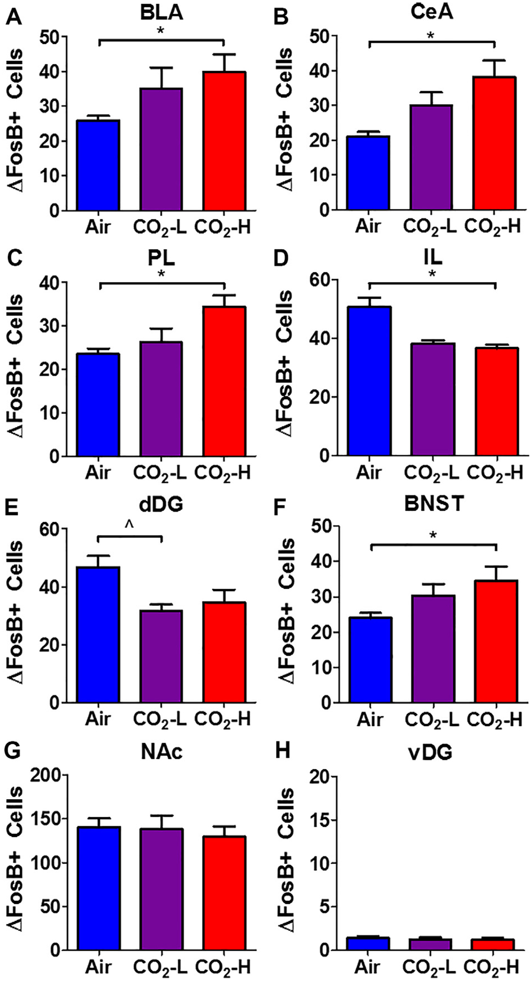
Fig. 5.

Regional ΔFosB+ cell counts in air, CO2-L and CO2-H mice. Significant alterations in ΔFosB+ cell counts were observed in multiple brain areas: Increased cell counts were observed in (A) basolateral amygdala (BLA), (B) central nucleus of amygdala (CeA), (C) prelimbic cortex (PL), (F) bed nucleus of stria terminalis (BNST) of CO2-H mice. (D) Significantly reduced cell counts were observed in CO2-H mice within the infralimbic cortex (IL). (E) Reductions were also observed within the dorsal dentate gyrus (dDG). No significant differences were noted within the (G) nucleus accumbens (NAc) and (H) ventral dentate gyrus (vDG). Data are mean ± SEM. *p < 0.05 CO2-H versus air, p < 0.05 CO2-L versus air.
