Abstract
There is ambiguity about the airborne transmission of the SARS-CoV-2. While a distance of 6 feet is considered a safe physical distance, new findings show that the virus can be transmitted more than that distance and cause infection. In hospitals, this may cause the virus to be transmitted from the treatment wards of COVID-19 patients to adjacent wards and infect medical staff, non-COVID-19 patients, and patient companions. The aim of this study was to investigate the presence of coronavirus in the air of ICU and adjacent wards. The low volume sampler (LVS) with two separate inlets for PM2.5 and PM10 was applied to collect indoor air of intensive care unit (ICU) with confirmed COVID- 19 patients and its surroundings. The samples were collected on 0.3μ PTFE filter fitted to the holder. Sampling was done at flow rate of 16.7 l/min for 24 h. The SRAS-CoV-2 virus was isolated using a SinaPure™ Virus Extraction Kit (SINACLON, Iran). The presence of SARS-CoV-2 genome was assessed using a commercially available SARS-CoV-2 Test Kit (Pishtaz-Iran), according to the manufacturer’s instructions using One Step plus Real-Time PCR system tool (Applied Biosystems, USA). A total of sixteen samples were taken, and the positive test rate for SRAS-CoV-2 was 12.5 % (2/16). All samples from surrounding (rest room and hallway) were negative, but two air samples from indoor of ICU (next to the patient bed and nursing station) were found to be positive. The results support the possibility of transmitting the SRAS-CoV-2 through the air at a greater distance than what is known as a safe physical distance. Therefore, in addition to maintaining a safe physical distance, other precautions including wearing a face mask, preventing air recirculation, and maximizing the use of natural ventilation should be considered, especially in crowded and enclosed environments.
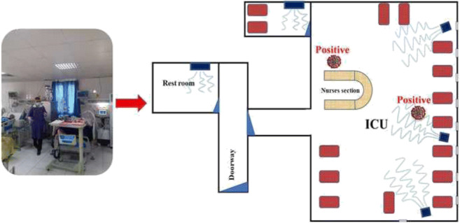
Graphical abstract

Supplementary Information
The online version contains supplementary material available at 10.1007/s11356-021-16010-x.
Keywords: Airborne transmission, Hospital, ICU, Particulate matter, SARS-CoV-2
Introduction
A novel human coronavirus, severe acute respiratory syndrome coronavirus 2 (SARS-CoV-2) which is considered the seventh member of the coronavirus family, was first detected in Wuhan, China, in late 2019 (Dindarloo et al. 2020, Faridi et al. 2020, Razzini et al. 2020, Yousefi et al. 2021). On 11 February 2020, the World Health Organization (WHO) formally named this disease as COVID-19 (Anonymous 2020). The disease has now spread to all countries of the world. Globally, as of 3 August 2021, more than 199,00,000 confirmed cases, and more than 4,240,000 deaths of COVID-19 in more than 200 countries, areas, and territories have been reported to WHO (Anonymous 2021). Coronaviruses are respiratory pathogens, and the SARS-CoV-2 has been identified in the samples taken from both upper and lower respiratory tract of patients (Bahl et al. 2020). Talking, sneezing, coughing, and singing can cause particles to emit with a wide range of sizes from 1 to 2000 μm, with the majority ranging from 2 to 100 μm (Carducci et al. 2020). The half-life of SARS-CoV-2 virus in airborne particles is more than 1 h. Therefore, it can cause more infection and further spreading of the disease upon inhalation by people (Morawska et al. 2020). Although transmission modes including direct (person-to-person) and indirect (fomite) contact, inhaling large respiratory droplet (> 5 μm), and scarcely fecal-oral route have been reported for the transmission of COVID-19 (da Silva et al. 2020, Dindarloo et al. 2020, Gholipour et al. 2020, Morawska et al. 2020, Wathore et al. 2020, Zuo et al. 2020), they still remain controversial (Orenes-Piñero et al. 2021). Depending on environmental conditions such as temperature, humidity, and particulate matter concentrations, viral particles can attach to aerosol particles and remain suspended in the air for longer durations (Zuo et al. 2020). In some circumstances, aerosol particles (< 5 μm) may be produced by infected individuals and travel more than the 1.50 m commonly known as safe physical distance (Birgand et al. 2020, Setti et al. 2020). It has even been stated that the small particles containing the virus can be transmitted up to 10 m from the source of the spread (Setti et al. 2020). Therefore, inhaling small airborne droplet (aerosol), especially in closed and crowded places, could be considered as another probable route of COVID-19 transmission (Al Huraimel et al. 2020, Morawska et al. 2020).
Another issue that has been less considered in research is the role of ventilation systems in transmission of COVID-19. The increasing number of people afflicted with COVID-19 in Iran, especially in warm climate regions where the air conditioners are required to be turned on in the summer, has intensified the hypothesis that these systems are a possible cause of increased COVID-19 patients in these areas. Due to the rapid spread of the coronavirus, there is an urgent need to investigate the possibility of airborne transmission [9]. Therefore, the aims of this study were to investigate (1) the presence of the SARS-CoV-2 in indoor air of COVID-19 patients ICU and its surroundings; (2) the role of cooling systems in transmission of SARS-CoV-2; and (3) the impact of environmental conditions such as particulate matter concentration, humidity, and temperature in the presence of SARS-CoV-2 in ICU and its surroundings.
Material and methods
Study design
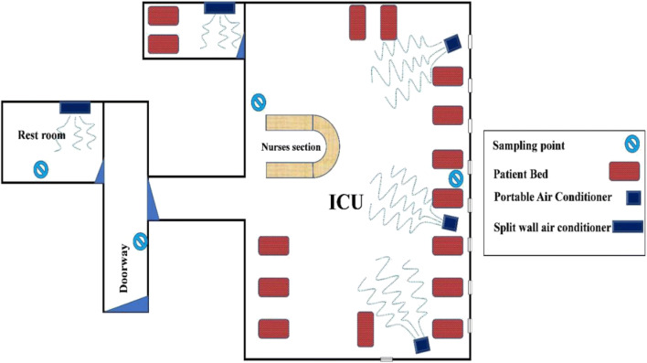
This cross-sectional study was conducted in intensive care unit (ICU) with confirmed COVID-19 at Shahid Mohammadi Hospital Complex, Bandar Abbas, Iran, in November and December 2020. The population of Bandar Abbas city is ∼ 680,000, and the city has a high rate of daily commute due to industrial and commercial activities. The first positive confirmed case of COVID-19 in Bandar Abbas city occurred on 23 February 2020. Subsequently, five hospitals were prepared for admission of COVID-19 patients with critical severe and symptoms. We investigated indoor air of COVID-19 ICU ward for detection of SARS-CoV-2. Detection of SARS-CoV-2 was conducted in the four sections of ICU including the patient section, nurse station, rest room, and doorway of ICU (Figure 1). The distance from the rest room and sampling point in doorway to the nearest patient bed was about 11 and 7 m, respectively. The area of ICU and rest room was about 220 and 20 m2, respectively.
Fig. 1.
Different locations of sampling in ICU ward of COVID-19 patient and its surroundings
Air sampling
The low volume sampler (LVS) (ESPS Model, Fanpaya) was applied to collect SARS-CO-2 virus bound to PM2.5 and PM10 (ESPS 2020). PTFE membrane filters with a pore size of 0.3 μm (Liu et al. 2020) were inserted in the PM2.5 and PM10 inlets of LVS. The sampling was done at a flow rate of 16.7 l/min for a period of 24 h with a total of 24048 L of air sucked for each sample (Marple et al. 1990). The LVS was installed in the above four mentioned sections of ICU at the height of 1.8 m from the floor (Figure 1). Sampling was conducted during 8 days (two samples per day). After sampling, PTFE filters were retaken from inlets of LVS and entered into a microtube with viral transport medium (VTM) culture at – 4 °C (McAuley et al. 2021). The samples were stored in – 20 °C and transferred to Professor Alborzi Clinical Microbiology Research Center in Namazi Hospital in Shiraz, Iran.
Samples were stored at this temperature before further analysis. During the sampling, indoor air concentration of PM2.5 and PM10, TSP, air temperature, and relative humidity were also monitored in time intervals of 3 min. In addition, other variables including the number and condition (closed or open) of doors and windows, area and volume of the four sections of the ICU, number of patients and nurses, and number of ventilation system were determined.
SARS-CoV-2 extraction and detection
All samples were shipped to PACMRC under cold conditions. In this study, samples were initially placed in 50 ml falcon containing 10 ml of phosphate-buffered saline (PBS) and stored in the refrigerator. Then, according to the manufacturer’s instructions, the SinaPure ™ Virus Extraction Kit (SINACLON, Iran) was applied to isolate the SARS-CoV-2 virus. Finally, the isolated RNA was tested using a one-step rRT-PCR for the SARS-CoV-2 virus. The presence of SARS-CoV-2 genome was assessed using a commercially available SARS-CoV-2 Kit (Pishtaz-Iran), according to the manufacturer’s instructions using One Step plus Real-Time PCR system tool (Applied Biosystems, USA). The SARS-CoV-2 Test Kit is a molecular in vitro diagnostic test that uses Taqman probe-based technology for the qualitative detection of SARS-CoV-2. The N and RdRp genes were the target for the detection of the virus, and RNase P was also used as an internal control. This also serves as the extraction control to ensure that samples resulting as negative contain extracted nucleic acid for testing. The positive control must be positive at a Ct value of 30.96 ± 2.00.
Results
Table 1 present the results of SARS-CoV-2 detection in ICU and its surroundings. This table additionally displays the environmental status, PM10, PM2.5, and TSP concentration in sampling location. A total of 16 samples were taken from the ICU and its surroundings (staff rest room and corridor leading to the ward). In fact, 4 samples were taken from each of the four selected locations. Out of 16 samples taken, 2 positive samples were observed, and the positive rate of the samples was found to be 12.5%. Of the two positive samples observed, one was taken from near the patient’s bed and another one from the nursing station. Data on the concentration of PM10, PM2.5, TSP, temperature, and relative humidity in the ICU is presented in the Appendix A.
Table 1.
The result of virus detection in different locations of ICU ward of COVID-19
| Day | Ward | bPM2.5 bound | PM10 bound | PM2.5 (μgm-3) | PM10 (μgm-3) | cTSP (μgm-3) | dT (° C) | fRH (%) |
|---|---|---|---|---|---|---|---|---|
| 1a | Patient bed | Negative | Positive | 25.7 ± 4.6 | 32.6 ± 5.9 | 36.0 ± 6.6 | 28.0 ± 1.0 | 48.3 ± 5.8 |
| 2 | Patient bed | Negative | Negative | 25.3 ± 5.9 | 31.9 ± 9.1 | 35.2 ± 10.2 | 26.5 ± 1.0 | 44.5 ± 5.5 |
| 3 | Nursing section | Negative | Negative | 24.1 ± 6.5 | 34.1 ± 7.9 | 38.1 ± 9.1 | 25.9 ± 0.7 | 42.1 ± 5.7 |
| 4 | Nursing section | Positive | Negative | 21.6 ± 4.6 | 29.1 ± 6.4 | 32.0 ± 7.1 | 25.6 ± 0.3 | 42.0 ± 3.6 |
| 5 | Hallway | Negative | Negative | 15.82 ± 9.27 | 27.06 ± 10.71 | 37.32 ± 20.33 | 27.43 ± 0.68 | 36.38 ± 7.01 |
| 6 | Hallway | Negative | Negative | 15.02 ± 4.20 | 20.83 ± 4.36 | 24.36 ± 6.56 | 27.78 ± 0.77 | 46.93 ± 5.39 |
| 7 | Rest room | Negative | Negative | 15.83 ± 2.34 | 20.42 ± 6.18 | 22.91 ± 10.31 | 27.33 ± 0.63 | 47.27 ± 3.87 |
| 8 | Rest room | Negative | Negative | 12.81 ± 5.52 | 20.69 ± 13.12 | 26.23 ± 23.38 | 27.52 ± 1.06 | 37.32 ± 6.16 |
aTwo samples per day
bParticulate matter
cTotal suspended particulate
dTemperature
fRelative humidity
Discussion
In the current study, we observed that SARS-CoV-2 bound to PM2.5 and PM10 were present in the indoor air of the ICU. The results of this study were similar to previous studies that observed SARS-CoV-2 in the indoor air (Guo et al. 2020, Jin et al. 2021, Kenarkoohi et al. 2020, Santarpia et al. 2020, Yuan et al. 2020) . The results of these studies confirmed the hypothesis of airborne transmission of SARS-CoV-2. In addition, many studies revealed that parameters including temperature and humidity can influence on the spread of virus in the indoor air (Chen et al. 2015, Guo et al. 2020, Santarpia et al. 2020, Yuan et al. 2020). When an infected person coughs or sneezes, respiratory droplets containing the virus are released into the indoor air along with high humidity. The size of droplets is reduced due to evaporation of the water droplet when surrounding humidity and temperature are low in the indoor air. Conversely, the size of droplet is not reduced in the high humidity and temperature (Chen et al. 2015, Wu et al. 2016, Yuan et al. 2020). At the same time, air humidity can act as a diluter and reduce viral activity. Therefore, it may have a beneficial role in controlling the virus. In our study, the relative humidity in the indoor air of ICU was at the range of 42.00 to 48.3% due to use of 3 portable air conditioners (Figure 1). Therefore, low relative humidity may increase the activity of SARS-CO-2 by reduction of droplets size. Peak size distribution of the SARS-CoV-2 is between 0.25 and 1.0 μm (Yuan et al. 2020). Particulate matter with aerodynamic diameters smaller than 0.5 μm can be more suspended in the indoor air (Ai et al. 2019). Therefore, the smaller the airborne particle size, the greater the chance of virus transmission via air. In addition, in our study, the volume of air sucked during sampling by LVS was 24048 L, which was much higher than many previous studies (Guo et al. 2020, Jin et al. 2021, Santarpia et al. 2020, Yuan et al. 2020). As the sampling rate increases, more virus-bound particles can be trapped onto the filter, thus increasing the chances of the samples to be positive in the PCR test.
While the distance from the nursing station to the confirmed COVID-19 patient was greater than the social distance (6 feet), a positive sample was observed in this area. The most probable explanation for the detected positive sample in the nursing station could be the transmission of the virus by airborne aerosol particles. Although there is controversial regarding airborne transmission of SARS-CoV-2, positive samples have been reported in indoor air of enclosed environments (Paules et al. 2020, Razzini et al. 2020). Transmission of coronavirus by airborne micro-droplets is suggested to be the third route of SARS-CoV-2 transmission (Morawska & Cao 2020). Airborne transmission of SARS-CO-2 in hospitals, healthcare facilities, and airplane has been reported in previous studies, and it was stated to be the most important route of SARS-CoV-2 transmission in the mentioned environments. After the droplets leave the source, the liquid begins to evaporate, and the droplets become so small that the air flow has a greater effect on them than the force of gravity. As a result, small particles containing the virus can travel even more than 10 m from the source of the spread (Setti et al. 2020). Van Doremalen’s study showed that SARS-CoV-2 virus is more stable in aerosols and surfaces than SARS-CoV-1 and can survive for hours in aerosols (Van Doremalen et al. 2020). In a study by Gholipour et al., viral RNA of SARS-CoV-2 was detected in 40% of air samples taken from around the wastewater treatment plant. Their results support the airborne transmission of SARS-CoV-2 (Gholipour et al. 2021). In a systematic review study, Carducci et al. reviewed three groups of articles, including air monitoring studies, laboratory studies, and epidemiological studies and airflow modeling. Airborne transmission of COVID-19 virus has been suggested by all three groups of studies reviewed by them. They concluded that while more evidence is needed, preventive precautions such as wearing a mask, keeping a safe physical distance, and air conditioning should be considered (Carducci et al. 2020). In a review study by Bahl, it was found that out of 10 studies, 8 studies showed that droplets can move up to a distance of more than 2 m and even 8 m. It has also been found that a large number of studies support the transmission of COVID-19 virus by aerosol, and one study has shown that the virus is transmitted more than 4 m from the source of transmission (Bahl et al. 2020).
With respect to the above, all precautions to reduce the transmission of the virus by air in closed environments including increasing the amount of ventilation, using natural ventilation, preventing air circulation, avoiding standing in the direct path of another person’s air flow, and reducing the number of people indoors should be taken into consideration (Morawska & Cao 2020). One of the most important measures is to maximize natural ventilation. These precautions are concentrated in public places where there is a higher risk of infection due to the greater likelihood of droplets carrying the virus in the air, a more stable virus in the indoor air, and a higher population density.
Although care for proper ventilation is a common practice in many hospitals, it is not often performed in all hospitals where new patients are admitted. Nursing homes, shops, malls, schools, restaurants, cruise ships, and, of course, public transportation are places where ventilation methods should be reviewed and ventilation maximized. Also, personal protective equipment (PPE), especially masks and respirators, should be recommended for use in public places where population density are high and ventilation is potentially inadequate (Morawska & Cao 2020).
Another issue that can contribute to a positive sample at the nursing station is the contribution of the cooling system to the transmission of the virus to this sampling point. As shown in the Figure 1, the sampling point in the nursing station is located in front of the wind path blowing from cooling systems. The outlet air from the cooling systems, especially the cooling system number one, passes over the patient and then flows to the sampling point in the nursing station. There are three hypothetical ways for the ventilation system to contribute to the spread of the corona virus, including circulating air in a closed environment where there are infected patients, circulating air by ventilation systems to adjacent floors and sections, and replacing polluted air with clean outside air that can pollute the environment around the ventilation system. Ventilation systems have been also reported as a route of transmission/spread of infectious diseases such as measles, tuberculosis, chickenpox, influenza, smallpox, and SARS (Morawska & Cao 2020).
Feng et al. studied the role of wind flow velocity and humidity in the transmission of the SARS-CoV-2 through the computational fluid-particle dynamics (CFPD) model. They found that the micro-droplets followed the airflow well and were observed at a distance of 3.05 m from the emission source. They stated that the rest of the droplets can be carried by the air at a distance of more than 3.05 m (10 feet) due to wind convection and pose a potential health risk to the nearby people. The main conclusion of this study was that the effect of wind on the transport and deposition of droplets is complex and highly depends on the wind flow patterns and localized secondary flow intensities between the two virtual humans, as well as the steadiness of the wind (Feng et al. 2020). Anchordoqui et al. used the fluid dynamic method to analyze the behavior of droplets containing the virus in the air at different sizes. They showed that vortices in the air could make a location remote from the source of the virus more dangerous than a place closer to it (e.g., 6 feet away) (Anchordoqui & Chudnovsky 2020). In another study by Somsen et al., it was found that droplets smaller than 5 microns produced at a height of 160 cm above the ground took 9 min to reach the ground. These tiny droplets are very important because they are associated with SARS-CoV-2 aerosol transmission. In their study, the role of ventilation in the retention of particles smaller than 5 microns in the air is also investigated. According to the results of their study, in unventilated conditions, single mechanical ventilation and mechanical ventilation with door and window openings respectively take 5 min, 1.4 min, and 30 s to reduce the number of droplets by half (Somsen et al. 2020).
Heating, ventilation, and air conditioning (HVAC) systems are used as the primary means of controlling infectious diseases. However, if these systems do not used properly, it may help transmit/spread airborne diseases as previously suggested for SARS. Two suggestions for reducing the spread of coronavirus in closed environments are to prevent air recirculation and to increase the entry of air from outside (Correia et al. 2020).
Most of the samples were negative despite the definite sources of SARS-CoV-2 virus in the ICU, no air exchange (windows closed), and also high-volume air for sampling. One of the reasons for the negativity of most samples is the detection limit of the RT-PCR method, which has a 30% error. For a sample to be positive, the virus load needs to reach the detection limit of the PCR method. Therefore, existence of negative samples does not indicate that there is no virus in the indoor air of ICU. In addition, fluctuations in the spread of the virus by patients can affect the number of negative samples (or simply: negative results). Additionally, the spread of the virus may have been reduced due to the clinical condition of patients, number of nurses and patients, and the use of protective equipment such as masks by patients during sampling (Morawska & Cao 2020).
T-test statistical analysis revealed that concentration of particulate matter (PM2.5, PM10, and TSP) in ICU was significantly (p < 0.05) higher than the other wards (rest room and doorway). The high concentration of PM in the indoor air of ICU may be due to sneezing and coughing, suction operations (evacuation of lung infections), movement of nurses, and the transfer of equipment and vehicles. Also, some factors such as the use of split air conditioner by continuous circulation of the indoor air of the ICU increase the retention time of PM in the indoor air of ICU. The study by Wang et al. showed that each 10 μg/m3 increase in the concentration of PM2.5 and PM10 was positively associated with the number of confirmed cases of COVID-19 (Somsen et al. 2020). Dunker et al. suggested that in area with high PM concentrations, virus transmission by PM can be considered an additional route (Dunker et al. 2021). In addition to the role of PM in transmitting the virus, PM can increase the infectivity of the virus and mortality among people, especially children and adults, by affecting the immune system (Wathore et al. 2020). Although more detailed studies are needed to prove the role of particulate matter in coronavirus transmission, with the observed two positive samples just in the ICU, this hypothesis that an increase in transmission of SARS-CoV-2 is probably associated with an increase in the concentration of PM can be supported.
Conclusion
In this study, SARS-CoV-2 was investigated in the ICU ward and its surroundings by considering environmental parameters including PM10, PM2.5, and TSP concentration; humidity and temperature; and the role of the cooling system. In total, 8 samples were taken from ICU environment and 8 samples from the surrounding wards. Two samples (2.5%) were found positive, both of which belonged to the ICU. These samples were taken from the places which had higher concentrations of PM. The results of this study support the transmission of SARS-CoV-2 by airborne aerosols and this hypothesis that higher concentrations of PM may be involved in the transmission of the SARS-CoV-2. Also, according to the results, the cooling system may be effective in transporting the virus to a distance greater than what is considered a safe physical distance. Therefore, in addition to maintaining a safe physical distance, other precautions, including wearing a mask, maximizing the use of natural ventilation, preventing air circulation in the closed environment, and using central ventilation system equipped with suspended particle control filters, should be considered.
Supplementary Information
(XLSX 192 kb)
Author’s contribution
Sampling and detection of COVID-19 were conducted by Hamid Reza Ghaffari, Yadolah Fakhri, and Marzieh Jamalidoust; analysis of data and preparing the manuscript were conducted by Hamid Reza Ghaffari, Hossein Farshidi, Vali Alipour, Kavoos Dindarloo, Mehdi Hassani Azad, Marzieh Jamalidoust, Abdolhossein Madani, Teamour Aghamolaei, Yaser Hashemi, Mehdi Fazlzadeh, and Yadolah Fakhri.
Funding
The authors would like to thank Hormozgan University of Medical Sciences for the financial grants of this study.
Data Availability
Data openly available in a public repository.
Declarations
Ethics approval
Ethical approval was approved from the Hormozgan University of Medical Sciences Ethics Committee (Ethical code: IR.HUMS.REC.1399.226).
Consent to participate
Not applicable.
Consent for publication
Not applicable.
Conflict of interest
The authors declare no competing interests.
Footnotes
Publisher’s note
Springer Nature remains neutral with regard to jurisdictional claims in published maps and institutional affiliations.
Contributor Information
Marzieh Jamalidoust, Email: mjamalidoust@gmail.com.
Yadolah Fakhri, Email: ya.fakhri@gmail.com.
References
- Ai Z, Huang T, Melikov A. Airborne transmission of exhaled droplet nuclei between occupants in a room with horizontal air distribution. Build Environ. 2019;163:106328. doi: 10.1016/j.buildenv.2019.106328. [DOI] [Google Scholar]
- Al Huraimel K, Alhosani M, Kunhabdulla S, Stietiya MH (2020) SARS-CoV-2 in the environment: modes of transmission, early detection and potential role of pollutions. Sci Total Environ 140946 [DOI] [PMC free article] [PubMed]
- Anchordoqui LA, Chudnovsky EM. A physicist view of COVID-19 airborne infection through convective airflow in indoor spaces. Sci Med J. 2020;2:68–72. [Google Scholar]
- Anonymous (2020): WHO Director-General's remarks at the media briefing on 2019-nCoV on 11 February 2020. WHO
- Anonymous (2021): WHO coronavirus disease (COVID-19) Dashboard. WHO
- Bahl P, Doolan C, De Silva C, Chughtai AA, Bourouiba L, MacIntyre CR (2020): Airborne or droplet precautions for health workers treating COVID-19? The Journal of infectious diseases [DOI] [PMC free article] [PubMed]
- Birgand G, Peiffer-Smadja N, Fournier S, Kerneis S, Lescure F-X, Lucet J-C. Assessment of air contamination by SARS-CoV-2 in hospital settings. JAMA Netw Open. 2020;3:e2033232–e2033232. doi: 10.1001/jamanetworkopen.2020.33232. [DOI] [PMC free article] [PubMed] [Google Scholar]
- Carducci A, Federigi I, Verani M. Covid-19 airborne transmission and its prevention: waiting for evidence or applying the precautionary principle? Atmosphere. 2020;11:710. doi: 10.3390/atmos11070710. [DOI] [Google Scholar]
- Chen C, Liu W, Lin C-H, Chen Q. Comparing the Markov chain model with the Eulerian and Lagrangian models for indoor transient particle transport simulations. Aerosol Sci Technol. 2015;49:857–871. doi: 10.1080/02786826.2015.1079587. [DOI] [Google Scholar]
- Correia G, Rodrigues L, Da Silva MG, Gonçalves T. Airborne route and bad use of ventilation systems as non-negligible factors in SARS-CoV-2 transmission. Med Hypo. 2020;141:109781. doi: 10.1016/j.mehy.2020.109781. [DOI] [PMC free article] [PubMed] [Google Scholar]
- da Silva PG, Nascimento MSJ, Soares RR, Sousa SI, Mesquita JR (2020) Airborne spread of infectious SARS-CoV-2: moving forward using lessons from SARS-CoV and MERS-CoV. Sci Total Environ 142802 [DOI] [PMC free article] [PubMed]
- Dindarloo K, Aghamolaei T, Ghanbarnejad A, Turki H, Hoseinvandtabar S, Pasalari H, Ghaffari HR. Pattern of disinfectants use and their adverse effects on the consumers after COVID-19 outbreak. J Environ Health Sci Eng. 2020;18:1301–1310. doi: 10.1007/s40201-020-00548-y. [DOI] [PMC free article] [PubMed] [Google Scholar]
- Dunker S, Hornick T, Szczepankiewicz G, Maier M, Bastl M, Bumberger J, Treudler R, Liebert UG, Simon J-C. No SARS-CoV-2 detected in air samples (pollen and particulate matter) in Leipzig during the first spread. Sci Total Environ. 2021;755:142881. doi: 10.1016/j.scitotenv.2020.142881. [DOI] [PMC free article] [PubMed] [Google Scholar]
- ESPS (2020): Environmental suspended particulate sampling (ESPS). https://cdn.ov2.com/content/fanpaya_ov2_com/wp-content_85/uploads/2020/10/%DA%A9%D8%A7%D8%AA%D8%A7%D9%84%D9%88%DA%AF-003.pdf. In: Fanpaya (Hrsg.)
- Faridi S, Niazi S, Sadeghi K, Naddafi K, Yavarian J, Shamsipour M, Jandaghi NZS, Sadeghniiat K, Nabizadeh R, Yunesian M (2020): a field indoor air measurement of SARS-CoV-2 in the patient rooms of the largest hospital in Iran. Science of the Total Environment 725, 138401 [DOI] [PMC free article] [PubMed]
- Feng Y, Marchal T, Sperry T, Yi H. Influence of wind and relative humidity on the social distancing effectiveness to prevent COVID-19 airborne transmission: a numerical study. Journal of aerosol science. 2020;147:105585. doi: 10.1016/j.jaerosci.2020.105585. [DOI] [PMC free article] [PubMed] [Google Scholar]
- Gholipour S, Nikaeen M, Manesh RM, Aboutalebian S, Shamsizadeh Z, Nasri E, Mirhendi H. Severe acute respiratory syndrome coronavirus 2 (SARS-CoV-2) contamination of high-touch surfaces in field settings. Biomed Environ Sci. 2020;33:925–929. doi: 10.3967/bes2020.126. [DOI] [PMC free article] [PubMed] [Google Scholar]
- Gholipour S, Mohammadi F, Nikaeen M, Shamsizadeh Z, Khazeni A, Sahbaei Z, Mousavi SM, Ghobadian M, Mirhendi H. COVID-19 infection risk from exposure to aerosols of wastewater treatment plants. Chemosphere. 2021;273:129701. doi: 10.1016/j.chemosphere.2021.129701. [DOI] [PMC free article] [PubMed] [Google Scholar]
- Guo Z-D, Wang Z-Y, Zhang S-F, Li X, Li L, Li C, Cui Y, Fu R-B, Dong Y-Z, Chi X-Y. Aerosol and surface distribution of severe acute respiratory syndrome coronavirus 2 in hospital wards, Wuhan, China, 2020. Emerg Infect Diseas. 2020;26:1586. doi: 10.3201/eid2607.200885. [DOI] [PMC free article] [PubMed] [Google Scholar]
- Jin T, Li J, Yang J, Li J, Hong F, Long H, Deng Q, Qin Y, Jiang J, Zhou X. SARS-CoV-2 presented in the air of an intensive care unit (ICU) Sustain Cities Soc. 2021;65:102446. doi: 10.1016/j.scs.2020.102446. [DOI] [PMC free article] [PubMed] [Google Scholar]
- Kenarkoohi A, Noorimotlagh Z, Falahi S, Amarloei A, Mirzaee SA, Pakzad I, Bastani E. Hospital indoor air quality monitoring for the detection of SARS-CoV-2 (COVID-19) virus. Sci Total Environ. 2020;748:141324. doi: 10.1016/j.scitotenv.2020.141324. [DOI] [PMC free article] [PubMed] [Google Scholar]
- Liu Y, Ning Z, Chen Y, Guo M, Liu Y, Gali NK, Sun L, Duan Y, Cai J, Westerdahl D. Aerodynamic analysis of SARS-CoV-2 in two Wuhan hospitals. Nature. 2020;582:557–560. doi: 10.1038/s41586-020-2271-3. [DOI] [PubMed] [Google Scholar]
- Marple VA, Liu BY, Burton RM. High-volume impactor for sampling fine and coarse particles. J Air Waste Manag Assoc. 1990;40:762–767. doi: 10.1080/10473289.1990.10466722. [DOI] [Google Scholar]
- McAuley J, Fraser C, Paraskeva E, Trajcevska E, Sait M, Wang N, Bert E, Purcell D, Strugnell R. Optimal preparation of SARS-CoV-2 viral transport medium for culture. Virol J. 2021;18:1–6. doi: 10.1186/s12985-021-01525-z. [DOI] [PMC free article] [PubMed] [Google Scholar]
- Morawska L, Cao J. Airborne transmission of SARS-CoV-2: The world should face the reality. Environ Intl. 2020;139:105730. doi: 10.1016/j.envint.2020.105730. [DOI] [PMC free article] [PubMed] [Google Scholar]
- Morawska L, Tang JW, Bahnfleth W, Bluyssen PM, Boerstra A, Buonanno G, Cao J, Dancer S, Floto A, Franchimon F. How can airborne transmission of COVID-19 indoors be minimised? Environ Intl. 2020;142:105832. doi: 10.1016/j.envint.2020.105832. [DOI] [PMC free article] [PubMed] [Google Scholar]
- Orenes-Piñero E, Baño F, Navas-Carrillo D, Moreno-Docón A, Marín JM, Misiego R, Ramírez P. Evidences of SARS-CoV-2 virus air transmission indoors using several untouched surfaces: a pilot study. Sci Total Environ. 2021;751:142317. doi: 10.1016/j.scitotenv.2020.142317. [DOI] [PMC free article] [PubMed] [Google Scholar]
- Paules CI, Marston HD, Fauci AS. Coronavirus infections—more than just the common cold. Jama. 2020;323:707–708. doi: 10.1001/jama.2020.0757. [DOI] [PubMed] [Google Scholar]
- Razzini K, Castrica M, Menchetti L, Maggi L, Negroni L, Orfeo NV, Pizzoccheri A, Stocco M, Muttini S, Balzaretti CM. SARS-CoV-2 RNA detection in the air and on surfaces in the COVID-19 ward of a hospital in Milan, Italy. Sci Total Environ. 2020;742:140540. doi: 10.1016/j.scitotenv.2020.140540. [DOI] [PMC free article] [PubMed] [Google Scholar]
- Santarpia JL, Rivera DN, Herrera V, Morwitzer MJ, Creager H, Santarpia GW, Crown KK, Brett-Major D, Schnaubelt E, Broadhurst MJ (2020): Transmission potential of SARS-CoV-2 in viral shedding observed at the University of Nebraska Medical Center. MedRxiv
- Setti L, Passarini F, De Gennaro G, Barbieri P, Perrone MG, Borelli M, Palmisani J, Di Gilio A, Piscitelli P, Miani A (2020): Airborne transmission route of COVID-19: why 2 meters/6 feet of inter-personal distance could not be enough. Multidisciplinary Digital Publishing Institute [DOI] [PMC free article] [PubMed]
- Somsen GA, van Rijn C, Kooij S, Bem RA, Bonn D. Small droplet aerosols in poorly ventilated spaces and SARS-CoV-2 transmission. Lancet Respiratory Med. 2020;8:658–659. doi: 10.1016/S2213-2600(20)30245-9. [DOI] [PMC free article] [PubMed] [Google Scholar]
- Van Doremalen N, Bushmaker T, Morris DH, Holbrook MG, Gamble A, Williamson BN, Tamin A, Harcourt JL, Thornburg NJ, Gerber SI. Aerosol and surface stability of SARS-CoV-2 as compared with SARS-CoV-1. New England J Med. 2020;382:1564–1567. doi: 10.1056/NEJMc2004973. [DOI] [PMC free article] [PubMed] [Google Scholar]
- Wathore R, Gupta A, Bherwani H, Labhasetwar N. Understanding air and water borne transmission and survival of coronavirus: insights and way forward for SARS-CoV-2. Sci Total Environ. 2020;749:141486. doi: 10.1016/j.scitotenv.2020.141486. [DOI] [PMC free article] [PubMed] [Google Scholar]
- Wu Y, Zhang X, Zhang X. Simplified analysis of heat and mass transfer model in droplet evaporation process. Appl Ther Eng. 2016;99:938–943. doi: 10.1016/j.applthermaleng.2016.01.020. [DOI] [Google Scholar]
- Yousefi M, Oskoei V, Jonidi Jafari A, Farzadkia M, Hasham Firooz M, Abdollahinejad B, Torkashvand J. Municipal solid waste management during COVID-19 pandemic: effects and repercussions. Environ Sci Pollut Res. 2021;28:32200–32209. doi: 10.1007/s11356-021-14214-9. [DOI] [PMC free article] [PubMed] [Google Scholar]
- Yuan L, Zhi N, Yu C, Ming G, Yingle L, Kumar GN, Li S, Yusen D, Jing C, Dane W (2020): Aerodynamic characteristics and RNA concentration of SARS-CoV-2 aerosol in Wuhan hospitals during COVID-19 outbreak. BioRxiv
- Zuo YY, Uspal WE, Wei T. Airborne transmission of COVID-19: aerosol dispersion, lung deposition, and virus-receptor interactions. ACS nano. 2020;14:16502–16524. doi: 10.1021/acsnano.0c08484. [DOI] [PubMed] [Google Scholar]
Associated Data
This section collects any data citations, data availability statements, or supplementary materials included in this article.
Supplementary Materials
(XLSX 192 kb)
Data Availability Statement
Data openly available in a public repository.


