Lowe discusses recent work from Welch et al. describing a central role for GOLPH3 in sorting Golgi proteins to maintain Golgi homeostasis.
Abstract
In this issue of JCB, Welch et al. (2021. J. Cell Biol. https://doi.org/10.1083/jcb.202106115) show that GOLPH3 mediates the sorting of numerous Golgi proteins into recycling COPI transport vesicles. This explains how many resident proteins are retained at the Golgi and reveals a key role for GOLPH3 in maintaining Golgi homeostasis.
The Golgi apparatus lies at the heart of the secretory pathway, where its major functions are the posttranslational modification of cargo proteins and lipids, particularly at the level of glycosylation, and the sorting of cargo to its correct onward destination. The Golgi is composed of stacked membrane compartments called cisternae, which contain numerous resident enzymes that act on the cargo as it passes through the organelle, from the entry or cis side to the exit or trans side. Each resident enzyme has its own distribution within the Golgi stack, resulting in the sequential modification of the secretory cargo as it moves through the Golgi.
Various mechanisms exist to ensure that Golgi residents are retained within the Golgi despite the huge flux of protein and lipid through this organelle (1). Major players are COPI vesicles, which recycle Golgi residents from later to earlier cisternae, at the same time as the cisternae are thought to slowly migrate across the stack, as on a conveyor belt, progressively changing composition in a process referred to as cisternal maturation (2). Unlike the Golgi resident enzymes, which enter recycling vesicles, cargo is thought to remain within the maturing cisternae as it moves through the Golgi. Certain Golgi enzymes can bind directly to the COPI coat, explaining their inclusion in COPI vesicles (3), but for other enzymes and resident proteins, their retention mechanism is less obvious.
Previous studies on the peripheral Golgi membrane protein GOLPH3 and its paralogue GOLPH3L (herein I will refer to both proteins as GOLPH3) indicated it can bind to certain Golgi enzymes and to the COPI coat, thereby acting as an adaptor to mediate sorting of these enzymes into COPI vesicles (4, 5). This was first shown for the yeast orthologue Vps74p (6, 7) and has also been demonstrated for the Drosophila version of the protein (8), consistent with a conserved function in Golgi enzyme retention. However, the extent to which GOLPH3 might participate in retention of different Golgi enzymes and other resident proteins, and its importance relative to other methods of protein retention in the Golgi, has remained unclear. Indeed, a recent study suggested that GOLPH3 selectively mediates the retention of enzymes involved in glycosphingolipid synthesis, consistent with a fairly selective role in retaining only a subset of resident Golgi enzymes (9). It should also be noted that GOLPH3 has been implicated in other functions, namely budding of exocytic vesicles from the Golgi, the DNA damage response, and mechanistic target of rapamycin signaling (10).
In their current paper, Welch et al. used a combination of approaches to reassess the role of GOLPH3 at the Golgi (11). Using proteomics, they could identify numerous GOLPH3 binding partners, which included COPI, as expected, and a large number of other Golgi residents, including numerous Golgi enzymes and other membrane proteins. The ability of GOLPH3 to retain enzymes at the Golgi was confirmed using microscopy and an innovative flow cytometry–based assay to quantify surface versus Golgi abundance. The large number of possible interactors suggested that GOLPH3 could mediate the Golgi retention of many proteins. To further assess this possibility, the authors took advantage of previous observations showing that Golgi enzymes may be misrouted to the lysosome and degraded upon their failure to be retained in the Golgi (6, 7, 9). Using mass spectrometry, they could show that numerous Golgi resident proteins were depleted in GOLPH3 knockout cells, many of which were also found in the GOLPH3 interactome. This included many enzymes involved in glycosylation, consistent with GOLPH3 playing an important role in maintaining Golgi-dependent glycosylation of proteins and lipids. This was supported by lectin analysis, which showed marked changes in a broad range of glycans in the GOLPH3 knockout cells.
The large number of GOLPH3 clients raises the question as to how it can recognize so many proteins. Previous work has shown binding to the cytoplasmic tails of Golgi enzymes and an interaction motif has been described for Vps74p and more recently for GOLPH3 (6, 9). However, bioinformatics analysis of the many GOLPH3 clients combined with mutational analysis, as performed in the current study, revealed the lack of a consensus sequence for GOLPH3 binding, with the common feature being a strong net positive charge combined with short cytoplasmic tail length. This would result in a high positive charge proximal to the membrane, which likely allows interaction with an acidic patch on the surface of GOLPH3. This mode of binding could mediate selective retention of many Golgi residents, while allowing for the forward trafficking of cargo proteins that have longer, less charged, or folded cytoplasmic domains.
GOLPH3 is an oncogene associated with many types of cancer (12). Several mechanisms have been proposed to account for the oncogenic properties of GOLPH3, but most compelling is that changes in glycosylation are responsible. It was recently shown that GOLPH3-dependent changes in glycosphingolipids affects cell growth by altering mitogenic signaling (9). Changes in glycosylation of surface receptors has also been reported, which can affect surface abundance and hence signaling (13). The new results from Welch et al. suggest that glycosylation of many proteins and lipids may be relevant in cancer and that potentially a broad range of downstream targets contribute to oncogenesis. Such targets could influence processes beyond signaling, including cell adhesion and migration, that are known to be sensitive to changes in the surface glycome and which have been reported in previous studies on GOLPH3 (12).
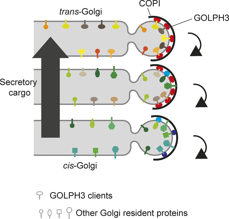
The study by Welch et al. indicates a major role for GOLPH3 in Golgi protein retention (Fig. 1). Clearly though, other retention mechanisms exist, including direct binding to COPI, and transmembrane domain length is also important, where the short transmembrane domain of resident proteins favors partitioning into recycling COPI vesicles and Golgi cisternal membranes of a similar thickness (1). Additional COPI adaptors are also likely, with TM9SF2 recently identified as a likely candidate, being present in Golgi vesicles and able to bind certain Golgi enzymes (1). It is possible that different resident proteins use different adaptors, or that a combination of retention mechanisms act in conjunction for certain residents, providing robustness to the retention process. However, any redundancy would seem incomplete given the strong phenotype seen upon loss of GOLPH3. GOLPH3 is localized toward the trans side of the Golgi, so it is possible that other adaptors, such as TM9SF2 and possibly others, might act earlier in the Golgi, or that direct coat binding is more important within the early Golgi. Hence different residents may be more likely to use different retention mechanisms depending on their location in the Golgi. Because GOLPH3 acts late in the Golgi and can bind many clients, we may think of it as a gatekeeper to prevent loss of numerous Golgi residents from the organelle.
Figure 1.
GOLPH3 plays a major role in Golgi protein retention. Golgi resident proteins, including many glycosylation enzymes, depicted by lollipops, are sorted into recycling COPI vesicles to maintain retention in the Golgi in the face of onward cisternal maturation and secretory cargo transport. Different enzymes are depicted by different lollipop shapes and colors, with GOLPH3 clients indicated by horizontal ovals. Enzymes retained by other mechanisms are depicted by lollipops with circles (transmembrane domain length), squares or vertical ovals (binding to other COPI adaptors, indicated in turquoise and purple), or hexagons (direct binding to the COPI coat). GOLPH3, which is more abundant toward the trans side of the Golgi, has many clients.
With regard to possible future studies, although we have a good idea of how GOLPH3 recognizes its clients, detailed structural analysis will prove informative in elucidating how it can bind so many proteins. Similarly, identification of additional adaptors linking Golgi residents to the COPI coat will be important to generate a more comprehensive view of Golgi protein retention. Finally, in the context of disease, further analysis of the glycoproteins and glycolipids whose levels are altered because of changes in GOLPH3 expression, of which there are likely to be many, should provide significant new insights into the mechanisms underlying GOLPH3-mediated tumorigenesis.
Acknowledgments
I apologize to those authors whose work could not be cited due to space constraints.
Work in the Lowe laboratory is funded by the Biotechnology and Biological Sciences Research Council (BB/S014799/1 and BB/T000945/1) and Leverhulme Trust (RPG-2019-134).
The author declares no competing financial interests.
References
- 1.Welch, L.G., and Munro S.. 2019. FEBS Lett. 10.1002/1873-3468.13553 [DOI] [PubMed] [Google Scholar]
- 2.Papanikou, E., and Glick B.S.. 2014. Curr. Opin. Cell Biol. 10.1016/j.ceb.2014.04.010 [DOI] [PMC free article] [PubMed] [Google Scholar]
- 3.Liu, L., et al. 2018. Proc. Natl. Acad. Sci. USA. 10.1073/pnas.1810291115 [DOI] [Google Scholar]
- 4.Eckert, E.S., et al. 2014. J. Biol. Chem. 10.1074/jbc.M114.608182 [DOI] [Google Scholar]
- 5.Pereira, N.A., et al. 2014. J. Biol. Chem. 10.1074/jbc.M114.548305 [DOI] [PMC free article] [PubMed] [Google Scholar]
- 6.Tu, L., et al. 2008. Science. 10.1126/science.1159411 [DOI] [Google Scholar]
- 7.Schmitz, K.R., et al. 2008. Dev. Cell. 10.1016/j.devcel.2008.02.016 [DOI] [PMC free article] [PubMed] [Google Scholar]
- 8.Chang, W.L., et al. 2013. Development. 10.1242/dev.087171 [DOI] [Google Scholar]
- 9.Rizzo, R., et al. 2021. EMBO J. 10.15252/embj.2020107238 [DOI] [Google Scholar]
- 10.Kuna, R.S., and Field S.J.. 2019. J. Lipid Res. 10.1194/jlr.R088328 [DOI] [PMC free article] [PubMed] [Google Scholar]
- 11.Welch, L.G., et al. 2021. J. Cell Biol. 10.1083/jcb.202106115 [DOI] [PMC free article] [PubMed] [Google Scholar]
- 12.Sechi, S., et al. 2020. Int. J. Mol. Sci. 10.3390/ijms21030933 [DOI] [Google Scholar]
- 13.Arriagada, C., et al. 2020. Int. J. Mol. Sci. 10.3390/ijms21228880 [DOI] [Google Scholar]


