Abstract
Background
The effect of intraindividual variability in lipid levels on the onset of acute coronary syndrome (ACS) remains uncertain. We evaluated the relationship between intraindividual variability in lipid levels and culprit lesion morphologies by optical coherence tomography (OCT).
Methods and Results
Seventy-four consecutive patients with ACS whose cholesterol levels were assessed ≥3 times during outpatient visits before the onset of ACS were enrolled in the study; 222 patients without significant stenotic lesions were used as a control group. Based on OCT findings of culprit lesions, ACS patients were categorized into a plaque rupture ACS (PR-ACS) group (n=44) or a non-plaque rupture ACS (NPR-ACS) group (erosion or calcified nodule; n=30). Visit-to-visit variability in lipid levels was evaluated using the corrected variability independent of the mean (cVIM). Patients with ACS had significantly higher low-density lipoprotein cholesterol (LDL-C) levels and cVIM in LDL-C than the control group. The PR-ACS group had significantly higher mean LDL-C levels and greater cVIM in LDL-C than the control group. The PR-ACS group had a significantly higher cVIM than the NPR-ACS group, despite similar mean LDL-C levels. Multivariate analysis revealed that higher cVIM of LDL-C was an independent predictor of PR-ACS (odds ratio 1.06; P=0.018).
Conclusions
In addition to the LDL-C level, greater visit-to-visit variability in LDL-C levels may be associated with the onset of ACS induced by plaque rupture.
Key Words: Acute coronary syndrome, Cholesterol variability, Plaque rupture
Visit-to-visit variability in low-density lipoprotein cholesterol (LDL-C) levels has recently gained attention as a possible risk factor of future cardiovascular events. Several clinical studies reported that visit-to-visit variability in atherogenic lipoprotein is associated with an increased risk of cardiovascular events, such as death, myocardial infarction (MI), and stroke.1–3 Although detailed mechanisms linking variability in atherogenic lipoprotein and increased cardiovascular risk remain unknown, Clark et al reported that greater visit-to-visit variability in atherogenic lipoprotein levels is significantly associated with the progression of coronary atheroma, as determined by intravascular ultrasonography (IVUS), thereby providing a plausible mechanism linking variability in atherogenic lipoprotein levels with the increased risk of cardiovascular events.4
According to previous autopsy studies, the 3 most common underlying mechanisms contributing to the onset of acute coronary syndrome (ACS) are plaque rupture, plaque erosion, and calcified nodule.5,6 A recent clinical study demonstrated that optical coherence tomography (OCT) can classify target plaques in ACS into these 3 types in vivo, and that the incidence of plaque rupture, erosion, and calcified nodule was 43.7%, 31%, and 7.9%, respectively.7 Although greater visit-to-visit variability in cholesterol is significantly associated with the progression of coronary atheroma and adverse cardiovascular events, the effect of visit-to-visit variability in cholesterol on the mechanism of ACS onset remains uncertain. Hence, the aim of this study was to evaluate the relationship between the morphology of culprit lesions in ACS as determined by OCT and intraindividual variability in lipids, including LDL-C and other atherogenic lipoproteins.
Methods
Study Design and Population
The present retrospective multicenter observational case-control study included patients who underwent percutaneous coronary intervention (PCI) for culprit lesions of ACS using OCT in 4 institutions (Kobe University, Kobe, Japan; Osaka Saiseikai Nakatsu Hospital, Osaka, Japan; Hyogo Prefectural Awaji Medical Center, Sumoto, Japan; and Hyogo Brain and Heart Center, Himeji, Japan) between January 2014 and June 2020. This study was registered with the University Hospital Medical Information Network (UMIN) Clinical Trials Registry (UMIN 000039835).
ACS was defined as unstable angina and MI. Unstable angina was defined as myocardial ischemia at rest or minimal exertion in the absence of cardiomyocyte necrosis.8 MI was defined as cardiomyocyte necrosis in a clinical setting consistent with acute myocardial ischemia.9 ST-elevation myocardial infarction (STEMI) was defined as MI with ST-segment elevation in at least 2 contiguous leads.10
As a control group, for each patient enrolled in the ACS group, 3 consecutive patients were selected from the catheter records of each institution, with those undergoing coronary angiography (CAG) and confirmed to have no significant coronary stenosis included in this study. Based on the OCT analysis of culprit lesions, patients in the ACS group were subsequently categorized into a plaque rupture ACS (PR-ACS) group or a non-plaque rupture ACS (NPR-ACS) group. The PR-ACS group consisted of patients with ACS whose culprit lesions had plaque rupture, whereas the NPR-ACS group consisted of patients with ACS whose culprit lesions were plaque erosion or calcified nodule.
Visit-to-visit variability in cholesterol was calculated for ≥3 visits using blood samples collected before the onset of ACS in the ACS group or before CAG in the control group. Cholesterol levels and variability were compared between the ACS and control groups, as well as among the PR-ACS, NPR-ACS, and control groups. All data were collected retrospectively from patients’ medical records.
The requirement for written informed consent was waived because this study only used data collected retrospectively. The study protocol complied with the Declaration of Helsinki and was approved by the ethics committee of Kobe University Hospital.
Inclusion and Exclusion Criteria
To be eligible for inclusion in the ACS group, patients had to have undergone PCI between January 2014 and June 2020 and OCT for de novo native culprit lesions of ACS, and to have had their cholesterol levels assessed ≥3 times during the 5-year period before the onset of ACS, with at least 1 month between each blood sampling session. The culprit lesion was identified based on findings from CAG as well as electrocardiography and transthoracic echocardiography.11 Patients with stent-related ACS (defined as a stent located within 10 mm from the culprit lesion), those whose culprit lesion was predilated with a balloon measuring ≥2.75 mm before OCT, and those from whom OCT image quality was insufficient for evaluation were excluded.
For each patient enrolled in the ACS group, 3 consecutive patients without significant coronary stenosis who underwent CAG after the PCI had been performed for the ACS patient were selected and enrolled in the control group. To be eligible for inclusion in the control group, patients had to have undergone CAG between January 2014 and June 2020, had to have non-obstructive coronary artery disease (CAD) confirmed by CAG (defined as coronary artery stenosis ≥20% but <50% in the left main coronary artery or stenosis ≥20% but <70% in any other epicardial coronary artery12), and to have had cholesterol values assessed ≥3 times before CAG. Patients with a history of ACS were excluded from the control group. In addition to the specific exclusion criteria for each group, patients who underwent PCI, endovascular treatment, or cardiovascular surgery between the first and last cholesterol measurements and those whose lipid-lowering therapy was changed during the time from the first to last cholesterol measurement were excluded from both groups.
OCT Image Acquisition and Analysis
In this study, OCT or optical frequency domain imaging (OFDI) was performed in patients in the ACS group. OCT images were acquired by a frequency-domain OCT system (ILUMIEN OCT Imaging System [Abbott Vascular, Santa Clara, CA, USA] or LUNAWAVE [Terumo, Tokyo, Japan]) with a Dragonfly Optis OCT imaging catheter (Abbott Vascular) or Fast View OFDI catheter (Terumo). A bolus intracoronary injection of nitroglycerin or isosorbide dinitrate was administered before OCT. After manual calibration, the OCT catheter was advanced more than 5 mm distally to the target lesion over a 0.014-inch conventional angioplasty guidewire. Following catheter placement, contrast medium was flushed through the guiding catheter. When blood was completely removed from the vessel segment by the contrast medium, OCT and OFDI were initiated and conducted throughout the entire target lesion at a rate of 36 or 40 mm/s using an automatic pullback device.
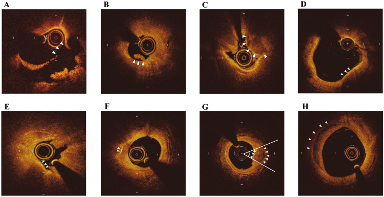
Offline OCT or OFDI analysis of the culprit lesions of ACS was performed using a dedicated image review system (Abbott Vascular) or LUNAWAVE Offline Viewer (Terumo). The analysis range was defined as the segments centered on the culprit lesion and extending bilaterally to 5 mm of a normal vessel segment.13 Plaque rupture was defined as the presence of fibrous cap discontinuity with a clear cavity formed inside the plaque.7 Plaque erosion was defined as the presence of an attached thrombus overlying an intact and visualized plaque, luminal surface irregularity at the culprit lesion in the absence of a thrombus, or attenuation of underlying plaque by a thrombus without superficial lipid or calcification immediately proximal or distal to the site of the thrombus.14,15 Calcified nodule was defined as fibrous cap disruption detected over a calcified plaque characterized by protruding calcification, superficial calcium, or the presence of substantial calcium proximal and/or distal to the lesion (Figure 1A–C).14 In addition, a standard OCT assessment was performed according to previously defined criteria (Figure 1D–H).15,17–21
Figure 1.
Representative optical coherence tomography images showing (A) plaque rupture, (B) plaque erosion, (C) calcified nodule, (D) thin cap fibroatheroma, (E) macrophage accumulation, (F) cholesterol crystal, (G) spotty calcification, and (H) healed plaque.
Culprit plaques of ACS were categorized using previously established criteria by OCT.7,14,16 A lipid plaque was defined as a plaque with a low signal region and a diffuse border with a lipid arc >90°.14 Calcification was defined as a signal-poor or heterogeneous region with sharply delineated borders.14 The minimum thickness of the fibrous cap, the maximum lipid arc, and lipid length were measured for each lipid plaque. The minimum thickness of the fibrous cap was defined as the thickness of the signal-rich layer overlying the lipid-rich plaque.15,17 The lipid index was calculated by multiplying lipid length by mean lipid arc. A thrombus was defined as a mass protruding into the vessel lumen discontinuously from the surface of the vessel wall.7 Thin-cap fibroatheroma was defined as a plaque with a lipid arc >90° and a minimum thickness of the fibrous cap <65 µm (Figure 1D).15,18 Macrophage accumulation was defined as areas of bright spots with high OCT backscattering signal variances (Figure 1E).17 Cholesterol crystals were defined as thin and linear structures with high backscattering without attenuation within the plaque (Figure 1F).17,19 Spotty calcification was defined as a calcification arc <90° and a calcification length <4 mm (Figure 1G).20 Healed plaques were defined as plaques with ≥1 layers with different optical densities and a clear demarcation from underlying components (Figure 1H).21
Measurement of Cholesterol Variability
Visit-to-visit variability in cholesterol was defined as the variability in cholesterol levels between outpatient visits. Measurement parameters of cholesterol were LDL-C, total cholesterol (TC), high-density lipoprotein cholesterol (HDL-C), triglyceride (TG), and non-HDL-C levels, and these parameters were measured after patients had fasted. The Friedewald equation was used to determine LDL-C levels. If the number of cholesterol measurements exceeded 7, the last 6 measurements were used in the calculations. Various measures of variability were used, namely the standard deviation (SD) of cholesterol levels, the coefficient of variation (CV), and the variability independent of the mean (VIM). The VIM was calculated as 100×SD/meanβ, where β is the regression coefficient based on the natural logarithm of the SD of the natural logarithm of the mean. In addition, this uncorrected VIM was corrected (cVIM) by using the following formula:22
cVIM = (uncorrected VIM × mean of CV) / (mean of uncorrected VIM)
cVIM is a transformation of SD that is designed to be uncorrelated with the mean follow-up cholesterol level. cVIM was chosen as the primary measurement.
Statistical Analysis
Continuous variables are presented as median values with the interquartile range. Variables were analyzed using the Mann-Whitney U test. The Kruskal-Wallis analysis of variance test was used to compare variables among 3 groups. Post hoc analysis was performed using the Mann-Whitney U test and Bonferroni correction. Categorical variables are presented as frequencies, and were compared using Chi-squared or Fisher’s exact tests. To identify parameters associated with ACS, univariate and multivariate analyses were performed between the ACS and control groups. In addition, to identify parameters associated with plaque rupture, univariate and multivariate analyses were performed between the PR-ACS group and a non-plaque rupture (NPR) group, defined as a combination of the NPR-ACS and control groups. Baseline variables with P<0.20 in the univariate regression analysis, age and male sex, were included in the multivariate logistic regression models.
In all analyses, 2-sided P<0.05 was considered significant. All confidence intervals (CIs) reported are 95% unless stated otherwise. Computations were performed using SPSS version 25.0 (IBM Corp., Armonk, NY, USA).
Results
Patient Population
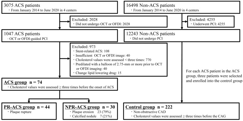
Of 3,075 patients with ACS, 1,047 underwent OCT-guided PCI in 4 centers between January 2014 and June 2020. Of these, 108 patients with stent-related ACS, 40 patients whose OCT image quality was insufficient, 770 patients whose cholesterol values were assessed <3 times before the onset of ACS, 40 patients whose lesions were predilated with a balloon of ≥2.75 mm prior to OCT imaging, and 15 patients whose medication affecting cholesterol levels was changed from the first to last cholesterol measurement were excluded. This left 74 patients with ACS who met the inclusion and exclusion criteria and were enrolled in the ACS group. Of these 74 patients, 44 were classified as PR-ACS by OCT and 30 were classified as NPR-ACS. Of the 12,243 non-ACS patients who underwent CAG during the same period, 222 non-ACS patients were assigned to the control group based on the inclusion and exclusion criteria. A detailed patient flowchart is shown in Figure 2.
Figure 2.
Study flowchart. ACS, acute coronary syndrome; CAD, coronary artery disease; NPR-ACS, non-plaque rupture acute coronary syndrome; OCT, optical coherence tomography; OFDI, optical frequency domain imaging; PCI, percutaneous coronary intervention; PR-ACS, plaque rupture acute coronary syndrome.
There were no significant differences in medical history between the PR-ACS, NPR-ACS, and control groups. Creatine kinase (CK), CK-MB, and C-reactive protein (CRP) concentrations on admission were significantly higher in the ACS than control group. CK, CK-MB, and CRP concentrations on admission were significantly higher in the PR-ACS and NPR-ACS groups than in the control group (Table 1).
Table 1.
Baseline Patient and Lesion Characteristics
| ACS (n=74) |
PR-ACS (n=44) |
NPR-ACS (n=30) |
Control (n=222) |
P value | ||
|---|---|---|---|---|---|---|
| ACS vs. control |
PR-ACS vs. NPR-ACS vs. control |
|||||
| Age (years) | 73 [65–80] | 75 [65–81] | 72 [60–80] | 75 [68–79] | 0.61 | 0.96 |
| Male sex | 55 (74.3) | 35 (79.5) | 20 (66.7) | 158 (71.2) | 0.66 | 0.42 |
| BMI (kg/m2) | 22.7 [21.0–25.4] | 22.7 [20.7–25.2] | 22.7 [21.1–26.0] | 23.3 [20.5–25.6] | 0.91 | 0.98 |
| Smoker | 43 (58.1) | 24 (54.5) | 19 (63.3) | 118 (53.2) | 0.50 | 0.58 |
| Medical history | ||||||
| Hypertension | 57 (77.0) | 33 (75) | 24 (80) | 183 (82.4) | 0.31 | 0.51 |
| Hyperlipidemia | 55 (74.3) | 33 (75) | 22 (73.3) | 163 (73.4) | 0.88 | 0.98 |
| Diabetes | 44 (59.5) | 22 (50) | 22 (73.3) | 119 (53.6) | 0.38 | 0.096 |
| Dialysis | 7 (9.5) | 4 (9.1) | 3 (10) | 17 (7.7) | 0.62 | 0.88 |
| History of PCI | 16 (21.6) | 10 (22.7) | 6 (20) | 71 (32.0) | 0.09 | 0.23 |
| History of PAD | 10 (13.5) | 4 (9.1) | 6 (20) | 27 (12.2) | 0.76 | 0.36 |
| History of HF | 8 (10.8) | 7 (15.9) | 1 (3.3) | 43 (19.4) | 0.091 | 0.089 |
| History of stroke | 1 (1.4) | 1 (2.3) | 0 (0) | 8 (3.6) | 0.46 | 0.53 |
| Family history of cardiovascular event | 4 (5.4) | 3 (6.8) | 1 (3.3) | 14 (6.3) | 1.00 | 0.80 |
| Medication on admission | ||||||
| Aspirin | 23 (31.1) | 16 (36.4) | 7 (23.3) | 95 (42.8) | 0.075 | 0.11 |
| Statin | 39 (52.7) | 23 (52.3) | 16 (53.3) | 136 (61.3) | 0.20 | 0.43 |
| EPA | 8 (10.8) | 4 (9.1) | 4 (13.3) | 19 (8.6) | 0.56 | 0.70 |
| Ezetimibe | 7 (9.5) | 2 (4.5) | 5 (16.7) | 13 (5.9) | 0.29 | 0.070 |
| Laboratory data | ||||||
| Hemoglobin level (g/dL) | 12.9 [12–14.7] | 12.8 [12.0–14.7] | 13.3 [12.0–14.8] | 13.0 [11.7–14.2] | 0.34 | 0.58 |
| Creatinine level (mg/dL) | 0.87 [0.69–1.18] | 0.88 [0.67–1.32] | 0.87 [0.69–1.05] | 0.95 [0.75–1.2] | 0.26 | 0.51 |
| eGFR (mL/min/1.73 m2) | 52.2 [36.1–73.0] | 50.0 [29.8–73.9] | 60.6 [39.1–70.7] | 55.8 [43.2–69.4] | 0.56 | 0.62 |
| BNP (pg/mL) | 92.9 [40.6–405.4] | 146.0 [46.3–533.9] | 65.1 [32.5–159.6] | 79.0 [34.5–216.5] | 0.097 | 0.086 |
| HbA1c (%) | 6.6 [5.9–7.5] | 6.3 [5.9–7.3] | 7.2 [5.9–7.8] | 6.3 [5.8–7.0] | 0.087 | 0.054 |
| CK (IU/L) | 150 [82–345] | 179 [74–380]* | 115 [84–257]* | 89 [61–121] | <0.001 | <0.001 |
| CK-MB (IU/L) | 11.4 [5.3–25] | 13.8 [6–24.7]* | 8 [4–25.6]* | 4 [4–6] | <0.001 | <0.001 |
| CRP (mg/dL) | 0.35 [0.13–0.86] | 0.45 [0.14–1.26]* | 0.27 [0.07–0.69]* | 0.09 [0.04–0.28] | <0.001 | <0.001 |
| LVEF (%) | 59 [50–63] | 54 [46–63] | 60 [55–66] | 60 [50–65] | 0.42 | 0.23 |
| STEMI | 43 (58.1) | 26 (59.1) | 17 (56.7) | 0.84† | ||
| Culprit lesion | 0.53† | |||||
| RCA | 22 (29.7) | 11 (25.0) | 11 (36.7) | |||
| LAD | 34 (45.9) | 21 (47.7) | 13 (43.3) | |||
| LCx | 18 (24.3) | 12 (27.3) | 6 (20.0) | |||
Values are presented as the median [interquartile range] or n (%). *P<0.05 compared with control; †P value for plaque rupture acute coronary syndrome (PR-ACS) vs. non-plaque rupture acute coronary syndrome (NPR-ACS). ACS, acute coronary syndrome; BMI, body mass index; BNP, B-type natriuretic peptide; CK, creatine kinase; CRP, C-reactive protein; eGFR, estimated glomerular filtration rate; EPA, eicosapentaenoic acid; HF, heart failure; LAD, left anterior descending coronary artery; LCx, left circumflex coronary artery; LVEF, left ventricular ejection fraction; NPR, non-plaque rupture; PAD, peripheral artery disease; PCI, percutaneous coronary intervention; PR, plaque rupture; RCA, right coronary artery; STEMI, ST-elevation myocardial infarction.
The images acquired were analyzed by 2 experienced observers (S.N., Y.T.). Investigation of intraobserver variability of the underlying plaque morphology revealed an almost perfect agreement (κ=0.95). Moreover, there was an almost perfect agreement in interobserver variability (κ=0.85). There was no significant difference in the target vessels between the PR-ACS and NPR-ACS groups. Plaque rupture was observed in 59% (44/74) of patients in the ACS group, compared with 41% (30/74) of patients in the NPR group. In the NPR-ACS group, plaque erosion was observed in the culprit lesions of 77% (23/30) of patients, and calcified nodule was observed in 23% (7/30) of patients. The frequency of cholesterol crystals was significantly higher in the PR-ACS than NPR-ACS group (Table 2).
Table 2.
OCT Findings of ACS Culprit Lesions
| Plaque rupture (n=44) |
Non-plaque rupture (n=30) |
P value | |
|---|---|---|---|
| Quantitative findings | |||
| Lesion length (mm) | 27 [18–33] | 24 [18–30] | 0.22 |
| Minimal lumen area (mm2) | 1.13 [0.98–1.52] | 1.25 [0.99–1.8] | 0.50 |
| Lipid length (mm) | 24 [16–31.6] | 24.3 [15.75–30.75] | 0.97 |
| Mean lipid arc (°) | 175.4 [152–186.4] | 168.4 [82.4–180.7] | 0.12 |
| Lipid index (mm×°) | 3,646 [2,411–5,265] | 3,069 [1,590–4,772] | 0.12 |
| Minimum fibrous cap thickness (μm) | 75 [60–90] | 90 [60–120] | 0.19 |
| Qualitative findings | |||
| Plaque erosion | 23 (76.7) | ||
| Calcified nodule | 7 (23.3) | ||
| TCFA | 16 (36.4) | 11 (36.7) | 0.98 |
| Cholesterol crystals | 26 (59.1) | 9 (30.0) | 0.014 |
| Macrophage | 31 (70.5) | 16 (53.3) | 0.13 |
| Calcification | 31 (70.5) | 23 (76.7) | 0.56 |
| Spotty calcification | 12 (27.3) | 4 (13.3) | 0.25 |
| Thrombus | 37 (84.1) | 21 (70.0) | 0.15 |
| Healed plaque | 18 (40.9) | 11 (36.7) | 0.71 |
Values are presented as the median [interquartile range] or n (%). ACS, acute coronary syndrome; OCT, optical coherence tomography; TCFA, thin cap fibroatheroma.
Cholesterol Measurement
The median duration between the first and last cholesterol measurements was not significantly different between the PR-ACS, NPR-ACS, and control groups.
Both the mean and the sum of variability indices of TC, LDL-C, and non-HDL-C were significantly higher in the ACS than control group. The mean and variability indices of HDL-C and TG were not significantly different between the 2 groups (Table 3).
Table 3.
Comparisons of Mean Values and Variability in Lipoproteins
| ACS (n=74) |
PR-ACS (n=44) |
NPR-ACS (n=30) |
Control (n=222) |
P value | ||
|---|---|---|---|---|---|---|
| ACS vs. control |
PR-ACS vs. NPR-ACS vs. control |
|||||
|
No. blood sampling sessions |
5 (3–6) | 4 (3–6) | 5 (4–6) | 5 (4–6) | 0.38 | 0.28 |
|
Duration between initial and final blood sampling (days) |
475 (258–812) | 511 (259–1,078) | 433 (240–686) | 550 (336–696) | 0.28 | 0.40 |
| TC | ||||||
| Mean (mg/dL) | 191 (155–215) | 191 (154–211)* | 193 (160–220)* | 169 (148–192) | 0.001 | 0.002 |
| SD (mg/dL) | 14.6 (9.54–21.11) | 17.4 (9.98–21.25)* | 13.2 (8.34–21.06) | 11.2 (7.48–16.26) | <0.001 | 0.001 |
| CV (%) | 8.61 (5.34–11.05) | 9.57 (6.25–12.94)* | 8.12 (4.8–9.85) | 6.92 (4.41–9.24) | 0.005 | 0.008 |
| cVIM (%) | 8.41 (5.6–12.11) | 9.82 (5.9–12.67)* | 7.53 (4.84–10.54) | 6.66 (4.43–9.34) | 0.001 | 0.001 |
| LDL-C | ||||||
| Mean (mg/dL) | 114 (85–136) | 116 (84–138)* | 111 (85–129)* | 90 (75–107) | <0.001 | <0.001 |
| SD (mg/dL) | 11.4 (6.6–18.4) | 14.1 (9.67–19.09)*,† | 8.5 (4.79–13.13) | 8.0 (5.35–10.86) | <0.001 | <0.001 |
| CV (%) | 10.5 (6.56–15.77) | 13.7 (10–17.48)*,† | 9.2 (5.85–10.5) | 8.8 (5.95–12.63) | 0.018 | 0.003 |
| cVIM (%) | 11.9 (7.46–18.04) | 14.0 (10.05–18.7)*,† | 9.2 (5.01–13.39) | 8.5 (5.89–11.43) | <0.001 | <0.001 |
| HDL-C | ||||||
| Mean (mg/dL) | 50 (42–59) | 47 (38–58) | 50 (45–61) | 52 (43–63) | 0.11 | 0.13 |
| SD (mg/dL | 4.45 (2.5–5.97) | 4.62 (2.49–6.77) | 4.19 (2.98–5.93) | 4.26 (2.8–6.91) | 0.77 | 0.91 |
| CV (%) | 8.95 (5.52–11.8) | 8.54 (5.39–13.28) | 9.04 (6–10.78) | 8.16 (5.25–12.23) | 0.79 | 0.96 |
| cVIM (%) | 13.8 (7.75–17.13) | 13.3 (7.55–20.1) | 14.0 (9.58–16.53) | 12.3 (8.13–19.75) | 0.91 | 0.90 |
| Non-HDL-C | ||||||
| Mean (mg/dL) | 136 (106–163) | 137 (104–162)* | 132 (116–167)* | 115 (95–134) | <0.001 | <0.001 |
| SD (mg/dL) | 14.5 (8.27–19.63) | 15.9 (9.44–21.33)* | 11.8 (6.87–18.93) | 9.86 (6.73–13.47) | <0.001 | <0.001 |
| CV (%) | 11.5 (6.86–14.69) | 12.5 (7.71–15.79)* | 9.09 (6.55–13.73) | 8.59 (5.75–11.67) | 0.015 | 0.004 |
| cVIM (%) | 5.52 (3.38–7.43) | 6.24 (3.52–7.93)* | 4.31 (3.1–6.6) | 4.04 (2.77–5.46) | 0.001 | <0.001 |
| Triglycerides | ||||||
| Mean (mg/dL) | 132 (96–181) | 130 (96–184) | 133 (97–173) | 118 (87–163) | 0.094 | 0.24 |
| SD (mg/dL) | 29.1 (17.36–54.9) | 30.1 (15.25–50.29) | 28.1 (17.98–56.17) | 27.3 (15.88–41.1) | 0.24 | 0.36 |
| CV (%) | 21.91 (16.08–31.89) | 21.88 (14.8–31.88) | 23.02 (17.65–34.62) | 21.78 (16.49–30.02) | 0.84 | 0.60 |
| cVIM (%) | 7.91 (5.49–11.6) | 7.57 (4.57–11.37) | 8.01 (5.68–12.14) | 7.42 (4.97–10.45) | 0.42 | 0.45 |
Values are presented as the median (interquartile range). *P<0.05 compared with control; †P<0.05 compared with non-plaque rupture acute coronary syndrome (NPR-ACS). ACS, acute coronary syndrome; CV, coefficient of variation; cVIM, corrected variability independent of the mean; HDL-C, high density lipoprotein cholesterol; LDL-C, low density lipoprotein cholesterol; PR-ACS, plaque rupture acute coronary syndrome; SD, standard deviation; TC, total cholesterol.
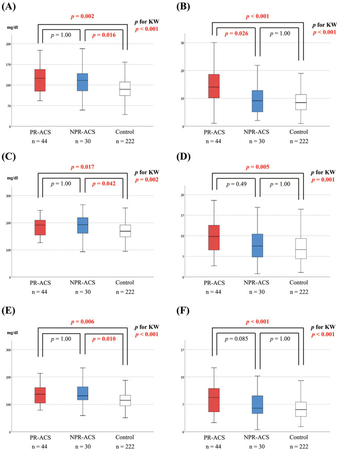
Both the mean and the sum of variability indices of LDL-C were significantly different between the PR-ACS, NPR-ACS, and control groups. Post hoc analysis showed that the PR-ACS and NPR-ACS groups had significantly higher mean LDL-C levels than the control group. The PR-ACS group had a mean LDL-C level comparable to that in the NPR-ACS group (116 [84–138] vs. 111 [85–129] mg/dL, respectively; P=1.00; Figure 3A; Table 3); however, the PR-ACS group had a significantly higher cVIM of LDL-C than the NPR-ACS group (14.0 [10.05–18.7] vs. 9.2 [5.01–13.39], respectively; P=0.026; Figure 3B; Table 3). The mean and cVIM of TC and non-HDL-C were significantly different between the PR-ACS, NPR-ACS, and control groups. Post hoc analysis showed that the PR-ACS group had significantly higher mean and cVIM values of TC and non-HDL-C than the control group (Figure 3C–F; Table 3). There were no significant differences in mean and cVIM values of TC and non-HDL-C between the PR-ACS and NPR-ACS groups (Figure 3C–F; Table 3).
Figure 3.
Comparisons of the (A,C,E) mean and (B,D,F) corrected variability independent of the mean (cVIM) of low-density lipoprotein cholesterol (LDL-C; A,B), total cholesterol (TC; C,D), and non-high-density lipoprotein cholesterol (non-HDL-C; E,F). The boxes show the interquartile range, with the median value indicated by the horizontal line; whiskers show the range. KW, Kruskal-Wallis; NPR-ACS, non-plaque rupture acute coronary syndrome; PR-ACS, plaque rupture acute coronary syndrome.
Predictors of ACS and Plaque Rupture
Of 13 parameters evaluated, higher CRP level (odds ratio [OR] 2.85, 95% CI 1.70–4.76, P<0.001), lower mean HDL-C level (OR 0.97, 95% CI 0.95–1.00, P=0.020), higher mean LDL-C level (OR 1.02, 95% CI 1.01–1.03, P<0.001), and higher cVIM of LDL-C (OR 1.04, 95% CI 1.00–1.08, P=0.049) were found to be independent parameters associated with ACS (Table 4).
Table 4.
Univariate and Multivariate Logistic Regression Analyses for ACS
| Univariate analysis | Multivariate analysis | |||||
|---|---|---|---|---|---|---|
| OR | 95% CI | P value | OR | 95% CI | P value | |
| Age | 0.99 | 0.97–1.02 | 0.99 | |||
| Male sex | 0.85 | 0.47–1.55 | 0.60 | |||
| History of PCI | 0.59 | 0.32–1.092 | 0.092 | |||
| History of HF | 0.51 | 0.23–1.13 | 0.096 | |||
| Aspirin use | 0.60 | 0.35–1.026 | 0.062 | |||
| BNP | 1.001 | 1.000–1.001 | 0.023 | |||
| HbA1c | 1.23 | 0.97–1.55 | 0.091 | |||
| CRP | 2.83 | 1.73–4.65 | <0.001 | 2.85 | 1.70–4.76 | <0.001 |
| Mean TC | 1.009 | 1.002–1.016 | 0.009 | |||
| Mean HDL-C | 0.98 | 0.96–1.00 | 0.034 | 0.97 | 0.95–1.00 | 0.020 |
| Mean LDL-C | 1.02 | 1.011–1.29 | <0.001 | 1.02 | 1.01–1.03 | <0.001 |
| cVIM of LDL-C | 1.07 | 1.022–1.12 | 0.004 | 1.04 | 1.00–1.08 | 0.049 |
| Mean non-HDL-C | 1.01 | 1.003–1.017 | 0.005 | |||
CAD, coronary artery disease; CI, confidence interval; OR, odds ratio. Other abbreviations as in Tables 1,3.
Of 12 parameters evaluated, higher CRP level, lower mean HDL-C level, higher mean LDL-C level, and higher cVIM of LDL-C were independently associated with plaque rupture (Table 5).
Table 5.
Univariate and Multivariate Logistic Regression Analyses for ACS Due to Plaque Rupture
| Univariate analysis | Multivariate analysis | |||||
|---|---|---|---|---|---|---|
| OR | 95% CI | P value | OR | 95% CI | P value | |
| Age | 1.01 | 0.98–1.044 | 0.56 | |||
| Male sex | 1.62 | 0.74–3.53 | 0.23 | |||
| BNP | 1.001 | 1.000–1.001 | 0.11 | |||
| CRP | 1.63 | 1.15–2.31 | 0.006 | 1.54 | 1.10–2.15 | 0.011 |
| LVEF | 0.98 | 0.95–1.004 | 0.094 | |||
| Mean TC | 1.006 | 0.99–1.013 | 0.13 | |||
| Mean HDL-C | 0.97 | 0.95–1.00 | 0.019 | 0.97 | 0.94–0.99 | 0.014 |
| cVIM of HDL-C | 1.006 | 0.99–1.015 | 0.15 | |||
| Mean LDL-C | 1.02 | 1.006–1.028 | 0.002 | 1.02 | 1.005–1.029 | 0.005 |
| cVIM of LDL-C | 1.06 | 1.019–1.11 | 0.005 | 1.06 | 1.01–1.10 | 0.018 |
| Mean non-HDL-C | 1.007 | 1.000–1.013 | 0.059 | |||
| cVIM of non-HDL-C | 1.10 | 1.03–1.18 | 0.003 | |||
Abbreviations as in Tables 1,3,4.
Discussion
The main findings of the present study can be summarized as follows: (1) the mean, SD, CV, and cVIM of LDL-C, TC, and non-HDL-C were significantly higher in the ACS than control group; (2) the PR-ACS group had significantly higher SD, CV, and cVIM of LDL-C than the NPR-ACS group, despite no significant difference in the mean LDL-C level between these 2 groups; and (3) visit-to-visit variability in LDL-C was independently associated with the onset of ACS, especially when induced by plaque rupture.
Relationship Between Cholesterol Variability, ACS Onset, and Plaque Rupture
A previous retrospective cohort study reported that visit-to-visit variability in LDL-C was an independent predictor of cardiovascular events, such as any coronary event and MI in patients with stable CAD.1 Regardless of the mean follow-up LDL-C and HDL-C levels, visit-to-visit variability in LDL-C and HDL-C was associated with the occurrence of major adverse cardiac events at >5 years after STEMI occurrence.22 Furthermore, in a recent post hoc patient-level analysis of 9 clinical trials using serial IVUS of 4,976 patients, Clark et al reported that greater visit-to-visit variability in atherogenic lipoprotein levels was significantly associated with the progression of coronary atheroma.4 However, the mechanisms linking visit-to-visit variability in LDL-C and the onset of ACS have yet to be elucidated. Herein, for the first time, we demonstrated that variability in LDL-C was associated with the onset of ACS, especially when induced by plaque rupture.
In an OCT study investigating the culprit lesions of 126 patients with ACS, Jia et al demonstrated that the incidences of plaque rupture, erosion, and calcified nodule were 43.7%, 31%, and 7.9%, respectively,7 which are in line with the incidences observed in the present study (59.5%, 31.1%, and 9.4%, respectively). Considering that approximately half of ACS events are caused by plaque rupture, the results of the present study can provide potential mechanistic insights into the relationship between cholesterol variability and the occurrence of cardiovascular events shown in previous studies. A further prospective study is warranted to investigate whether an additional intervention to reduce cholesterol variability can reduce the incidence of plaque rupture-induced ACS.
Mechanism of Linking Variability in LDL-C and Plaque Rupture
Although mechanisms linking variability in LDL-C and plaque rupture remain unknown, several hypotheses have been reported. First, greater visit-to-visit variability in LDL-C may change the composition of atherosclerotic plaques. A previous experimental study demonstrated an association between cholesterol crystals and plaque rupture.23 By using the coronary arteries of patients who died of ACS, Abela et al showed that all patients with ACS had cholesterol crystals that perforated the intima, but no cholesterol crystals were present in patients without ACS.23 There were strong associations of cholesterol crystals with plaque rupture, thrombus, symptoms, and plaque size. In the present study, the frequency of cholesterol crystals was significantly higher in the PR-ACS than NPR-ACS group. Interestingly, Abela et al also demonstrated that the structure of cholesterol crystals could be altered from pointed tip to blunt and dissolved by lipid-lowering therapy with a statin, and that the density of cholesterol crystals was markedly decreased and the crystals appeared dissolved in human plaques incubated with statins compared with those incubated with a saline solution.24 Based on such findings, we currently speculate that greater visit-to-visit variability in LDL-C may induce a repeated process of crystallization and dissolution of the cholesterol within the confined space of coronary plaques, resulting in melted globs and piles of fragmented crystals. These fragmented crystals may cut through fibrous membranes and increase the risk of plaque rupture.3,24
Second, variability in cholesterol may be linked to endothelial dysfunction. Smit et al reported that higher variability in LDL-C was associated with lower cognitive performance, lower cerebral blood flow, and greater white matter hyperintensity load, which have been linked to endothelial dysfunction.2 Endothelial dysfunction involves many atheroprotective mechanisms and progresses to vulnerable plaque, potentially leading to plaque rupture.25
Third, variability of cholesterol may be linked to inflammatory stress. Previously, Chen et al reported that inflammatory stress can disrupt 3-hydroxy-3-methylglutaryl coenzyme A reductase-mediated cholesterol synthesis, resulting in intracellular lipid accumulation and statin resistance.26 For this reason, it is possible that inflammatory stress affects the variability of cholesterol and leads to ACS with plaque rupture. Therefore, tailoring patients’ lifestyle and controlling other risk factors in patients with dyslipidemia may improve variability in LDL-C, and consequently reduce the incidence of ACS with plaque rupture.
Clinical Implication
The present study demonstrated that variability in LDL-C was associated with the onset of ACS, especially when induced by plaque rupture. The relationship between cholesterol variability and ACS onset has been reported in many studies; however, the mechanism is still not well understood. The distinct pathologic features and clinical characteristics associated with plaque rupture, erosion, and calcified nodule suggest that they may be caused by different pathophysiologic processes and need to be considered separately with regard to potential contributing factors and their prevention. In particular, because plaque rupture is the most common pathogenetic mechanism of culprit lesions of ACS, it is important to investigate the potential factors associated with the development of plaque rupture.
In a recent OCT study that enrolled 318 patients with ACS, Yonetsu et al demonstrated that culprit lesions of ACS with plaque rupture were associated with poor long-term outcomes compared with lesions with an intact fibrous cap, such as erosion.27 Thus, the findings of the present study show that cholesterol variability may be a new therapeutic target for preventing high-risk ACS with poor prognosis.
We could not find factors associated with the variability in LDL-C levels due to the retrospective design of the study; nevertheless, inflammatory stress, systemic conditions, and statin adherence may potentially contribute to higher LDL-C variability. Chen et al reported that inflammatory stress increased intracellular lipid accumulation and statin resistance.26 This may also affect the visit-to-visit variability in cholesterol levels. Furthermore, generalized frailty in patients with systemic conditions is associated with variability in several biological parameters, and therefore may be associated with LDL-C.1 Finally, the visit-to-visit variability of LDL-C levels is reportedly associated with statin prescription compliance.28 Intervening in these factors may reduce the variability in LDL-C levels. Further studies are warranted to evaluate whether controlling LDL-C variability can be a new preventive measure for ACS due to plaque rupture.
Study Limitations
This study has several limitations. First, the number of patients with ACS was small and the analysis was retrospective; however, the patients enrolled in the ACS group were valuable, because all of them underwent OCT or OFDI for their culprit plaque at the time of primary PCI and had ≥3 serial cholesterol measurements before the onset of ACS. Second, there is a possibility for selection bias, because the present study included patients who had multiple lipid measurements before the onset of ACS. According to the recent results of the Japanese ACS registry, 27.3% of patients with ACS received statins before the onset of ACS,29 whereas the frequency of statin pretreatment in the present study was 52.7%. Thus, the proportion of patients who received statin treatment in our study population is higher than that in the general population of ACS patients in Japan, which is probably because we included patients who attended hospital regularly and who underwent multiple blood sampling for lipid assessment. Further studies are warranted to confirm the generalizability of our results to the ACS population with a lower proportion of patients on statin pretreatment. Third, because of the retrospective design of this study, the intervals between cholesterol measurement were not consistent. However, the mean interval did not differ significantly among groups. Fourth, OCT or OFDI was not performed in the control group. However, OCT or OFDI was used to classify the culprit lesion of ACS; it is unlikely that OCT or OFDI in the control group had any effect on the present results. Hence, we believe that our findings will provide important insights into the potential relationship between intraindividual variability of lipids and the mechanism of ACS onset. Fifth, the resolution and depth of OCT imaging are not sufficient to perfectly detect plaque morphology. When red thrombi are present, OCT imaging may miss minor ruptured plaques behind the thrombi. Finally, it is possible that iatrogenic luminal injuries caused by intracoronary thrombectomies or predilation were incorrectly identified as plaque ruptures.
Conclusions
Greater visit-to-visit variability in LDL-C may be associated with the onset of ACS events with plaque rupture independent of LDL-C levels. Further large-scale studies are required to confirm this finding and to gain a full understanding of the mechanism linking variability in LDL-C with plaque rupture.
Sources of Funding
This study did not receive any specific funding.
Disclosures
None declared.
IRB Information
This study was approved by the Ethics Committee of Kobe University Hospital (B200305).
Acknowledgments
None.
Data Availability
The deidentified participant data will not be shared.
References
- 1. Bangalore S, Breazna A, DeMicco DA, Wun CC, Messerli FH, Committee TNTS, et al.. Visit-to-visit low-density lipoprotein cholesterol variability and risk of cardiovascular outcomes: Insights from the TNT trial. J Am Coll Cardiol 2015; 65: 1539–1548. [DOI] [PubMed] [Google Scholar]
- 2. Smit RA, Trompet S, Sabayan B, le Cessie S, van der Grond J, van Buchem MA, et al.. Higher visit-to-visit low-density lipoprotein cholesterol variability is associated with lower cognitive performance, lower cerebral blood flow, and greater white matter hyperintensity load in older subjects. Circulation 2016; 134: 212–221. [DOI] [PubMed] [Google Scholar]
- 3. Kim MK, Han K, Kim HS, Park YM, Kwon HS, Yoon KH, et al.. Cholesterol variability and the risk of mortality, myocardial infarction, and stroke: A nationwide population-based study. Eur Heart J 2017; 38: 3560–3566. [DOI] [PMC free article] [PubMed] [Google Scholar]
- 4. Clark D 3rd, Nicholls SJ, St John J, Elshazly MB, Kapadia SR, Tuzcu EM, et al.. Visit-to-visit cholesterol variability correlates with coronary atheroma progression and clinical outcomes. Eur Heart J 2018; 39: 2551–2558. [DOI] [PubMed] [Google Scholar]
- 5. Farb A, Tang AL, Burke AP, Sessums L, Liang Y, Virmani R.. Sudden coronary death: Frequency of active coronary lesions, inactive coronary lesions, and myocardial infarction. Circulation 1995; 92: 1701–1709. [DOI] [PubMed] [Google Scholar]
- 6. Burke AP, Taylor A, Farb A, Malcom GT, Virmani R.. Coronary calcification: Insights from sudden coronary death victims. Z Kardiol 2000; 89: S049–S053. [DOI] [PubMed] [Google Scholar]
- 7. Jia H, Abtahian F, Aguirre AD, Lee S, Chia S, Lowe H, et al.. In vivo diagnosis of plaque erosion and calcified nodule in patients with acute coronary syndrome by intravascular optical coherence tomography. J Am Coll Cardiol 2013; 62: 1748–1758. [DOI] [PMC free article] [PubMed] [Google Scholar]
- 8. Roffi M, Patrono C, Collet JP, Mueller C, Valgimigli M, Andreotti F, et al.. 2015 ESC guidelines for the management of acute coronary syndromes in patients presenting without persistent ST-segment elevation: Task Force for the Management of Acute Coronary Syndromes in Patients Presenting without Persistent ST-Segment Elevation of the European Society of Cardiology (ESC). Eur Heart J 2016; 37: 267–315. [DOI] [PubMed] [Google Scholar]
- 9. Thygesen K, Alpert JS, Jaffe AS, Simoons ML, Chaitman BR, White HD, et al.. Third universal definition of myocardial infarction. J Am Coll Cardiol 2012; 60: 1581–1598. [DOI] [PubMed] [Google Scholar]
- 10. Ibanez B, James S, Agewall S, Antunes MJ, Bucciarelli-Ducci C, Bueno H, et al.. 2017 ESC guidelines for the management of acute myocardial infarction in patients presenting with ST-segment elevation: The Task Force for the Management of Acute Myocardial Infarction in Patients Presenting with ST-Segment Elevation of the European Society of Cardiology (ESC). Eur Heart J 2018; 39: 119–177. [DOI] [PubMed] [Google Scholar]
- 11. Kubo T, Imanishi T, Takarada S, Kuroi A, Ueno S, Yamano T, et al.. Assessment of culprit lesion morphology in acute myocardial infarction: Ability of optical coherence tomography compared with intravascular ultrasound and coronary angioscopy. J Am Coll Cardiol 2007; 50: 933–939. [DOI] [PubMed] [Google Scholar]
- 12. Gu J, Yin ZF, Pan JA, Zhang JF, Wang CQ.. Visit-to-visit variability in low-density lipoprotein cholesterol is associated with adverse events in non-obstructive coronary artery disease. Anatol J Cardiol 2019; 22: 117–124. [DOI] [PMC free article] [PubMed] [Google Scholar]
- 13. Sheng Z, Zhou P, Liu C, Li J, Chen R, Zhou J, et al.. Relationships of coronary culprit-plaque characteristics with duration of diabetes mellitus in acute myocardial infarction: An intravascular optical coherence tomography study. Cardiovasc Diabetol 2019; 18: 136. [DOI] [PMC free article] [PubMed] [Google Scholar]
- 14. Higuma T, Soeda T, Abe N, Yamada M, Yokoyama H, Shibutani S, et al.. A combined optical coherence tomography and intravascular ultrasound study on plaque rupture, plaque erosion, and calcified nodule in patients with ST-segment elevation myocardial infarction: Incidence, morphologic characteristics, and outcomes after percutaneous coronary intervention. JACC Cardiovasc Interv 2015; 8: 1166–1176. [DOI] [PubMed] [Google Scholar]
- 15. Yamamoto E, Yonetsu T, Kakuta T, Soeda T, Saito Y, Yan BP, et al.. Clinical and laboratory predictors for plaque erosion in patients with acute coronary syndromes. J Am Heart Assoc 2019; 8: e012322. [DOI] [PMC free article] [PubMed] [Google Scholar]
- 16. Virmani R, Kolodgie FD, Burke AP, Farb A, Schwartz SM.. Lessons from sudden coronary death: A comprehensive morphological classification scheme for atherosclerotic lesions. Arterioscler Thromb Vasc Biol 2000; 20: 1262–1275. [DOI] [PubMed] [Google Scholar]
- 17. Tearney GJ, Regar E, Akasaka T, Adriaenssens T, Barlis P, Bezerra HG, et al.. Consensus standards for acquisition, measurement, and reporting of intravascular optical coherence tomography studies: A report from the International Working Group for Intravascular Optical Coherence Tomography Standardization and Validation. J Am Coll Cardiol 2012; 59: 1058–1072. [DOI] [PubMed] [Google Scholar]
- 18. Soeda T, Uemura S, Saito Y, Mizuno K, Jang IK.. Optical coherence tomography and coronary plaque characterization. J Jpn Coronary Assoc 2013; 19: 307–314. [Google Scholar]
- 19. Nishimura S, Ehara S, Hasegawa T, Matsumoto K, Yoshikawa J, Shimada K.. Cholesterol crystal as a new feature of coronary vulnerable plaques: An optical coherence tomography study. J Cardiol 2017; 69: 253–259. [DOI] [PubMed] [Google Scholar]
- 20. Afolabi A, Mustafina I, Zhao L, Li L, Sun R, Hu S, et al.. Does spotty calcification attenuate the response of nonculprit plaque to statin therapy?: A serial optical coherence tomography study. Catheter Cardiovasc Interv 2018; 91: 582–590. [DOI] [PubMed] [Google Scholar]
- 21. Fracassi F, Crea F, Sugiyama T, Yamamoto E, Uemura S, Vergallo R, et al.. Healed culprit plaques in patients with acute coronary syndromes. J Am Coll Cardiol 2019; 73: 2253–2263. [DOI] [PubMed] [Google Scholar]
- 22. Boey E, Gay GM, Poh KK, Yeo TC, Tan HC, Lee CH.. Visit-to-visit variability in LDL- and HDL-cholesterol is associated with adverse events after ST-segment elevation myocardial infarction: A 5-year follow-up study. Atherosclerosis 2016; 244: 86–92. [DOI] [PubMed] [Google Scholar]
- 23. Abela GS, Aziz K, Vedre A, Pathak DR, Talbott JD, Dejong J.. Effect of cholesterol crystals on plaques and intima in arteries of patients with acute coronary and cerebrovascular syndromes. Am J Cardiol 2009; 103: 959–968. [DOI] [PubMed] [Google Scholar]
- 24. Abela GS, Vedre A, Janoudi A, Huang R, Durga S, Tamhane U.. Effect of statins on cholesterol crystallization and atherosclerotic plaque stabilization. Am J Cardiol 2011; 107: 1710–1717. [DOI] [PubMed] [Google Scholar]
- 25. Teng N, Maghzal GJ, Talib J, Rashid I, Lau AK, Stocker R.. The roles of myeloperoxidase in coronary artery disease and its potential implication in plaque rupture. Redox Rep 2017; 22: 51–73. [DOI] [PMC free article] [PubMed] [Google Scholar]
- 26. Chen Y, Ku H, Zhao L, Wheeler DC, Li LC, Li Q, et al.. Inflammatory stress induces statin resistance by disrupting 3-hydroxy-3-methylglutaryl-CoA reductase feedback regulation. Arterioscler Thromb Vasc Biol 2014; 34: 365–376. [DOI] [PubMed] [Google Scholar]
- 27. Yonetsu T, Lee T, Murai T, Suzuki M, Matsumura A, Hashimoto Y, et al.. Plaque morphologies and the clinical prognosis of acute coronary syndrome caused by lesions with intact fibrous cap diagnosed by optical coherence tomography. Int J Cardiol 2016; 203: 766–774. [DOI] [PubMed] [Google Scholar]
- 28. Mann DM, Glazer NL, Winter M, Paasche-Orlow MK, Muntner P, Shimbo D, et al.. A pilot study identifying statin nonadherence with visit-to-visit variability of low-density lipoprotein cholesterol. Am J Cardiol 2013; 111: 1437–1442. [DOI] [PMC free article] [PubMed] [Google Scholar]
- 29. Nakamura M, Ako J, Arai H, Hirayama A, Murakami Y, Nohara A, et al.. Investigation into lipid management in acute coronary syndrome patients from the EXPLORE-J Study. J Atheroscler Thromb 2019; 26: 559–572. [DOI] [PMC free article] [PubMed] [Google Scholar]
Associated Data
This section collects any data citations, data availability statements, or supplementary materials included in this article.
Data Availability Statement
The deidentified participant data will not be shared.


